. 1998 Dec 14;143(6):1427-36.
doi: 10.1083/jcb.143.6.1427.

Chromosomal proteins HMG-14 and HMG-17 are released from mitotic chromosomes and imported into the nucleus by active transport

Affiliations

Chromosomal proteins HMG-14 and HMG-17 are released from mitotic chromosomes and imported into the nucleus by active transport

R Hock et al. J Cell Biol. .

Abstract

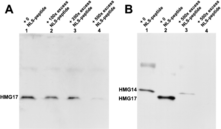
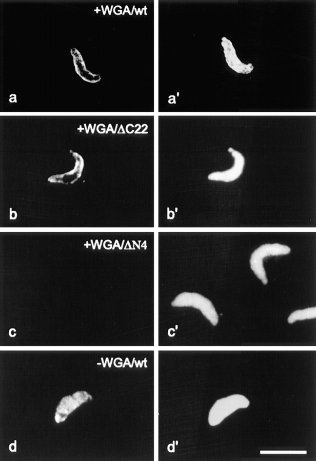
The high mobility group 14/17 (HMG-14/-17) proteins form specific complexes with nucleosome core particles and produce distinct footprints on nucleosomal DNA. Therefore, they could be an integral part of the chromatin fiber. Here we show that during the cell cycle these proteins are transiently dissociated from chromatin. They colocalize with the nuclear DNA in interphase and prophase but not in metaphase and anaphase. They relocate into the nucleus and colocalize again with the DNA in late telophase, concomitantly with the appearance of the nuclear envelope. Thus, these nucleosomal binding proteins are not always associated with chromatin. Using reconstituted nuclei and permeabilized cells, we demonstrate that these two small proteins, with a molecular mass <10 kD, are actively imported into the nucleus. We identify the major elements involved in the nuclear import of these chromosomal proteins: HMG-14/-17 proteins contain an intrinsic bipartite nuclear localization signal, and their entry into the nucleus through nuclear pores requires energy and the participation of importin alpha. These findings suggest that the cell cycle-related association of HMG-14/-17 with chromatin is dependent on, and perhaps regulated by, nuclear import processes.


Figures

Figure 1
Figure 1
HMG proteins are absent from mitotic chromosomes. (A) Immunofluorescence analysis of the distribution of HMG-17 in cultured Hep2 cells with affinity pure antibodies to HMG-17 is shown in the upper panels. The lower panels show staining for DNA with Hoechst. Cell cycle stages are indicated below the panels. Staining for HMG proteins shows a punctuate staining in interphase. In metaphase or anaphase (arrow, early; arrowhead, late) the protein is present in the cytoplasm and not associated with chromatin. Nuclear staining reappears in the late telophase cells. (B) Control with antibodies to histone H1 showing antibody binding in metaphase (a) and corresponding Hoechst (a′). Arrows point to a cell in metaphase. (C) Confocal microscopy depicting immunofluorescence in human Hep2 cells stained with antibodies to HMG-17.
Figure 2
Figure 2
Nuclear staining of HMG-17 protein correlates with the presence of the nuclear envelope. Human Hep2 cells were double labeled with affinity pure anti–HMG-17 (a and b) and with the monoclonal antibody PI1, specific for nucleoporin p62 (a′ and b′), and counterstained for DNA with Hoechst (a′′ and b′′). Bound anti–HMG-17 and PI1 were detected with an Tx-red anti– rabbit and a FITC-coupled anti–mouse, respectively. The arrows in a, a′, and a′′ point to a cell in mitosis. The arrows in b, b′, and b′′ point to cells in late telophase. Bar represents 10 μm.
Figure 3
Figure 3
Recombinant HMG-14/-17 proteins accumulate in reconstituted nuclei. (A) Immunofluorescence analysis of reconstituted nuclei after addition of recombinant HMG-17 (a) or HMG-14 (b). Corresponding DNA stain with Hoechst and phase are shown in a′ and b′, and a′′ and b′′, respectively. Bar represents 10 μm. (B) Coomassie stained SDS-gel depicting proteins isolated from purified, reconstituted nuclei (105 nuclei/lane) which were either incubated with (lanes 1 and 2) or without (lane 3) recombinant HMG-17 (arrow). As a marker, 200 ng recombinant HMG-17 protein was loaded in lane 4. The nuclei were incubated in the presence of HMG-17 for either 30 min (lane 1) or 15 min (lane 2).
Figure 5
Figure 5
Nuclear transport of HMG-17 and HMG-14 can be competed with the SV-40 T-antigen NLS. Isolated reconstituted nuclei were resuspended in extracts containing various amounts of the NLS peptide (indicated on the top of the lanes as molar excess of peptide over protein) for 3–5 min before the addition of HMG-14/-17 protein. After a 10-min incubation with HMG-14/-17, the nuclei were isolated and the amount of HMG-14/-17 in the nuclei was analyzed by Western blotting. 30,000 nuclei were loaded per lane. Increasing amounts of NLS peptide lead to decreased nuclear HMG-14/-17. (A) Dose-dependent inhibition of HMG-17 transport by the NLS peptide. (B) The transport of HMG-14 (lanes 1 and 3) or HMG-17 (lanes 2 and 4) in the presence of the indicated amounts of NLS peptide.
Figure 4
Figure 4
Identification of the HMG-17 NLS. a–e show staining with antibodies directed against the nucleosomal binding region (domain B) which is present in all the truncation mutants diagrammed on the right. a′–e′ show the corresponding Hoechst images. Bar is 10 μm. The following mutants are used: ΔN4, lacking the first four NH2-terminal amino acids; ΔN16, lacking the first 16 NH2-terminal amino acids; ΔC37, lacking the last 37 COOH-terminal amino acids; ΔC22, lacking the last 22 COOH-terminal amino acids.
Figure 6
Figure 6
The NH2-terminal, first element of HMG-NLS, is essential for docking of HMGs to the nuclear periphery. Wild-type (wt) recombinant HMG-17 or truncated versions of HMG-17 were added to Xenopus extract preincubated with WGA (see Materials and Methods). The localization of the added proteins was visualized by immunofluorescence (a–d). In a′–d′ corresponding DNA staining is shown. In the presence of WGA, wtHMG-17 and a truncation mutant containing both elements of the NLS dock to the pores as visualized by the ring-like staining around the nucleus (a and b). In contrast, a truncation mutant lacking the first four amino acids (i.e., the fist NLS element) fails to bind to the nuclear pores (c). In the absence of WGA, HMG17 enters the nucleus (d). Bar represents 10 μm.
Figure 7
Figure 7
Active nuclear import of fluorescein-labeled HMG-14 in permeabilized cells. Nuclear import of the protein is facilitated by incubation with egg extract (b) but not by buffer (a). The nuclear import is energy dependent (c) and is inhibited by WGA (d) and by a peptide corresponding to the SV-40-NLS (f). It is not inhibited by the reverse SV-40-NLS (e). The pictures were taken on a Leica confocal laser scanning microscope. Bar represents 10 μm.
Figure 8
Figure 8
Both elements of HMG-14 are required for nuclear import. The import of various APC constructs (see Materials and Methods) in permeabilized cells was analyzed by fluorescence microscopy. APC conjugated with the entire HMG-14 (a), with a peptide containing both elements of the HMG NLS (b), or with the SV-40 NLS (f), enters the nucleus. In contrast, APC conjugated with only the first HMG-14 NLS element (b), with a peptide whose sequence is the reverse of the two HMG-14 NLS elements (c), or with the nonfunctional, reverse SV-40 NLS, does not enter the nucleus of the permeabilized cells. Bars represent 10 μm.

Similar articles

Cited by

References

    1. Adam S, Lobi T, Mitchell M, Gerace L. Identification of specific binding proteins for a nuclear localization sequence. Nature. 1989;337:276–279. - PubMed
    1. Adam S, Sterne-Marr R, Gerace L. Nuclear protein import in permeabilized mammalian cells requires solubilized cytoplasmic factors. J Cell Biol. 1990;111:807–816. - PMC - PubMed
    1. Alfonso PJ, Crippa MP, Hayes JJ, Bustin M. The footprint of chromosomal proteins HMG-14 and HMG-17 on chromatin subunits. J Mol Biol. 1994;236:189–198. - PubMed
    1. Bell P, Mais C, McStay B, Scheer U. Association of the nucleolar transcription factor UBF with the transcriptionally inactive rRNA genes of pronuclei and early Xenopusembryos. J Cell Sci. 1997;110:2053–2063. - PubMed
    1. Blow J, Laskey R. Initiation of replication in nuclei and purified DNA by a cell free extract of Xenopuseggs. Cell. 1986;47:577–587. - PubMed

Publication types