. 1998 Dec 14;143(6):1437-46.
doi: 10.1083/jcb.143.6.1437.

pEg7, a new Xenopus protein required for mitotic chromosome condensation in egg extracts

Affiliations

pEg7, a new Xenopus protein required for mitotic chromosome condensation in egg extracts

F Cubizolles et al. J Cell Biol. .

Abstract

We have isolated a cDNA, Eg7, corresponding to a Xenopus maternal mRNA, which is polyadenylated in mature oocytes and deadenylated in early embryos. This maternal mRNA encodes a protein, pEg7, whose expression is strongly increased during oocyte maturation. The tissue and cell expression pattern of pEg7 indicates that this protein is only readily detected in cultured cells and germ cells. Immunolocalization in Xenopus cultured cells indicates that pEg7 concentrates onto chromosomes during mitosis. A similar localization of pEg7 is observed when sperm chromatin is allowed to form mitotic chromosomes in cytostatic factor-arrested egg extracts. Incubating these extracts with antibodies directed against two distinct parts of pEg7 provokes a strong inhibition of the condensation and resolution of mitotic chromosomes. Biochemical experiments show that pEg7 associates with Xenopus chromosome-associated polypeptides C and E, two components of the 13S condensin.


Figures

Figure 1
Figure 1
Cloning of Eg7 cDNA. (A) The open box represents the coding region (1,360 amino acids). Eg7.1–Eg7.4 correspond to partial cDNAs obtained by screening a Xenopus egg library. Eg7.5 and Eg7.6 were obtained by PCR from an oocyte library. The positions of primers used for nested PCRs are indicated by arrows. (B) Comparison of the deduced amino-acid sequences of Xenopus pEg7 (Xl-Eg7) and the human gene product KIAA0159 (Hs-ORF). The P (amino acids 489–710) and G (amino acids 720–960) regions used to construct the His-tagged recombinant polypeptides are delimited by arrows above the Xl–Eg7 sequence.
Figure 1
Figure 1
Cloning of Eg7 cDNA. (A) The open box represents the coding region (1,360 amino acids). Eg7.1–Eg7.4 correspond to partial cDNAs obtained by screening a Xenopus egg library. Eg7.5 and Eg7.6 were obtained by PCR from an oocyte library. The positions of primers used for nested PCRs are indicated by arrows. (B) Comparison of the deduced amino-acid sequences of Xenopus pEg7 (Xl-Eg7) and the human gene product KIAA0159 (Hs-ORF). The P (amino acids 489–710) and G (amino acids 720–960) regions used to construct the His-tagged recombinant polypeptides are delimited by arrows above the Xl–Eg7 sequence.
Figure 2
Figure 2
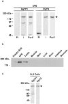
Expression of pEg7. (a) Pattern of the proteins revealed by anti–pEg7 P1 and anti–pEg7 G antibodies. Protein samples corresponding to one unfertilized egg were separated by SDS-PAGE and transferred onto nitrocellulose. The blots were probed with preimmune serums, immune serums, or purified antibodies as indicated. Positions of the molecular weight markers are indicated at the left. The position of pEg7 is indicated by an arrow. (b) Tissue expression of pEg7. Protein samples (40 μg) from each different tissue were separated by SDS-PAGE, transferred to a nitrocellulose filter, and probed with purified anti– pEG7 P1 antibodies. (c) Expression of pEg7 in Xl2 culture cells. Protein samples corresponding to 1.6 × 10⁵ cells were processed as described above and probed with purified anti–pEg7 P1 or anti–pEg7 G antibodies as indicated.
Figure 3
Figure 3
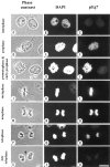
Immunolocalization of pEg7 in Xenopus Xl2 culture cells. The cells were observed in phase contrast (A, D, G, J, M, P, S), and DNA was stained with DAPI (B, E, H, K, N, Q, T). pEg7 was detected by indirect immunofluorescence using purified anti–pEg7 G antibodies (C, F, I, L, O, R, U). (S) Arrow indicates the midbody. Bar, 10 μm.
Figure 4
Figure 4
Immunolocalization of pEg7 on mitotic chromosomes assembled in vitro. Chromosomes were assembled under standard conditions (see Materials and Methods) and aliquots were taken to analyze DNA at different time points. Samples were fixed and DNA was stained with ethidium bromide (a, c, e, g, i, and k) and pEg7 was detected by indirect immunofluorescence using anti–pEg7 G immune serum (b, d, f, h, and j) or preimmune serum as a control (l). (a and b) 0 min, (c and d) 15 min, (e and f) 30 min, (g and h) 60 min, (i–l) 120 min. Bar, 5 μm.
Figure 5
Figure 5
pEg7 is required for assembly of mitotic chromosomes in vitro. (Left) Samples from high-speed mitotic extracts (1 μl) were separated by SDS-PAGE, transferred to a nitrocellulose filter, and probed with nonimmune immunoglobulins, purified anti–pEg7 P1 or purified anti–pEg7 G antibodies as indicated. (Right) Chromosomes were assembled in the presence of IgG (a–c), purified anti–pEg7 G antibodies (d–f), or purified anti–pEg7 P1 antibodies (g–i). Samples were taken at different time points and fixed. DNA was stained with ethidium bromide: (a, d, and g) 30 min, (b, e, and h) 60 min, (c, f, and i) 120 min. Bar, 5 μm.
Figure 6
Figure 6
pEg7 is a component of the 13S condensin complex. (a) Immunoprecipitation of XCAP-E was performed using preimmune serum (lane 2) or immune serum (lane 3). Immunoprecipitates were analyzed on SDS-PAGE, transferred to a nitrocellulose filter, and probed with anti–XCAP-E (top), anti–XCAP-C (middle), or anti–pEg7 P1 (bottom). An unfertilized egg was used as an internal standard (lane 1). (b) Immunoprecipitation of pEg7 was performed using anti–pEg7 P1 immune serum (lane 2) or preimmune serum (lane 3). Immunoprecipitates were analyzed on SDS-PAGE, transferred to a nitrocellulose filter, and probed with anti–pEg7 P1, anti–XCAP-E, anti–XCAP-C, and anti–topoisomerase IIα (top to bottom).

Similar articles

Cited by

References

    1. Adachi Y, Luke M, Laemmli UK. Chromosome assembly in vitro: topoisomerase II is required for condensation. Cell. 1991;64:137–148. - PubMed
    1. Bhat MA, Philp AV, Glover DM, Bellen HJ. Chromatid segregation at anaphase requires the barren product, a novel chromosome-associated protein that interacts with Topoisomerase II. Cell. 1996;87:1103–1114. - PubMed
    1. Bravo R, Knowland J. Classes of proteins synthesized in oocytes, eggs, embryos, and differentiated tissues of Xenopus laevis. . Differentiation. 1979;13:101–108. - PubMed
    1. Earnshaw WC, Halligan B, Cooke CA, Heck MM, Liu LF. Topoisomerase II is a structural component of mitotic chromosome scaffolds. J Cell Biol. 1985;100:1706–1715. - PMC - PubMed
    1. Gasser SM. Chromosome structure. Coiling up chromosomes. Curr Biol. 1995;5:357–360. - PubMed

Publication types

MeSH terms

Associated data