. 1998 Dec 14;143(6):1457-70.
doi: 10.1083/jcb.143.6.1457.

Recruitment and the role of nuclear localization in polyglutamine-mediated aggregation

Affiliations

Recruitment and the role of nuclear localization in polyglutamine-mediated aggregation

M K Perez et al. J Cell Biol. .

Abstract

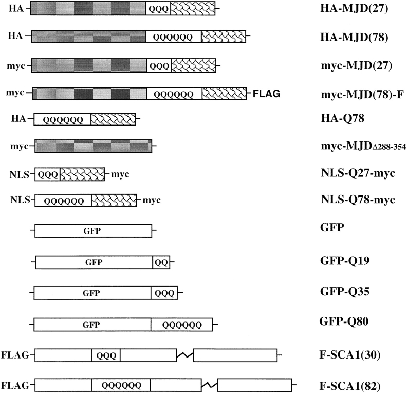
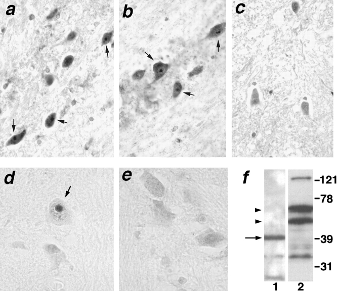
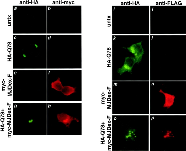
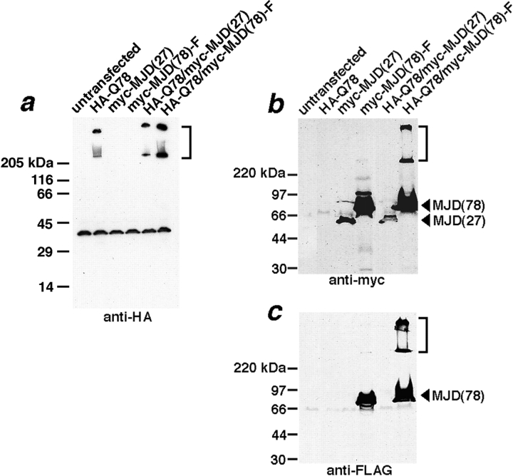
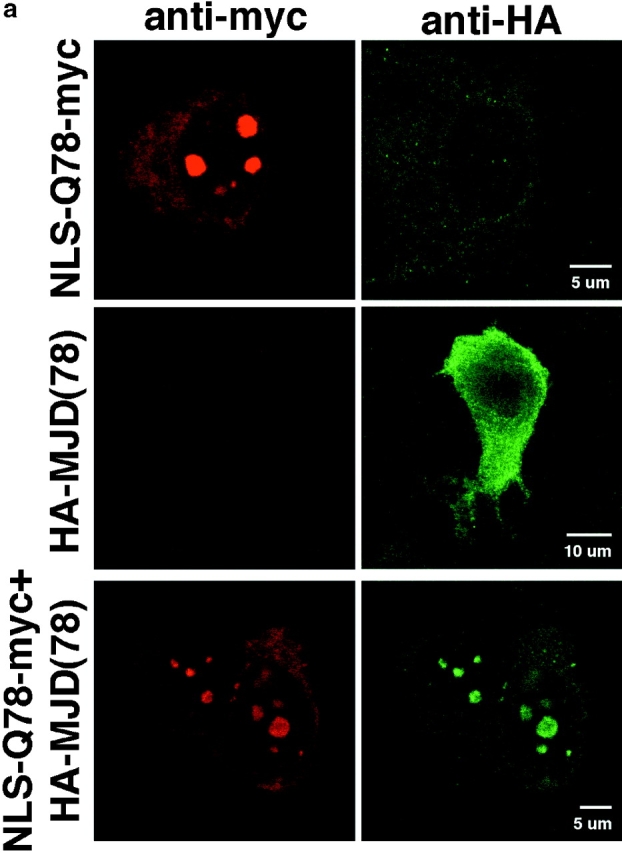
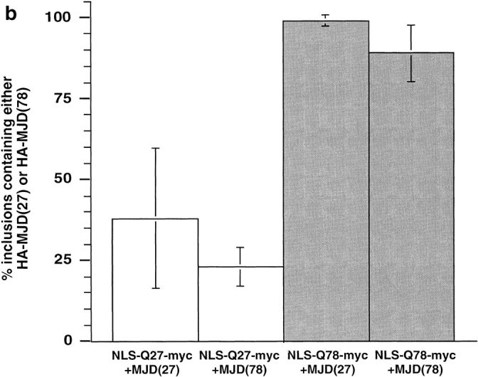
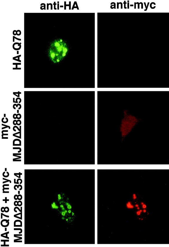
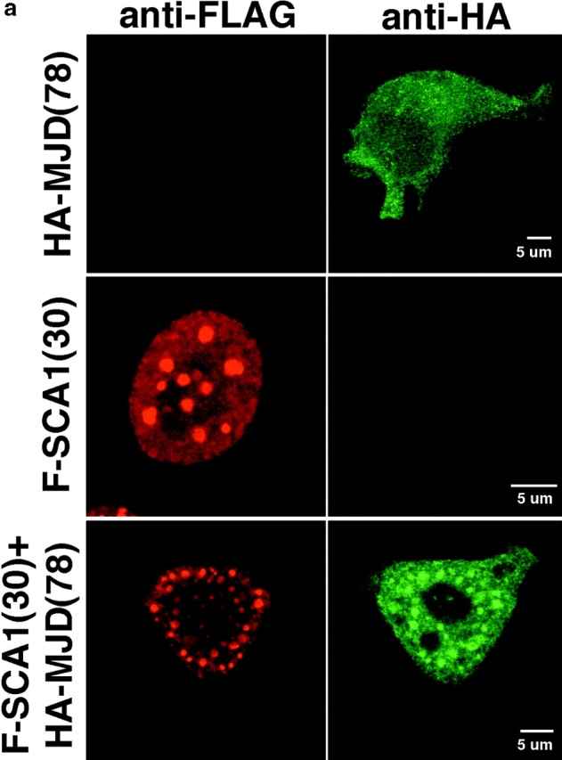
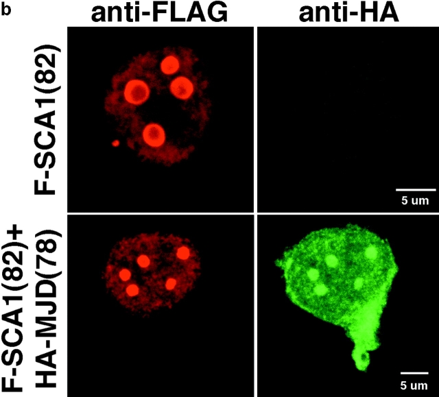
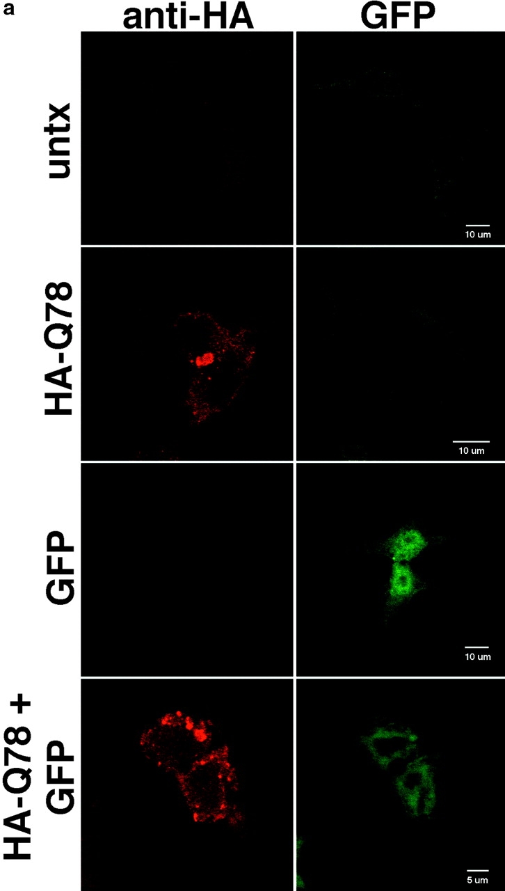
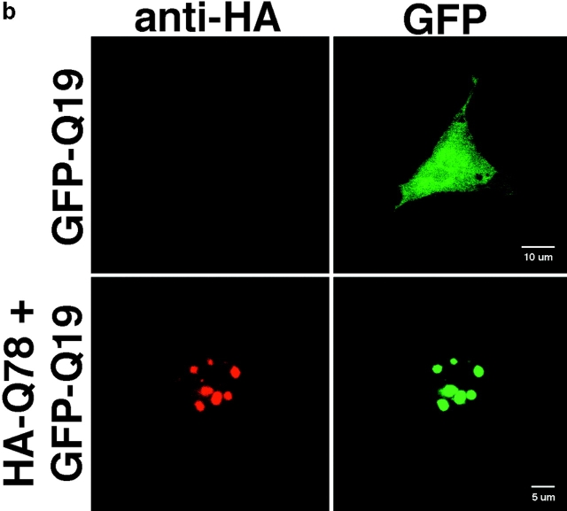
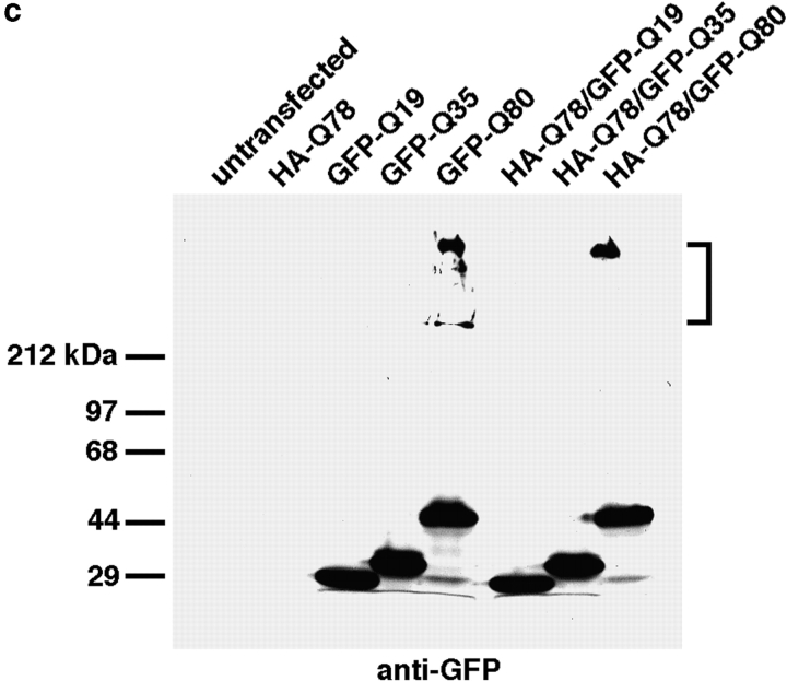
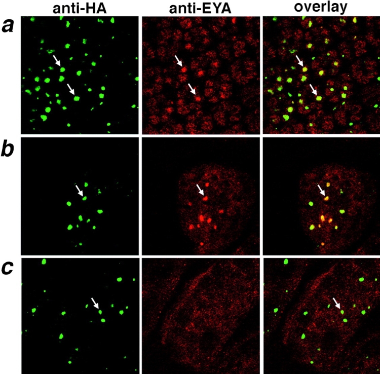
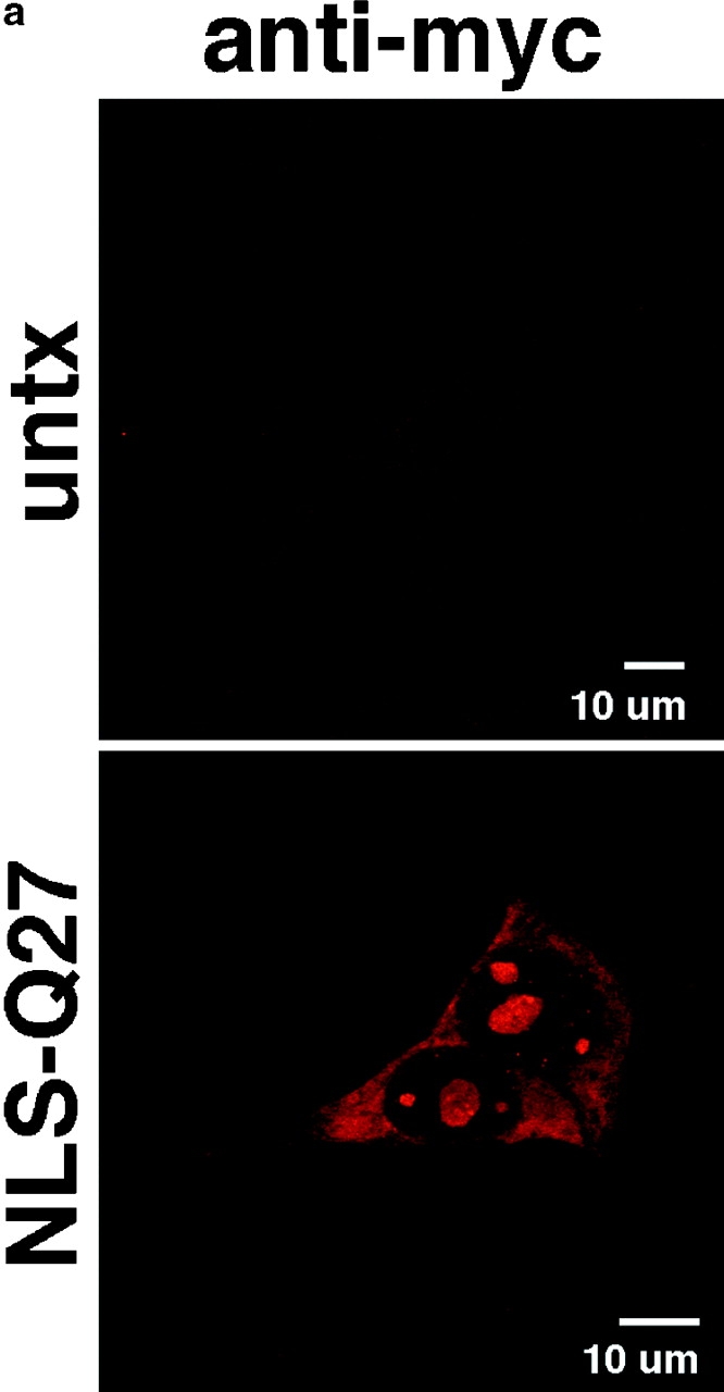
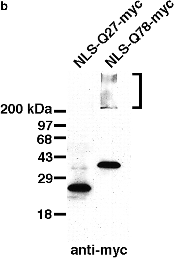
The inherited neurodegenerative diseases caused by an expanded glutamine repeat share the pathologic feature of intranuclear aggregates or inclusions (NI). Here in cell-based studies of the spinocerebellar ataxia type-3 disease protein, ataxin-3, we address two issues central to aggregation: the role of polyglutamine in recruiting proteins into NI and the role of nuclear localization in promoting aggregation. We demonstrate that full-length ataxin-3 is readily recruited from the cytoplasm into NI seeded either by a pathologic ataxin-3 fragment or by a second unrelated glutamine-repeat disease protein, ataxin-1. Experiments with green fluorescence protein/polyglutamine fusion proteins show that a glutamine repeat is sufficient to recruit an otherwise irrelevant protein into NI, and studies of human disease tissue and a Drosophila transgenic model provide evidence that specific glutamine-repeat-containing proteins, including TATA-binding protein and Eyes Absent protein, are recruited into NI in vivo. Finally, we show that nuclear localization promotes aggregation: an ataxin-3 fragment containing a nonpathologic repeat of 27 glutamines forms inclusions only when targeted to the nucleus. Our findings establish the importance of the polyglutamine domain in mediating recruitment and suggest that pathogenesis may be linked in part to the sequestering of glutamine-containing cellular proteins. In addition, we demonstrate that the nuclear environment may be critical for seeding polyglutamine aggregates.


Figures

Figure 1
Figure 1
Expression constructs used in this study. Some constructs contain an NH2-terminal HA, myc, or FLAG epitope tag as indicated, whereas others have an NH2-terminal NLS and a COOH-terminal myc tag. For the first eight constructs, the gray region represents the NH2-terminal portion of ataxin-3 up to the glutamine domain and the hatched region represents the COOH-terminal 43 amino acids after the glutamine domain. Individual nomenclature for the constructs is as follows: HA-MJD(27) and HA-MJD(78), full-length ataxin-3 containing 27 or 78 glutamines with NH2-terminal HA epitope tag; myc-MJD(27), full-length ataxin-3 containing 27 glutamines with NH2-terminal myc epitope tag; myc-MJD(78)-F, full-length ataxin-3 containing 78 glutamines with NH2-terminal myc epitope tag and COOH-terminal FLAG epitope tag; HA-Q78, NH2-terminal HA tagged truncated ataxin-3 containing 12 amino acids NH2-terminal and 43 amino acids COOH-terminal of the 78 glutamine residues; myc-MJDΔ288-354, truncated ataxin-3 containing the first 287 amino acids with an NH2-terminal myc tag; NLS-Q27-myc and NLS-Q78-myc, COOH-terminal myc tagged, nuclear-targeted fragment of ataxin-3 containing 12 amino acids NH2-terminal and 43 amino acids COOH-terminal of 27 or 78 glutamine residues; GFP, GFP-Q19, GFP-Q35, or GFP-Q80, GFP either alone of in fusion with 19, 35, or 80 glutamine residues; F-SCA1(30) and F-SCA1(82), full-length ataxin-1 containing 30 or 82 glutamines and an NH2-terminal FLAG epitope tag.
Figure 9
Figure 9
TBP immunoreactivity in NI in SCA3/MJD disease brain. Sections from control and disease pons were immunohistochemically stained with two different anti-TBP antisera or anti– ataxin-3 antiserum. (a) Anti–ataxin-3 immunostains NI within neurons of SCA3/MJD brain. (b) An adjacent section stained with the anti-TBP antibody SI-1 demonstrates TBP present within a subset of NI. (c) Control brain immunolabeled with SI-1. (d) Higher power magnification of SCA3/MJD pons showing immunolabeling of a NI with a second anti-TBP antibody, N-12. (e) Control brain immunolabeled with N-12. (f) Western blot of lysate from SCA3/MJD pons probed with the anti-TBP antibody N-12 (lane 1) or ataxin-3 antisera (lane 2) confirms the specificity of the TBP antibody. Arrows in a, b, and d indicate neurons that have NI. Original magnification, ×630 (a–c) and ×1,260 (d and e). The arrow indicates TBP in lane 1 and arrowheads indicate ataxin-3 protein from normal and expanded alleles in lane 2.
Figure 2
Figure 2
Immunofluorescence studies confirm that the full-length disease protein is recruited into aggregates formed by the polyglutamine-containing ataxin-3 fragment, HA-Q78. When expressed alone in transfected 293T cells, HA-Q78 forms perinuclear inclusions detectable by anti-HA (c and k). In contrast, full-length mutant ataxin-3 [myc-MJD(78)-F] is diffuse in the cell, detectable with antibodies to epitope tags placed at the NH2 and COOH termini (myc and FLAG, respectively, shown in f and n). When coexpressed with HA-Q78, full-length mutant ataxin-3 [myc-MJD(78)-F] is efficiently recruited into the inclusions, again detectable with antibodies to tags at both ends of the protein (h and p show the myc and FLAG staining, respectively). This demonstrates that the full protein, not simply a proteolytic fragment, is incorporated into polyglutamine inclusions. Background staining in untransfected 293T cells (untx) is shown for anti-HA (a and i), anti-myc (b), or anti-FLAG (j).
Figure 3
Figure 3
Intact, full-length expanded ataxin-3 is recruited into SDS-insoluble polyglutamine aggregates. Western blots of 293T cells transfected with the indicated constructs (above blots) and probed with anti-HA (a), anti-myc (b), or anti-FLAG (c). Blots represent identical samples run in parallel. (a) HA-Q78 migrates as a high molecular weight complex that remains in the stacking gel (bracket). On Western blots, anti-HA also cross-reacts with a cellular protein at 35 kD. This protein is not detected under more native conditions using immunofluorescence (see Fig. 2 a). (b) Coexpression of myc-MJD (78)-F with HA-Q78 leads to recruitment of the NH2 terminus (myc epitope) of myc-MJD(78)-F into a high molecular weight SDS-insoluble complex in the stacking gel; myc-MJD(27) is not recruited into an SDS-insoluble complex. (c) Blots probed with anti-FLAG demonstrate that the COOH terminus of myc-MJD(78)-F is also present in the high molecular weight SDS-insoluble complex. Similar results were obtained when blots were stripped and sequentially probed with each antibody.
Figure 4
Figure 4
Addition of a NLS to the COOH-terminal fragment of mutant ataxin-3 (NLS-Q78-myc) results in the formation of NI in 293T cells and is capable of redistributing mutant ataxin-3 from a predominately cytoplasmic to nuclear localization. (a) Expression of NLS-Q78-myc results in nuclear inclusions which are labeled with anti-myc (top). The normally cytoplasmic full-length, expanded repeat ataxin-3 [HA-MJD(78), middle] is recruited into these nuclear inclusions (bottom). (b) Nuclear inclusions formed by NLS-Q78-myc recruit MJD(27) and MJD(78) more efficiently than NLS-Q27-myc (see Fig. 9 a for inclusions formed by NLS-Q27-myc). Recruitment was quantified in a blinded manner by counting cotransfected cells with nuclear inclusions in which the ataxin-3 label (anti-HA) colocalized with the inclusions.
Figure 4
Figure 4
Addition of a NLS to the COOH-terminal fragment of mutant ataxin-3 (NLS-Q78-myc) results in the formation of NI in 293T cells and is capable of redistributing mutant ataxin-3 from a predominately cytoplasmic to nuclear localization. (a) Expression of NLS-Q78-myc results in nuclear inclusions which are labeled with anti-myc (top). The normally cytoplasmic full-length, expanded repeat ataxin-3 [HA-MJD(78), middle] is recruited into these nuclear inclusions (bottom). (b) Nuclear inclusions formed by NLS-Q78-myc recruit MJD(27) and MJD(78) more efficiently than NLS-Q27-myc (see Fig. 9 a for inclusions formed by NLS-Q27-myc). Recruitment was quantified in a blinded manner by counting cotransfected cells with nuclear inclusions in which the ataxin-3 label (anti-HA) colocalized with the inclusions.
Figure 5
Figure 5
Ataxin-3 lacking a glutamine domain is incorporated into polyglutamine inclusions. Inclusions formed in 293T cells by HA-Q78 are detected by anti-HA antibody (top). An NH2-terminal fragment of ataxin-3 lacking the glutamine domain, myc-MJDΔ288-354, maintains the diffuse localization seen with the full-length protein (middle). When coexpressed with HA-Q78, myc-MJDΔ288-354 becomes redistributed into the inclusions seeded by HA-Q78 (bottom), suggesting that regions other than the glutamine domain of ataxin-3 may also play a role in recruitment into inclusions.
Figure 6
Figure 6
Cytoplasmic ataxin-3 is redistributed into NI formed by normal and expanded repeat ataxin-1. (a) FLAG-tagged normal ataxin-1, SCA1(30), forms small intranuclear structures (middle) which are able to recruit HA-tagged MJD(27) (not shown) and HA-MJD(78) (bottom). (b) FLAG-tagged expanded repeat ataxin-1, SCA1(82), forms larger NI (top) which also recruit HA-MJD(27) (not shown) and HA-MJD(78) (bottom).
Figure 6
Figure 6
Cytoplasmic ataxin-3 is redistributed into NI formed by normal and expanded repeat ataxin-1. (a) FLAG-tagged normal ataxin-1, SCA1(30), forms small intranuclear structures (middle) which are able to recruit HA-tagged MJD(27) (not shown) and HA-MJD(78) (bottom). (b) FLAG-tagged expanded repeat ataxin-1, SCA1(82), forms larger NI (top) which also recruit HA-MJD(27) (not shown) and HA-MJD(78) (bottom).
Figure 7
Figure 7
A glutamine stretch on the control protein, GFP, is sufficient for recruitment into polyglutamine inclusions but not SDS-insoluble protein complexes. Untransfected 293T cells (untx) do not label for anti-HA or GFP (a, top). Expression of GFP (a, third row) or GFP-Q19 (b, top) alone shows a diffuse staining throughout the cell. However, when coexpressed with HA-Q78, GFP-Q19 becomes redistributed into inclusions (b, bottom) whereas GFP itself does not (a, bottom). (c) GFP-Q80 forms a high molecular weight SDS-insoluble protein complex (bracket) when expressed alone, whereas GFP-Q19 and GFP-Q35 do not. When coexpressed with HA-Q78, neither GFP-Q19 nor GFP-Q35 becomes recruited into the HA-Q78 SDS-insoluble protein complex, showing that recruitment into this complex is dependent on glutamine repeat length.
Figure 7
Figure 7
A glutamine stretch on the control protein, GFP, is sufficient for recruitment into polyglutamine inclusions but not SDS-insoluble protein complexes. Untransfected 293T cells (untx) do not label for anti-HA or GFP (a, top). Expression of GFP (a, third row) or GFP-Q19 (b, top) alone shows a diffuse staining throughout the cell. However, when coexpressed with HA-Q78, GFP-Q19 becomes redistributed into inclusions (b, bottom) whereas GFP itself does not (a, bottom). (c) GFP-Q80 forms a high molecular weight SDS-insoluble protein complex (bracket) when expressed alone, whereas GFP-Q19 and GFP-Q35 do not. When coexpressed with HA-Q78, neither GFP-Q19 nor GFP-Q35 becomes recruited into the HA-Q78 SDS-insoluble protein complex, showing that recruitment into this complex is dependent on glutamine repeat length.
Figure 8
Figure 8
In vivo recruitment of EYA in a Drosophila model of polyglutamine disease is dependent on the presence of its glutamine domain. The left column represents sections labeled with anti-HA, detecting the HA-Q78 protein. The middle column is labeled with 10H6, recognizing a conserved peptide domain present in the expressed EYA proteins. The first two columns are merged into the right column. (a) NI (arrows) formed by truncated ataxin-3 (HA-Q78) are visualized with anti-HA labeling. Anti-Eya label demonstrates that in the transgenic fly expressing an expanded glutamine domain, the endogenous EYA protein is primarily localized throughout the nucleus and in many cells has a distinct punctate appearance. The overlay indicates that the EYA protein colocalizes with polyglutamine inclusions. (b) Ectopic expression of the NH2-terminal half of the EYA protein, which contains the polyglutamine repeat. Coexpression of the HA-Q78 protein with the EYA NH2 terminus shows that EYA containing the glutamine domain is concentrated in the NI (arrow). (c) Ectopic expression of the COOH-terminal half of the EYA protein, which contains the highly conserved Eya domain, but not the glutamine repeat. Coexpression of the HA-Q78 protein (arrow) with the EYA COOH-terminal domain shows that the COOH-terminal domain lacking the glutamine repeat does not become recruited into NI. Photographs in a are from the developing eye field of an eye-antennal imaginal disc from a third-instar larva expressing HA-Q78 with gmr-GAL4; those in b and c are from the antennal field of developing eye-antennal imaginal discs of larvae expressing HA-Q78 and the eya constructs with dpp-GAL4.
Figure 10
Figure 10
Expression of a nonpathologic glutamine stretch in the nucleus leads to formation of nuclear inclusions but not SDS-insoluble complexes in 293T cells. (a) Nuclear localization of a COOH-terminal fragment of ataxin-3 containing 27 glutamines (NLS-Q27-myc) leads to the formation of NI detected by anti-myc epitope tag. Untransfected cells (untx) are not labeled with anti-myc. (b) Western blot of 293T cells transfected with a COOH-terminal fragment of ataxin-3 containing 27 glutamines (NLS-Q27-myc) or 78 glutamines (NLS-Q78-myc) targeted to the nucleus. Anti-myc labeling shows that NLS-Q27-myc forms a major band at 25 kD, whereas NLS-Q78-myc forms a 40-kD band as well as a high molecular weight, SDS-insoluble complex which remains in the stacking gel (bracket).
Figure 10
Figure 10
Expression of a nonpathologic glutamine stretch in the nucleus leads to formation of nuclear inclusions but not SDS-insoluble complexes in 293T cells. (a) Nuclear localization of a COOH-terminal fragment of ataxin-3 containing 27 glutamines (NLS-Q27-myc) leads to the formation of NI detected by anti-myc epitope tag. Untransfected cells (untx) are not labeled with anti-myc. (b) Western blot of 293T cells transfected with a COOH-terminal fragment of ataxin-3 containing 27 glutamines (NLS-Q27-myc) or 78 glutamines (NLS-Q78-myc) targeted to the nucleus. Anti-myc labeling shows that NLS-Q27-myc forms a major band at 25 kD, whereas NLS-Q78-myc forms a 40-kD band as well as a high molecular weight, SDS-insoluble complex which remains in the stacking gel (bracket).

Similar articles

Cited by

References

    1. Banif S, Servadio A, Chung M, Kwiatkowski TJ, McCall AE, Duvick LA, Shen Y, Roth EJ, Orr HT, Zoghbi HY. Identification and characterization of the gene causing type 1 spinocerebellar ataxia. Nat Genet. 1994;7:513–520. - PubMed
    1. Bonini NM, Leiserson WM, Benzer S. The eyes absent gene: genetic control of cell survival and differentiation in the developing Drosophilaeye. Cell. 1993;72:379–395. - PubMed
    1. Bonini NM, Leiserson WM, Benzer S. Expression and multiple roles of the eyes absent gene in Drosophila. . Dev Biol. 1998;129:42–57. - PubMed
    1. Burright EN, Clark HB, Servadio A, Matilla T, Feddersen RM, Yunis WS, Duvick LA, Zoghbi HY, Orr HT. SCA1 transgenic mice: a model for neurodegeneration caused by an expanded CAG trinucleotide repeat. Cell. 1995;82:937–948. - PubMed
    1. Cooper JK, Schilling G, Peters MF, Herring WJ, Sharp AH, Kaminsky Z, Masone J, Khan FA, Delanoy M, Borchelt DR, et al. Truncated N-terminal fragments of huntingtin with expanded glutamine repeats form nuclear and cytoplasmic aggregates in cell culture. Hum Mol Genet. 1998;7:783–790. - PubMed

Publication types

MeSH terms