. 1998 Dec 14;143(6):1535-45.
doi: 10.1083/jcb.143.6.1535.

The localization of myosin VI at the golgi complex and leading edge of fibroblasts and its phosphorylation and recruitment into membrane ruffles of A431 cells after growth factor stimulation

Affiliations

The localization of myosin VI at the golgi complex and leading edge of fibroblasts and its phosphorylation and recruitment into membrane ruffles of A431 cells after growth factor stimulation

F Buss et al. J Cell Biol. .

Abstract

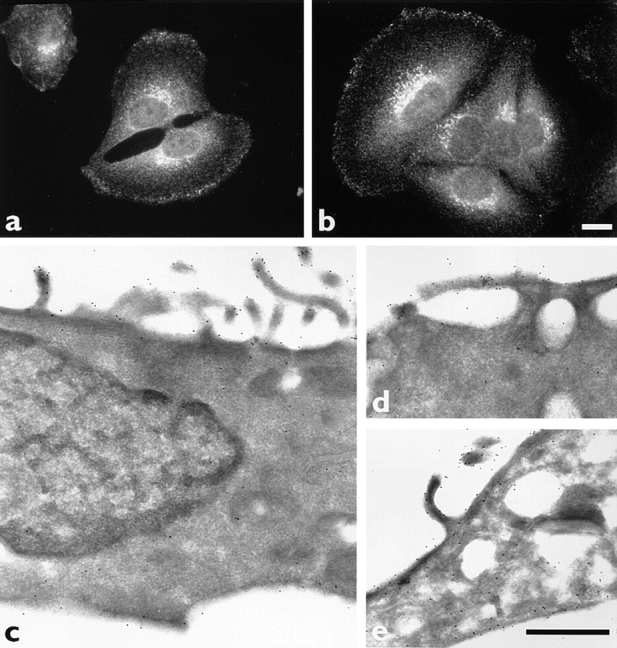
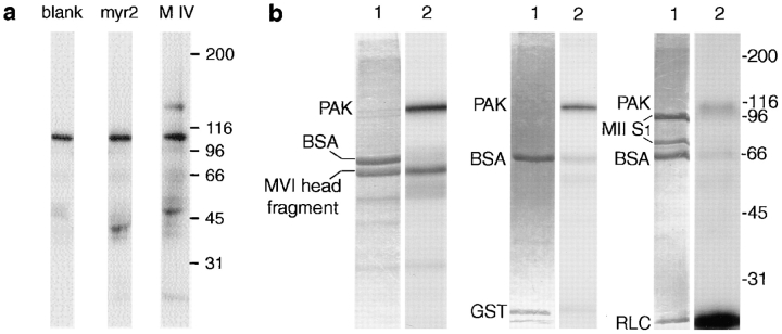
Myosin VI is an unconventional myosin that may play a role in vesicular membrane traffic through actin rich regions of the cytoplasm in eukaryotic cells. In this study we have cloned and sequenced a cDNA encoding a chicken intestinal brush border myosin VI. Polyclonal antisera were raised to bacterially expressed fragments of this myosin VI. The affinity purified antibodies were highly specific for myosin VI by immunoblotting and immunoprecipitation and were used to study the localization of the protein by immunofluorescence and immunoelectron microscopy. It was found that in NRK and A431 cells, myosin VI was associated with both the Golgi complex and the leading, ruffling edge of the cell as well as being present in a cytosolic pool. In A431 cells in which cell surface ruffling was stimulated by EGF, myosin VI was phosphorylated and recruited into the newly formed ruffles along with ezrin and myosin V. In vitro experiments suggested that a p21-activated kinase (PAK) might be the kinase responsible for phosphorylation in the motor domain. These results strongly support a role for myosin VI in membrane traffic on secretory and endocytic pathways.


Figures

Figure 1
Figure 1
Amino acid sequence of the chicken brush border myosin VI. (a) Schematic representation of the domain organization and special features in the sequence. (b) Alignment of the chicken brush border myosin VI sequence (top) with porcine myosin VI (bottom). Dots indicate identical amino acids, whereas capital letters indicate a different amino acid. Features are highlighted as follows (starting from the NH2-terminus): box surrounded by dashed line, 22 amino acid insert in the head domain; *threonine406 a possible phosphorylation site for a specific heavy chain kinase; box surrounded by solid line with sequence dashed underlined, spacer region COOH-terminal of the motor domain; dashed underlined, IQ motif; box surrounded by solid line, region of predicted coiled coil; bold letters and underlined within this coiled coil region are charged repeats; shaded box, 23-aa insert in the myosin VI tail.
Figure 2
Figure 2
Specificity of the myosin VI tail antibody (PGT). (a) Schematic diagram of the structure of myosin VI showing the different domains which were expressed in E. coli as GST-fusion proteins and used to raise antibodies. A BamHI fragment encoding aa 308– 631 of the head domain was used to raise the H ab; a HindIII fragment of the myosin VI tail encoding aa 742–1,030 was used to raise the CCT ab, a PstI fragment encoding the COOH-terminal globular tail was used to raise the PGT ab and an ab was raised against the whole tail (T ab) encoding aa 846–1,254. (b) Immunoblot of whole cell extracts of NRK and A431 cells showing the specificity of the affinity purified PGT ab. (c) Immunoprecipitation of myosin VI from [35S]methionine/ cysteine-labeled NRK and A431 cells. Lanes 1 and 3 show immunoprecipitations using the preimmune serum, whereas for lanes 2 and 4, 10 μg of the affinity purified PGT ab was used. (d) Immunoblot of immunoprecipitations from A431 cells of myosin VI probed with antibodies to different myosins. Lane 1, immunoprecipitation using the preimmune serum; lane 2, with the tail ab (PGT) to myosin VI; lane 3, with the tail ab (T) to myosin VI. The immunoprecipitates were blotted onto nitrocellulose and probed with the polyclonal antibodies to myosin I (myr2), myosin VI (MVI), myosin II (MII), and myosin V (MV).
Figure 3
Figure 3
Localization of myosin VI in NRK cells. (a and b) Indirect immunofluorescence localization of myosin VI using the affinity purified tail ab (PGT) at 10 μg/ml. Myosin VI is especially enriched in the juxtanuclear area and in the dynamic leading edge of the fibroblast. There is also a cytosolic pool. (c–e) Immunoelectron microscopic localization of myosin VI on frozen thin sections with affinity purified tail ab (PGT) followed by protein A–gold. The highest concentration of myosin VI is visible at plasma membrane profiles with dynamic membrane protrusions. Bars: (b) 20 μm; (e) 500 nm.
Figure 4
Figure 4
Association of myosin VI with the Golgi complex. NRK cells were double labeled by incubation with the affinity purified tail ab (PGT) (a) together with an mAb to TGN38 (b). Extended focus projections of a z-series of images obtained by confocal microscopy are shown. (c) Immunoblot of 15 μg of purified rat liver Golgi membranes (LG) or rat kidney Golgi membranes (KG) probed with antibodies to TGN38 or myosin VI. (d) Immunoblot of rat kidney factions. 10 μg each of cytosol (S, 100,000 g, 60 min supernatant), pellet (P, 100,000 g, 60 min pellet) and purified rat kidney Golgi membranes (G) were probed with the antibodies to TGN38 or myosin VI.
Figure 5
Figure 5
Localization of myosin VI, V, and ezrin in A431 cells after stimulation with EGF. Cells were fixed at 0, 2, 5, and 15 min after addition of EGF and stained with rabbit polyclonal antibodies to myosin VI (affinity-purified PGT ab), myosin V (affinity-purified tail domain ab), and ezrin (affinity-purified ab) followed by fluorescently labeled anti–rabbit IgG. Extended focus projections of a z-series of images obtained by confocal microscopy are shown. Bar, 20 μm.
Figure 6
Figure 6
Phosphorylation of myosin VI in vivo. (a) A431 cells were first labeled with 33P for 1 h and then used unstimulated or were stimulated with EGF before immunoprecipitation of myosin VI and ezrin. Phosphorylation of myosin VI and ezrin increased after stimulation with EGF. (b) Time course of phosphorylation of myosin VI and ezrin in A431 cells after stimulation with EGF. 33P-labeled cells were stimulated before lysing the cells for immunoprecipitation with antibodies to myosin VI or ezrin after 2, 5, or 15 min. The amount of 33P incorporation into ezrin (▪) or myosin VI (□) was quantified using a phosphorImager and plotted as a function of time. (c) Chymotryptic digest of 33P-phosphorylated myosin VI. The 33P-labeled myosin VI was immunoprecipitated from A431 cells and digested with chymotrypsin before blotting with the antibody to the head domain of myosin VI, lanes 1 and 2. Lane 3 shows the autoradiogram of the blot shown in lane 2. An asterisk marks the bands recognized by the ab to the head domain (H) in lane 2 and also labeled with 33P (lane 3), indicating that phosphorylation of myosin VI occurs in the head domain.
Figure 7
Figure 7
Phosphorylation in vitro by PAK. (a) Myosin VI and myosin I (myr 2) were immunoprecipitated from A431 cells and then incubated with recombinant GST-PAK and γ-[32P]ATP for 30 min. Myosin VI (148 kD) but not myosin I (myr 2, 118 kD) was clearly phosphorylated by PAK, which also autophosphorylates and results in a band at 105 kD (b). Myosin VI head fragment (aa 308–631) was expressed in E. coli as a GST-fusion protein, purified and incubated with recombinant GST-PAK and γ-[32P]ATP for 30 min. As controls bacterially expressed GST or the motor domain (subfragment 1) of smooth muscle myosin II (MII S1) were incubated with PAK. The major heavy chain fragment in the smooth muscle myosin subfragment 1 runs at 90 kD with a minor heavy chain fragment at 70 kD. Lane 1 shows the Coomassie stained gel and lane 2 the corresponding autoradiogram of the same gel.
Figure 8
Figure 8
Binding of myosin VI to actin filaments. Myosin VI was immunoprecipitated from A431 cells under native conditions (a) and incubated with 10 μM of F-actin in the presence (lane 2) or absence (lane 3) of 2.5 mM MgATP. The immunobeads containing bound myosin VI were briefly centrifuged and washed with or without MgATP to remove nonbound actin. The washed beads were run on SDS-PAGE and stained with Coomassie blue. Lane 1 shows actin alone and lane 4 an immunoprecipitation with preimmune serum incubated with actin. In b the immunoprecipitated myosin VI was incubated with (lane 2) or without (lane 1) recombinant PAK before assessing binding to actin filaments under the same conditions as described in a.

Similar articles

Cited by

References

    1. Allan V. Role of motor proteins in organizing the endoplasmic reticulum and Golgi apparatus. Semin Cell Devel Biol. 1996;7:335–342.
    1. Avraham KB, Hasson T, Steel KP, Kingsley DM, Russell LB, Mooseker MS, Copeland NG, Jenkins NA. The mouse Snell's waltzerdeafness gene encodes an unconventional myosin required for structural integrity of inner ear hair cells. Nat Genet. 1995;11:369–375. - PubMed
    1. Ayscough KR, Stryker J, Pokala N, Sanders M, Crews P, Drubin DG. High rates of actin filament turnover in budding yeast and roles for actin in establishment and maintenance of cell polarity revealed using the actin inhibitor latrunculin-A. J Cell Biol. 1997;137:399–416. - PMC - PubMed
    1. Beck KA, Burchanan JA, Malhotra V, Nelson WJ. Golgi spectrin: identification of an erythroid β-spectrin homolog associated with the Golgi complex. J Cell Biol. 1994;127:707–723. - PMC - PubMed
    1. Bement WM, Hasson T, Wirth JA, Cheney RE, Mooseker MS. Identification and overlapping expression of multiple unconventional myosin genes in vertebrate cell types. Proc Natl Acad Sci USA. 1994;91:6549–6553. - PMC - PubMed

Publication types

MeSH terms