. 1998 Dec 14;143(6):1547-58.
doi: 10.1083/jcb.143.6.1547.

Heterotrimeric kinesin II is the microtubule motor protein responsible for pigment dispersion in Xenopus melanophores

Affiliations

Heterotrimeric kinesin II is the microtubule motor protein responsible for pigment dispersion in Xenopus melanophores

M C Tuma et al. J Cell Biol. .

Abstract

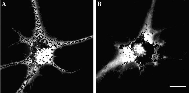
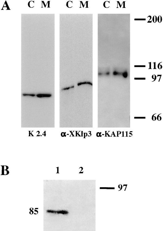
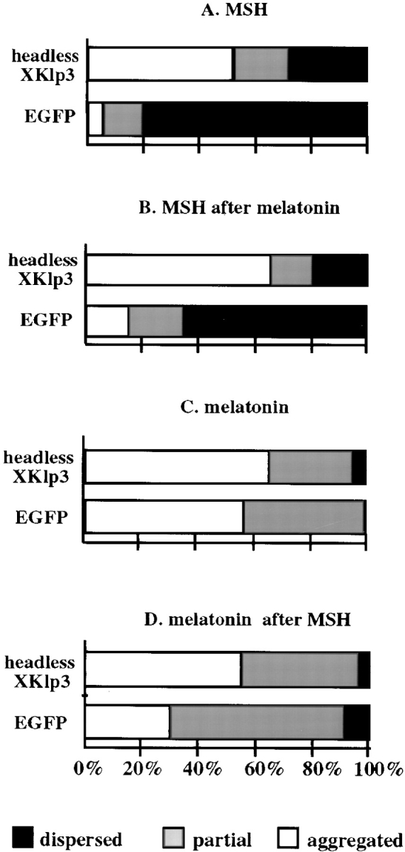
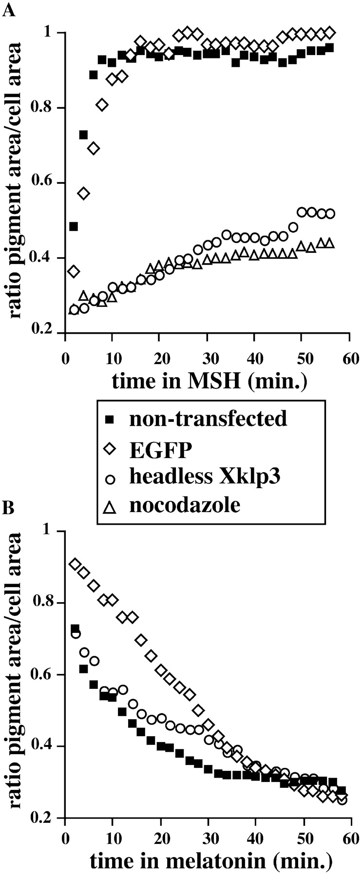
Melanophores move pigment organelles (melanosomes) from the cell center to the periphery and vice-versa. These bidirectional movements require cytoplasmic microtubules and microfilaments and depend on the function of microtubule motors and a myosin. Earlier we found that melanosomes purified from Xenopus melanophores contain the plus end microtubule motor kinesin II, indicating that it may be involved in dispersion (Rogers, S.L., I.S. Tint, P.C. Fanapour, and V.I. Gelfand. 1997. Proc. Natl. Acad. Sci. USA. 94: 3720-3725). Here, we generated a dominant-negative construct encoding green fluorescent protein fused to the stalk-tail region of Xenopus kinesin-like protein 3 (Xklp3), the 95-kD motor subunit of Xenopus kinesin II, and introduced it into melanophores. Overexpression of the fusion protein inhibited pigment dispersion but had no effect on aggregation. To control for the specificity of this effect, we studied the kinesin-dependent movement of lysosomes. Neither dispersion of lysosomes in acidic conditions nor their clustering under alkaline conditions was affected by the mutant Xklp3. Furthermore, microinjection of melanophores with SUK4, a function-blocking kinesin antibody, inhibited dispersion of lysosomes but had no effect on melanosome transport. We conclude that melanosome dispersion is powered by kinesin II and not by conventional kinesin. This paper demonstrates that kinesin II moves membrane-bound organelles.


Figures

Figure 3
Figure 3
Overexpression of headless Xklp3 inhibits pigment dispersion. Cell nuclei were injected with plasmids pEGFP-C1 (A) or pEGFP-headless Xklp3 (B). Cells were allowed to recover and express exogenous protein for 48 h and then treated with melatonin for 1 h to induce pigment aggregation, followed by 1 h in MSH, to disperse pigment. A cell expressing EGFP (A) dispersed its pigment normally, while a cell expressing the EGFP-headless Xklp3 fusion protein (B) dispersed melanosomes only partially. Bright field and fluorescence images of cells were overlaid to show pigment distribution in cells expressing EGFP-tagged proteins. Bar, 20 μm.
Figure 1
Figure 1
(A) All subunits of the heterotrimeric motor protein, kinesin II, are present on melanosomes. Western blots of cell extracts and purified melanosomes probed with antibodies against the motor subunits of kinesin II. Lanes C are cell extracts, and lanes M are purified melanosomes. The first two lanes were probed with K2.4 to detect the 85-kD motor subunit, the following two lanes were probed with a polyclonal anti-Xklp3 to detect the 95-kD motor subunit, and the last two lanes were probed with a polyclonal anti–sea urchin KAP 115 to detect the 115-kD accessory subunit. Notice that all the subunits are enriched in the melanosome fraction. (B) The overexpressed EGFP-headless Xklp3 protein forms a complex with the endogenous 85-kD motor subunit. Melanophore extracts were prepared from cells transfected with pEGFP-C1 or pEGFP-headless Xklp3, immunoprecipitated with an anti-GFP antibody, and probed with K2.4 to detect the 85-kD motor subunit. Shown are Western blots from both immunoprecipitates. The 85-kD subunit coimmunoprecipitates with EGFP-headless Xklp3 (lane 1), but not with EGFP alone (lane 2).
Figure 2
Figure 2
Quantitative analysis of pigment distribution in cells transfected with either control DNA (pEGFP-C1) or headless Xklp3 (pEGFP-headless Xklp3). Melanophores were transfected by electroporation and plated on coverslips, and after 72 h of expression, cells were incubated in serum-free medium containing MSH (A) or melatonin (C) for 1 h. In sequential treatments (B and D), cells were incubated for 1 h in melatonin followed by 1 h in MSH (B) or vice versa (D). The horizontal axis shows the percentage of cells that were scored as aggregated (white), partially dispersed (gray), or dispersed (black). For each treatment, 100 cells were scored. Data shown here are representative from one of four independent experiments.
Figure 4
Figure 4
Distribution of microtubules is not affected by expression of headless Xklp3. Immunofluorescence staining with an antitubulin primary antibody followed by a Texas red secondary in a melanophore overexpressing the headless Xklp3. Microtubules radiate from the cell center, forming a highly organized polar array. The inset is a fluorescence image taken at the green channel, showing that the cell in the large picture expresses the EGFP-headless Xklp3 fusion protein. Bar, 20 μm.
Figure 5
Figure 5
Headless Xklp3 affects the kinetics of pigment dispersion but not aggregation. Video-microscopic analysis of pigment movement was conducted on nontransfected melanophores, on melanophores 72 h after transfection, or immediately after treatment with nocodazole (see detailed description in Materials and Methods). Representative plots of changes in pigment distribution, determined by the ratio of pigment area to total cell area over time: (A) dispersion; (B) aggregation.
Figure 6
Figure 6
Distribution of lysosomes is not affected in melanophores overexpressing headless Xklp3. Cells exhibit normal lysosomal movement in response to changes in pH (dispersed in acidic conditions, clustered in alkaline conditions). Upon treatment with low pH, lysosomes redistribute through the entire cytoplasm (A). Normal perinuclear distribution is reestablished upon treatment with high pH (B). Transfected cells expressing the EGFP-headless Xklp3 fusion protein are shown in the insets, while in the large pictures the cell outlines are traced, and lysosomes stained with Texas red dextran are shown. Bar, 20 μm.
Figure 7
Figure 7
SUK4 inhibits dispersion of lysosomes but does not affect melanosomes. Melanophores with labeled lysosomes were injected with SUK4 and then exposed to acidic conditions in presence of MSH. Injected cells were identified by staining with a rhodamine anti–mouse antibody. (A) Overlay of bright field and fluorescence images, showing that melanosomes are normally dispersed in an injected cell. (B) Clustered distribution of lysosomes, labeled with FITC-dextran, in acidic conditions. Instead of normally dispersed through the cytoplasm, they remain clustered in the cell center. Bar, 20 μm.

References

    1. Arhets P, Olivo JC, Gounon P, Sansonetti P, Guillen N. Virulence and functions of myosin II are inhibited by overexpression of light meromyosin in Entamoeba histolytica. . Mol Biol Cell. 1998;9:1537–1547. - PMC - PubMed
    1. Bi GQ, Morris RL, Liao G, Alderton JM, Scholey JM, Steinhardt RA. Kinesin- and myosin-driven steps of vesicle recruitment for Ca2+-regulated exocytosis. J Cell Biol. 1997;138:999–1008. - PMC - PubMed
    1. Blose SH, Meltzer DI, Feramisco JR. 10-nm filaments are induced to collapse in living cells microinjected with monoclonal and polyclonal antibodies against tubulin. J Cell Biol. 1984;98:847–858. - PMC - PubMed
    1. Burns CG, Larochelle DA, Erickson H, Reedy M, De Lozanne A. Single-headed myosin II acts as a dominant negative mutation in Dictyostelium. . Proc Natl Acad Sci USA. 1995;92:8244–8248. - PMC - PubMed
    1. Cole DG, Scholey JM. Structural variations among the kinesins. Trends Cell Biol. 1995;5:259–262. - PubMed

Publication types

MeSH terms