. 1998 Dec 14;143(6):1603-16.
doi: 10.1083/jcb.143.6.1603.

Role of polo kinase and Mid1p in determining the site of cell division in fission yeast

Affiliations

Role of polo kinase and Mid1p in determining the site of cell division in fission yeast

J Bähler et al. J Cell Biol. .

Abstract

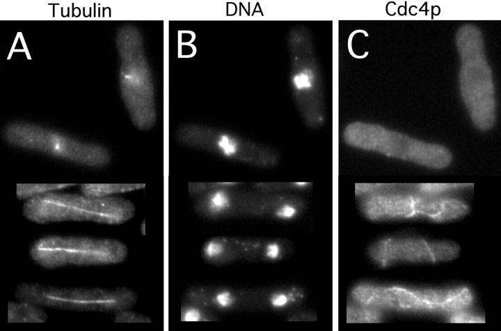
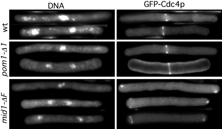
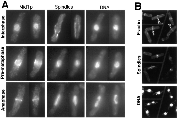
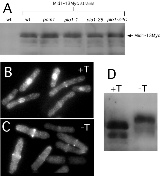
The fission yeast Schizosaccharomyces pombe divides symmetrically using a medial F-actin- based contractile ring to produce equal-sized daughter cells. Mutants defective in two previously described genes, mid1 and pom1, frequently divide asymmetrically. Here we present the identification of three new temperature-sensitive mutants defective in localization of the division plane. All three mutants have mutations in the polo kinase gene, plo1, and show defects very similar to those of mid1 mutants in both the placement and organization of the medial ring. In both cases, ring formation is frequently initiated near the cell poles, indicating that Mid1p and Plo1p function in recruiting medial ring components to the cell center. It has been reported previously that during mitosis Mid1p becomes hyperphosphorylated and relocates from the nucleus to a medial ring. Here we show that Mid1p first forms a diffuse cortical band during spindle formation and then coalesces into a ring before anaphase. Plo1p is required for Mid1p to exit the nucleus and form a ring, and Pom1p is required for proper placement of the Mid1p ring. Upon overexpression of Plo1p, Mid1p exits the nucleus prematurely and displays a reduced mobility on gels similar to that of the hyperphosphorylated form observed previously in mitotic cells. Genetic and two-hybrid analyses suggest that Plo1p and Mid1p act in a common pathway distinct from that involving Pom1p. Plo1p localizes to the spindle pole bodies and spindles of mitotic cells and also to the medial ring at the time of its formation. Taken together, the data indicate that Plo1p plays a role in the positioning of division sites by regulating Mid1p. Given its previously known functions in mitosis and the timing of cytokinesis, Plo1p is thus implicated as a key molecule in the spatial and temporal coordination of cytokinesis with mitosis.


Figures

Figure 1
Figure 1
Medial ring and septum defects in plo1-1 mutant cells and comparison of the plo1-1 and mid1-18 defects. Wild-type strain JB13 (A and B), plo1-1 mutant strain YDM110 (C–E), and mid1-18 strain YDM296 (F), all expressing GFP-Cdc4p fusion protein, were grown at 25°C and shifted to 36°C for 2 h before examination. (A–D) Cells were fixed and stained with Calcofluor to visualize septal material; GFP-Cdc4p fluorescence (A and C) and Calcofluor fluorescence (B and D) of the same cells are shown. (E and F) Living cells were mounted on a microscope slide and overlaid with an agar slab, and GFP-Cdc4p was viewed by fluorescence microscopy at 36°C. Images were collected every 0.25 μm and processed by deconvolution methods to generate a two-dimensional projection of the three-dimensional image (see Materials and Methods).
Figure 2
Figure 2
Spindle formation defects in plo1-24C mutant cells. Cells of strain YDM114 were grown at 25°C to exponential phase, shifted to 36°C for 2 h, and triple stained for tubulin (A), DNA (B), and Cdc4p (C). The upper two cells were blocked at an early stage of spindle formation with highly condensed chromatin and no Cdc4p rings, whereas the lower three cells had leaked past the block and formed spindles and aberrant medial rings.
Figure 3
Figure 3
Time-lapse analysis of medial ring formation in mutant cells. Wild-type (JB13), pom1-Δ1 (JB110), plo1-1 (YDM110), and mid1-18 (YDM296) cells, all expressing GFP-Cdc4p fusion protein, were grown at 25°C, then shifted to 36°C for 1 h in liquid culture. The living cells were then mounted on a microscope slide overlaid with an agar slab and viewed by fluorescence microscopy at 36°C. For wild-type, plo1-1, and mid1-18 cells, images were collected every 0.5 μm at 5-min intervals and processed by deconvolution methods to generate two-dimensional projections of the three-dimensional images (see Materials and Methods). Images of pom1-Δ1 cells were taken in a single focal plane. Asterisks and the arrowhead indicate structures described in the text.
Figure 4
Figure 4
Medial ring formation in elongated cells. cdc25-22 (strain YDM152; wt), cdc25-22 pom1-Δ1 (strain JB120), and cdc25-22 mid1-ΔF (strain JB43) cells, all expressing GFP-Cdc4p fusion protein and growing exponentially at 25°C, were shifted to 36°C for 4 hr, returned to 25°C, and observed 45 min later.
Figure 5
Figure 5
Timing of Mid1p ring formation and F-actin ring formation in wild-type cells. (A) Mid1p-GFP expressing cells (strain YDM403) growing exponentially at 32°C were triple stained for Mid1p (using GFP-specific antibodies), spindles, and DNA. Representative interphase, pre-metaphase, and anaphase cells are shown. Very similar results were obtained when strain 972 (wild-type) cells were stained with Mid1p-specific antibodies or when Mid1p-13Myc-expressing cells (strain YDM603) were stained with antibodies to the Myc epitope. (B) Strain 972 cells growing exponentially at 36°C were triple stained for F-actin, spindles, and DNA. The fixation protocols used visualize Mid1p, Mid1p-fusion proteins, F-actin, and spindle microtubules well, but preserve cytoplasmic microtubules poorly. Large arrowheads, pre-metaphase cells with short spindles; arrow, cell with an anaphase spindle.
Figure 6
Figure 6
Failure of Mid1p nuclear exit and ring formation in the plo1-1 mutant. (A) mid1p-GFP plo1-1 cells (strain JB250) growing exponentially at 25°C were fixed and triple stained for Mid1p, spindles, and DNA as in Fig. 5. Very similar results were obtained when plo1-1 cells (strain YDM110) were stained with Mid1p-specific antibodies or when mid1-13Myc plo1-1 cells (strain YDM607) were stained with Myc-specific antibodies. (B) plo1-1 cells expressing Mid1p-GFP (strain JB250) or Mid1p-13Myc (strain YDM607) cells growing exponentially at 25°C were shifted to 36°C for 2.5 h before fixation. The fixed cells were triple stained for Mid1p-GFP (top) or Mid1p-13Myc (bottom) using GFP-specific or Myc-specific antibodies as appropriate, for spindles, and for DNA as in Fig. 5. Very similar results were obtained when plo1-1 cells (strain YDM110) were stained with Mid1p-specific antibodies. Note that the anti-tubulin antibodies used to stain the Mid1p-13Myc cells (see Materials and Methods) do not give the background nuclear staining like the TAT-1 anti-tubulin monoclonal antibody which is used above.
Figure 7
Figure 7
Mislocalization of Mid1p rings in a pom1 mutant. Cells of pom1-Δ1 strain JB110 growing exponentially at 30°C were double stained for Mid1p using Mid1p-specific antibodies (A) and DNA (B) or for Mid1p (C) and F-actin (D). Arrows indicate the positions of mislocalized Mid1p and actin rings.
Figure 8
Figure 8
Mid1p levels in plo1 and pom1 mutants, and the effect of Plo1p overproduction on Mid1p. (A) Strains expressing either normal Mid1p (no Myc) (YDM105) or Mid1p-13Myc (wt, YDM603; pom1, YDM609; plo1-1, YDM607; plo1-25, YDM604; and plo1-24C, YDM608) were grown at 25°C, shifted to 36°C for 2.5 h, and harvested. Protein lysates were prepared from each strain and equal amounts of protein were analyzed by Western blotting using anti-Myc antibodies. (B-D) Mid1p-13Myc-expressing cells (YDM603) containing a plasmid (pREP1Plo1) for expression of plo1 from the thiamine-repressible nmt1 promoter, were grown at 30°C in the presence of thiamine. This culture was then used to inoculate two new cultures either with (promoter off) or without (promoter on) thiamine, and growth was continued for 16 h. Portions of each culture were then either fixed and stained for Mid1p-13Myc using Myc-specific antibodies or used to prepare lysates for analysis by western blotting. (B and C) Mid1p-13Myc staining in cells either not overexpressing (B) or overexpressing (C) Plo1p. (D) Western blot analysis of Mid1p-13Myc in cells either not overexpressing (+T) or overexpressing (−T) Plo1p.
Figure 9
Figure 9
Localization of Plo1p-GFP in wild-type, cdc25-22, and cdc25-22 mid1-ΔF cells. (A) plo1-GFP strain JB206 was grown at 30°C, and cells were photographed at various stages in the cell cycle as described in the text. (B and C) plo1-GFP cdc25-22 cells (strain YDM457; B) and plo1-GFP cdc25-22 mid1-ΔF cells (strain JB214; C) were blocked at 36°C for 4 h and then shifted to 25°C for 45 min. The arrowheads indicate medial Plo1p rings.

Similar articles

Cited by

References

    1. Ausubel, F.M., R. Brent, R.E. Kingston, D.D. Moore, J.G. Seidman, J.A. Smith, and K. Struhl. 1995. Current Protocols in Molecular Biology. John Wiley and Sons Ltd., New York.
    1. Bähler J, Pringle JR. Pom1p, a fission yeast protein kinase that provides positional information for both polarized growth and cytokinesis. Genes Dev. 1998;12:1356–1370. - PMC - PubMed
    1. Bähler J, Wu J-Q, Longtine MS, Shah NG, McKenzie A, III, Steever AB, Wach A, Philippsen P, Pringle JR. Heterologous modules for efficient and versatile PCR-based gene targeting in Schizosaccharomyces pombe. . Yeast. 1998;14:943–951. - PubMed
    1. Balasubramanian MK, McCollum D, Gould KL. Cytokinesis in the fission yeast Schizosaccharomyces pombe. . Methods Enzymol. 1997;283:494–506. - PubMed
    1. Balasubramanian MK, McCollum D, Chang LC, Wong KCY, Naqvi NI, He X, Sazer S, Gould KL. Isolation and characterization of new fission yeast cytokinesis mutants. Genetics. 1998;149:1265–1275. - PMC - PubMed

Publication types

MeSH terms