. 1998 Dec 14;143(6):1635-46.
doi: 10.1083/jcb.143.6.1635.

AIR-2: An Aurora/Ipl1-related protein kinase associated with chromosomes and midbody microtubules is required for polar body extrusion and cytokinesis in Caenorhabditis elegans embryos

Affiliations

AIR-2: An Aurora/Ipl1-related protein kinase associated with chromosomes and midbody microtubules is required for polar body extrusion and cytokinesis in Caenorhabditis elegans embryos

J M Schumacher et al. J Cell Biol. .

Abstract

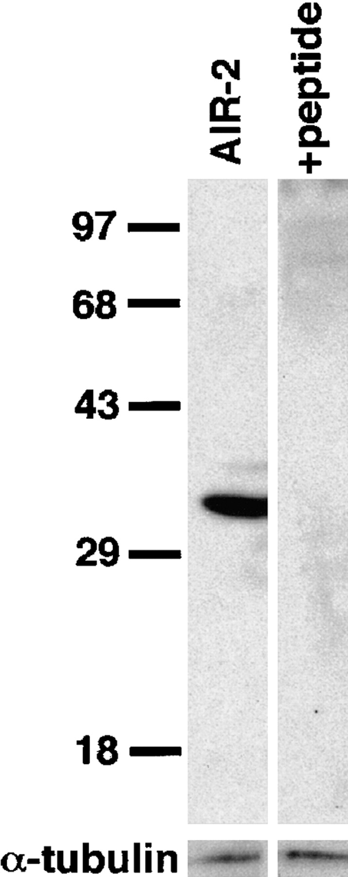
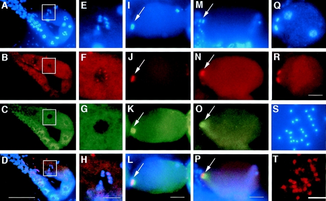
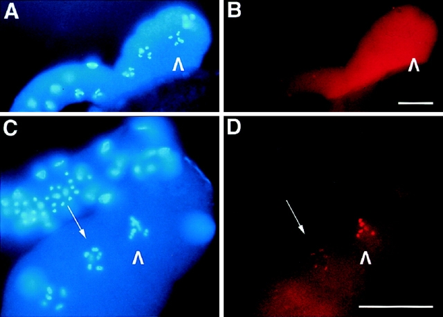
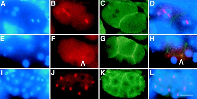
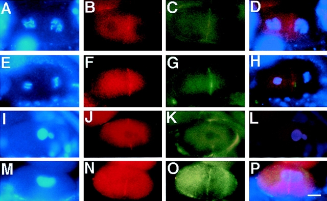
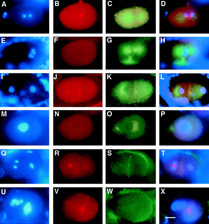
An emerging family of kinases related to the Drosophila Aurora and budding yeast Ipl1 proteins has been implicated in chromosome segregation and mitotic spindle formation in a number of organisms. Unlike other Aurora/Ipl1-related kinases, the Caenorhabditis elegans orthologue, AIR-2, is associated with meiotic and mitotic chromosomes. AIR-2 is initially localized to the chromosomes of the most mature prophase I-arrested oocyte residing next to the spermatheca. This localization is dependent on the presence of sperm in the spermatheca. After fertilization, AIR-2 remains associated with chromosomes during each meiotic division. However, during both meiotic anaphases, AIR-2 is present between the separating chromosomes. AIR-2 also remains associated with both extruded polar bodies. In the embryo, AIR-2 is found on metaphase chromosomes, moves to midbody microtubules at anaphase, and then persists at the cytokinesis remnant. Disruption of AIR-2 expression by RNA- mediated interference produces entire broods of one-cell embryos that have executed multiple cell cycles in the complete absence of cytokinesis. The embryos accumulate large amounts of DNA and microtubule asters. Polar bodies are not extruded, but remain in the embryo where they continue to replicate. The cytokinesis defect appears to be late in the cell cycle because transient cleavage furrows initiate at the proper location, but regress before the division is complete. Additionally, staining with a marker of midbody microtubules revealed that at least some of the components of the midbody are not well localized in the absence of AIR-2 activity. Our results suggest that during each meiotic and mitotic division, AIR-2 may coordinate the congression of metaphase chromosomes with the subsequent events of polar body extrusion and cytokinesis.


Figures

Figure 1
Figure 1
AIR-2 is member of a highly conserved family of protein kinases. An alignment of the predicted protein product sequences for C. elegans AIR-2 and AIR-1, mammalian AIM-1 and IAK-1, Drosophila Aurora, and budding yeast Ipl1 is shown. Black shading, identical residues; gray shading, similar residues; underline, predicted kinase domain. The AIR-2 kinase domain has the following amino acid identities and similarities with the kinase domains of (a) AIM-1: 63% identical, 76% similar; (b) IAK-1: 62% identical, 75% similar; (c) Aurora: 55% identical, 71% similar; (d) AIR-1: 51% identical, 69% similar; and (e) Ipl1: 50% identical, 70% similar. Nucleotide and predicted protein sequences for the air-2 cDNA have been submitted to GenBank/EMBL/DDBJ under accession number AF071207.
Figure 2
Figure 2
Specificity of the AIR-2 antibody. Western blots of protein lysates from C. elegans embryos were prepared as described in Materials and Methods. Affinity-purified AIR-2 antiserum specifically recognizes a protein of the predicted size of 34.7 kD. Antibody recognition of the 34.7-kD band is completely eliminated by addition of the antigenic peptide to the primary antibody incubation. α-Tubulin was used as a protein loading control.
Figure 3
Figure 3
AIR-2 is localized to meiotic chromosomes. Gonads were dissected from wild-type adult hermaphrodites, fixed, and then stained with DAPI (A, E, I, M, and Q), AIR-2 antisera (B, F, J, N, R, and T), and α-tubulin antisera (C, G, K, and O). Merged images are shown in D, H, L, and P. (A–D) AIR-2 is diffuse throughout the distal and proximal gonad, but is specifically localized to chromosomes in the oocyte most proximal to the spermatheca (white box). The spermatheca is to the left of the white box in A–D, but is out of the focal plane. (E–H) High magnification of the most proximal oocyte (boxed region in A–D). AIR-2 is specifically localized to meiotic chromosomes in the proximal oocyte. (I–L) Meiosis resumes after the oocyte is ovulated into the spermatheca and is fertilized. AIR-2 is associated with the chromosomes undergoing meiotic divisions (arrows). The sperm chromatin is visible at the posterior in I. Left, anterior. (M–P) AIR-2 is localized between the chromosomes during meiotic anaphase (arrows). Left, anterior. (Q and R) Polar bodies are extruded after each meiotic division at the anterior of the embryo. AIR-2 staining remains on the polar bodies throughout their presence during embryogenesis. AIR-2 staining is not associated with maternal or paternal pronuclear chromatin (the two nuclei within the embryo, Q). Left, anterior. (S and T) AIR-2 stains the mature sperm found in hermaphrodites but appears to be distinct from the chromatin. AIR-2 was also found in mature sperm from males (data not shown). Bars: (A–D) 50 μm; (E–H, I–L, M–P, Q, and R) 10 μm.
Figure 4
Figure 4
The localization of AIR-2 to meiotic chromosomes requires the presence of sperm in the spermatheca. (A and B) Gonads dissected from virgin tra-2(gf) females were fixed and stained with DAPI (A) and AIR-2–specific antisera (B). AIR-2 staining is diffuse throughout the cellularized oocytes and does not localize to chromosomes in the proximal oocyte (arrowheads). (C and D) Gonads dissected from mated tra-2(gf) females were fixed and stained with DAPI (C) and AIR-2–specific antisera (D). AIR-2 strongly stains the chromosomes of the proximal oocyte (arrowheads) and fainter staining of chromosomes is also seen in a neighboring oocyte (arrow). Bars, 20 μm.
Figure 5
Figure 5
AIR-2 is localized to metaphase chromosomes and midbody microtubules in C. elegans embryos. (A–L) Embryos were dissected from wild-type adult hermaphrodites, fixed, and then stained with DAPI (A, E, and I), AIR-2 (B, F, and J), and α-tubulin antibodies (C, G, and K). Merged images are shown in (D, H, and L). (A–D) AIR-2 is concentrated at the metaphase plate, and (E–H) moves to the midbody microtubules as anaphase progresses. (I–L) AIR-2 staining (arrowhead) persists at the cytokinesis remnant as seen in this four-cell embryo. The other remnant is out of the focal plane. Bar, 10 μm.
Figure 6
Figure 6
AIR-2 is retained at the cytokinesis remnant. (A– L) Embryos were dissected from wild-type adult hermaphrodites, fixed, and then stained with DAPI (A, E, and I), AIR-2 (B, F, and J), and actin antibodies (C, G, and K). Merged images are shown in (D, H, and L). (A–D) AIR-2 is concentrated between separating chromatin at anaphase. The AB cell in anterior (left) is in telophase, the P1 cell in posterior (right) is in anaphase. (E– H) AIR-2 staining persists on the cell membrane once cytokinesis is complete (arrowhead). The dot of staining that is not associated with the membrane is a polar body that is out of the focal plane. (I–L) AIR-2 appears to behave similarly throughout embryogenesis in all cell lineages. Bar, 10 μm.
Figure 7
Figure 7
air-2(RNAi) embryos execute multiple cell cycles in the absence of cytokinesis. (A–C) Embryos were dissected from hermaphrodites that had been injected with double-stranded RNA corresponding to the entire air-2 cDNA. air-2 (RNAi) embryos were fixed and stained with DAPI (A), AIR-2 (B), and α-tubulin antibodies (C). (A) air-2(RNAi) embryos accumulate DNA, (B) do not express a detectable level of AIR-2, and (C) contain multiple microtubule asters. The embryos appear to execute multiple cell cycles without completing cytokinesis. As revealed by tubulin staining (C), some of the embryos appear to be in interphase whereas others are in mitosis. Bar, 20 μm.
Figure 8
Figure 8
Polar bodies are not extruded in air-2(RNAi) embryos. (A–L) air-2(RNAi) embryos at various stages of development were dissected from injected mothers, fixed, and then stained with DAPI (left) and an antibody to α-tubulin (right). (A and B) An air-2(RNAi) embryo completing meiosis. (A) The maternal meiotic chromatin is apparent at the anterior (arrowhead), whereas the sperm nucleus can be seen at the posterior of the embryo. (B) By tubulin staining, a morphologically normal meiotic spindle is apparent surrounding the meiotic chromatin at the anterior of the embryo (arrowhead). (C and D) A one-cell air-2 (RNAi) embryo undergoing anaphase. (C) Note the chromatin bridges and hypercondensation of the chromatin. The second chromatin body appears to be a replicating unextruded polar body (arrowhead). (D) The spindle is morphologically normal. (E and F) An air-2(RNAi) embryo that has gone through multiple rounds of DNA replication and centrosome duplication without completing cytokinesis is shown. (E) The chromatin is condensed and (F) multiple microtubule asters are apparent. (G and H) air-2 (RNAi) embryos cycle through interphase. (G) Chromosomes are decondensed and (H) an “interphase array” of microtubules is observed. (I–L) Polar bodies in some air-2(RNAi) embryos proceed through the cell cycle asynchronously from the rest of the chromatin in the embryo (arrowheads). (I) This polar body appears to be in anaphase. (K) The polar body (arrowhead) has microtubule asters surrounding it whereas the nuclear chromatin appears to be interphase (L). Bar, 10 μm.
Figure 9
Figure 9
The cleavage furrow is unstable in air-2 (RNAi) embryos. Wild-type and air-2(RNAi) embryos were dissected from adult hermaphrodites, fixed, and then stained with DAPI (A, E, I, and M), NMY-2 (B, F, J, and N), actin (C, G, and K), and α-tubulin antibodies (O). Merged images are shown in D, H, L, and P. (A–D) A wild-type two-cell embryo in prophase. (B) NMY-2 is faintly seen on the cell membrane and at the cytokinesis remnant. (C) Actin is apparent at the cell membrane. (D) Both proteins overlap at the cell membrane. (E–H) A wild-type two-cell embryo where the AB cell (left) is in anaphase and the P1 (right) is in metaphase. (F) NMY-2 and (G) actin colocalize at the cell membrane (H). (I–L) An air-2(RNAi) embryo undergoing the first cell cycle. (J) NMY-2 and (K) actin colocalize to an incomplete cleavage furrow (L). (M–P) An air-2(RNAi) embryo undergoing the first cell cycle. A transient cleavage furrow bisecting the cell is apparent by (N) NMY-2 and (O) α-tubulin staining that overlaps (P). Bar, 10 μm.
Figure 10
Figure 10
C. elegans MKLP-1/ZEN-4 protein does not localize properly in the absence of AIR-2. Wild-type and air-2(RNAi) embryos were dissected from adult hermaphrodites, fixed, and then stained with DAPI (A, E, I, M, Q, and U), MKLP-1 (B, F, J, N, R, and V), α-tubulin (C, G, K, and O) and actin antibodies (S and W). Merged images are shown in D, H, L, P, T, and X. (A–D) Wild-type one-cell embryo undergoing anaphase. (B) MKLP-1 localizes to the midbody microtubules. (C) Tubulin staining and (D) merged DAPI, MKLP-1, and tubulin. (E–H) air-2 (RNAi) embryo in metaphase/anaphase. (F) No MKLP-1 staining is apparent on the (G) tripolar spindle. (H) merged DAPI, MKLP-1, and tubulin. (I–L) Wild-type two-cell embryo in prophase. (J) MKLP-1 staining is localized to the midbody remnant on the cell membrane. (K) Tubulin staining, (L) merged DAPI, MKLP-1, and tubulin. (M–P) air-2(RNAi) embryo in prophase. (N) No MKLP-1 staining is found in this embryo. (O) Tubulin staining, (P) merged DAPI, MKLP-1, and tubulin staining. (Q–T) Wild-type two-cell embryo where the AB cell (left) is in anaphase, and the P1 cell (right) is in metaphase. (R) MKLP-1 staining is found between the separating chromosomes in the anaphase cell and is found at low levels on the cell membrane and surrounding the metaphase plate in the P1 cell. (S) Actin staining of the cell membrane, (T) merged DAPI, MKLP-1, and actin staining. (U– X) A multinucleate air-2(RNAi) embryo. (V) MKLP-1 staining is found in a “cloud” around the chromatin. (W) Actin staining reveals no cleavage furrows in this embryo, (X) merged DAPI, MKLP-1, and actin staining. Bar, 10 μm.

Similar articles

Cited by

References

    1. Ault JG, Rieder CL. Centrosome and kinetochore movement during mitosis. Curr Opin Cell Biol. 1994;6:41–49. - PubMed
    1. Blangy A, Arnaud L, Nigg EA. Phosphorylation by p34cdc2 protein kinase regulates binding of the kinesin-related motor HsEg5 to the dynactin subunit p150. J Biol Chem. 1997;272:19418–19424. - PubMed
    1. Blumenthal T. Trans-splicing and polycistronic transcription in Caenorhabditis elegans. . Trends Genet. 1995;11:132–136. - PubMed
    1. Boleti H, Karsenti E, Vernos I. Xklp2, a novel Xenopuscentrosomal kinesin-like protein required for centrosome separation during mitosis. Cell. 1996;84:49–59. - PubMed
    1. Brenner S. The genetics of Caenorhabditis elegans. . Genetics. 1974;77:71–94. - PMC - PubMed

Publication types

MeSH terms