. 1998 Dec 14;143(6):1691-703.
doi: 10.1083/jcb.143.6.1691.

p53 is essential for developmental neuron death as regulated by the TrkA and p75 neurotrophin receptors

Affiliations

p53 is essential for developmental neuron death as regulated by the TrkA and p75 neurotrophin receptors

R S Aloyz et al. J Cell Biol. .

Abstract

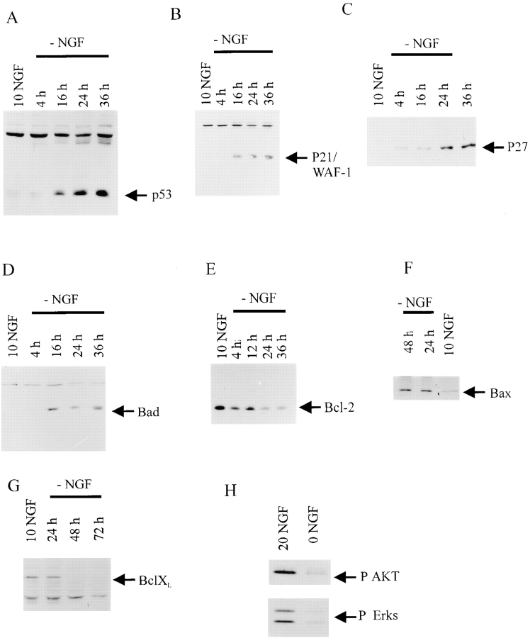
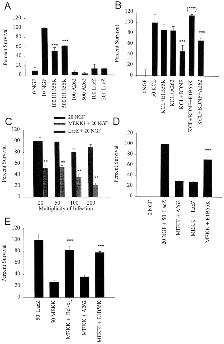
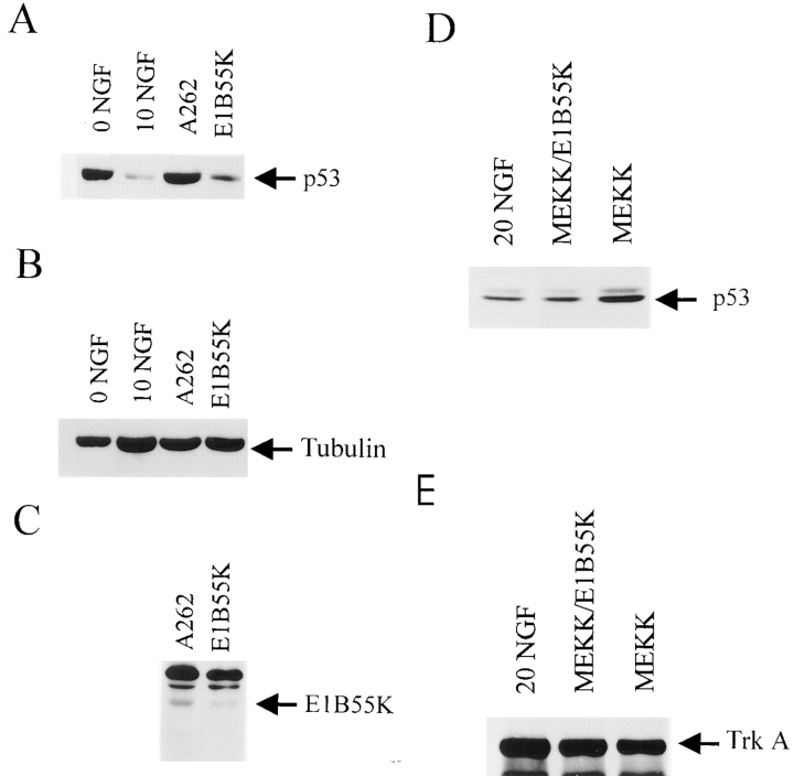
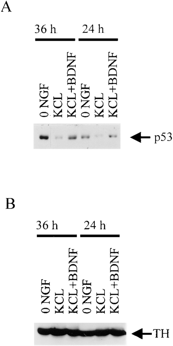
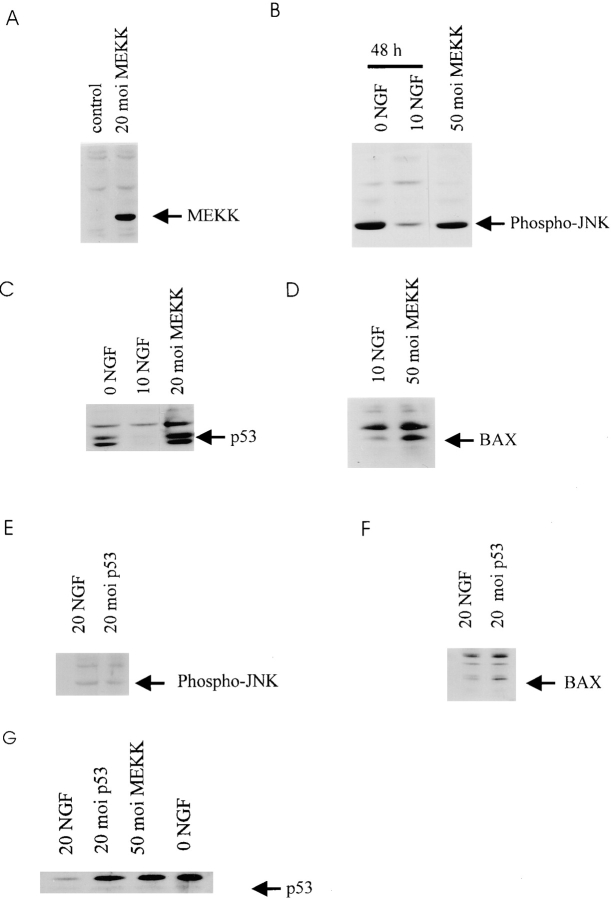
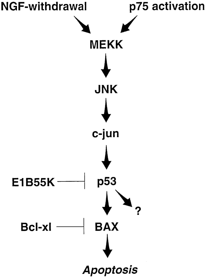
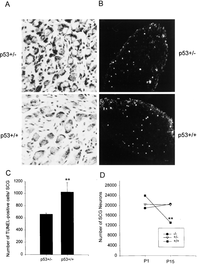
Naturally occurring sympathetic neuron death is the result of two apoptotic signaling events: one normally suppressed by NGF/TrkA survival signals, and a second activated by the p75 neurotrophin receptor. Here we demonstrate that the p53 tumor suppressor protein, likely as induced by the MEKK-JNK pathway, is an essential component of both of these apoptotic signaling cascades. In cultured neonatal sympathetic neurons, p53 protein levels are elevated in response to both NGF withdrawal and p75NTR activation. NGF withdrawal also results in elevation of a known p53 target, the apoptotic protein Bax. Functional ablation of p53 using the adenovirus E1B55K protein inhibits neuronal apoptosis as induced by either NGF withdrawal or p75 activation. Direct stimulation of the MEKK-JNK pathway using activated MEKK1 has similar effects; p53 and Bax are increased and the subsequent neuronal apoptosis can be rescued by E1B55K. Expression of p53 in sympathetic neurons indicates that p53 functions downstream of JNK and upstream of Bax. Finally, when p53 levels are reduced or absent in p53+/- or p53-/- mice, naturally occurring sympathetic neuron death is inhibited. Thus, p53 is an essential common component of two receptor-mediated signal transduction cascades that converge on the MEKK-JNK pathway to regulate the developmental death of sympathetic neurons.


Figures

Figure 1
Figure 1
p53 and its transcriptional targets, p21 and Bax, are increased during NGF-withdrawal induced apoptosis of sympathetic neurons. (A–G) Western blot analysis of equal amounts of protein derived from sympathetic neurons either maintained in 10 ng/ml NGF (NGF), or at various timepoints from 4 to 48 h after withdrawal of NGF (−NGF).Western blots were probed with antibodies specific to p53 (A), p21 (B), p27 (C), Bad (D), Bcl-2 (E), Bax (F), or Bcl-xl (G). Note that p53, p21, p27, Bad, and Bax all increase after NGF withdrawal, whereas Bcl-2 and Bcl-xl decrease. (H) Western blot analysis of equal amounts of protein derived from sympathetic neurons either maintained in 20 ng/ml NGF, or withdrawn from NGF for 12 h (0 NGF). Western blots were probed with antibodies specific to phosphorylated forms of Akt (P AKT) or ERKS (P Erks). Note that phosphorylation of these TrkA targets was significantly decreased after NGF withdrawal.
Figure 2
Figure 2
E1B55K-mediated inhibition of p53 rescues sympathetic neuron apoptosis as induced by NGF withdrawal (A), p75NTR activation (B), and activated MEKK (C–E). (A) p53 and NGF withdrawal-induced apoptosis. Sympathetic neurons were infected in parallel with recombinant adenovirus expressing E1B55K, the mutant E1B55K A262, or β-galactosidase (LacZ) at titers of 100 or 500 moi and NGF was withdrawn from the media 2 d later. After 48 h, cell survival was measured by MTT assays. 0 NGF represents uninfected sympathetic neurons that were also withdrawn from NGF, whereas 10 NGF represents uninfected sympathetic neurons maintained in the presence of 10 ng/ml NGF for the course of the experiment. Results are those obtained in a representative experiment performed in triplicate, and represent the mean ± the standard error. Similar results were obtained in five separate experiments. ***Indicates those values that are significantly different from the 0 NGF control (P < 0.005). Note that only those neurons transduced with E1B55K were rescued from NGF withdrawal-induced apoptosis. (B) p53 and p75-induced apoptosis. Sympathetic neurons were infected with recombinant adenovirus expressing E1B55K or the mutant E1B55K A262 at a titer of 500 moi and 2 d later were switched into 50 mM KCl (KCL+E1B55K and KCL+A262) or 50 mM KCl plus 100 ng/ml BDNF (KCL+BDNF+E1B55K and KCL+BDNF+ A262). As controls, uninfected neurons were switched into 50 mM KCl (50 KCL), into 50 mM KCl plus 100 ng/ml BDNF (KCL+BDNF), or were withdrawn from NGF (0 NGF). After 48 h, cell survival was measured using MTT assays. Results are those obtained in a representative experiment performed in triplicate, and represent the mean ± standard error. Similar results were obtained in four separate experiments. ***Indicates those values that are significantly different from 50 mM KCl alone, and (***) indicates those values amongst the points that received BDNF that are significantly different from KCL+BDNF (P < 0.005). Note that, as previously reported (Bamji et al., 1998), BDNF reduces neuronal survival in the presence of KCl, and that E1B55K but not A262 is able to completely rescue this BDNF- mediated apoptosis. (C) MEKK1-induced apoptosis. Sympathetic neurons were infected with recombinant adenovirus expressing activated MEKK1 or β-galactosidase (LacZ) at concentrations of 20 to 200 moi, and were maintained in 20 ng/ml NGF for 4 d after infection. As controls, uninfected neurons were maintained for the entire experiment in 20 ng/ml NGF. Cell survival was measured using MTT assays, and results are those obtained in a representative experiment performed in triplicate, and represent the mean ± standard error. Similar results were obtained in 4 separate experiments. **Indicates those values that are significantly different between MEKK1 and the β-galactosidase control at a given moi (P < 0.005). Note that activated MEKK1 decreases sympathetic neuron survival in a concentration-dependent fashion. (D) p53 and MEKK1-induced apoptosis. Sympathetic neurons were infected with 50 moi of MEKK1 plus 500 moi of E1B55K (MEKK +E1B55K), A262 (MEKK + A262) or β-galactosidase (MEKK + LacZ), were maintained in 20 ng/ml NGF for 4 d after infection, and survival was then measured using MTT assays. As further controls, neurons were infected with 50 moi β-galactosidase virus (20 NGF+ 50 LacZ) for the same time period, or were withdrawn from NGF for the final two days. Results are those obtained in a representative experiment performed in triplicate, and represent the mean ± standard error. Results are normalized so that 0 NGF is 0% survival, and 20 ng/ml NGF plus 50 moi β-galactosidase (20 NGF+ 50 LacZ) is 100% survival. Similar results were obtained in three separate experiments. ***Indicates that E1B55K significantly rescues MEKK1-induced killing of sympathetic neurons (P < 0.005). (E) p53 in JNK-Bax cell death pathway. Sympathetic neurons were infected with 50 moi of the activated MEKK1 adenovirus (50 MEKK) plus or minus 50 moi Bcl-xl (MEKK+ Bcl-xl), 500 moi E1B55K (MEKK+ E1B55K) or 500 moi A262 (MEKK + A262) adenoviruses. As a control, neurons were infected with 50 moi β-galactosidase adenovirus (50 LacZ). 4 d after the initial infection, during which time neurons were maintained in 10 ng/ml NGF, survival was measured using MTT assays. Results are those obtained in a representative experiment performed in triplicate, and represent the mean ± standard error. Results are normalized so that 0 NGF is 0%, and 10 ng/ml NGF plus 50 moi β-galactosidase adenovirus is 100% survival. ***Indicates those values that are significantly different from MEKK alone (P < 0.005).
Figure 3
Figure 3
E1B55K reduces p53 levels in sympathetic neurons withdrawn from NGF. (A) Western blot analysis for p53 in sympathetic neurons that were withdrawn from NGF for 36 h (0 NGF), that were maintained in 10 ng/ml NGF (10 NGF), or that were infected with 500 moi of recombinant adenovirus expressing A262 or E1B55K, and 2 d later were withdrawn from NGF for 24 h. Equivalent amounts of protein from lysed neurons were electrophoresed, transferred to nitrocellulose filters, and p53 protein levels were assessed using anti-p53. Note that p53 levels increase after NGF withdrawal, and that expression of E1B55K reverses this increase, while expression of A262 has no effect. (B) The same blot as in panel (A) reprobed for the cytoskeletal protein tubulin. (C) Western blot analysis for E1B55K using anti-E1B55K in equal amounts of protein derived from sympathetic neurons infected for 36 h with adenoviruses expressing E1B55K or the E1B55K mutant A262. The antibody used for these studies recognizes both the wild-type and mutant proteins. (D) Western blot analysis for p53 in sympathetic neurons that were maintained in 20 ng/ml NGF, and that were infected with 50 moi of recombinant adenovirus expressing activated MEKK1 (MEKK) with or without 200 moi of E1B55K for 36 h. Equivalent amounts of protein from lysed neurons were electrophoresed, transferred to nitrocellulose filters, and p53 protein levels were assessed using anti-p53. (E) The same blot as in D reprobed for TrkA.
Figure 4
Figure 4
p53 levels increase during apoptosis of sympathetic neurons as induced by BDNF-mediated activation of p75NTR. (A) Western blot analysis for p53 in equal amounts of protein derived from sympathetic neurons that were cultured in 50 ng/ ml NGF for 4 d, and then were washed free of NGF and switched into 50 mM KCl (KCL), 50 mM KCl plus 100 ng/ml BDNF (KCL + BDNF), or media containing no NGF or KCl (0 NGF) for 24 or 36 h. Note that p53 levels in the neurons treated with KCl and BDNF are similar to those in 0 NGF, and are greater than those maintained in KCl alone. (B) The same blot as in A reprobed for the neurotransmitter enzyme, tyrosine hydroxylase (TH) to demonstrate that equal amounts of protein were present in each of the lanes.
Figure 5
Figure 5
(A–D) p53 and Bax protein levels increase after activation of the MEKK-JNK pathway in sympathetic neurons. (A) Western blot analysis for c-myc in equal amounts of protein derived from sympathetic neurons infected with 20 moi myc-tagged MEKK1 (20 moi MEKK) or β-galactosidase (control) adenovirus for 48 h. In both cases, neurons were maintained in 20 ng/ml NGF for the entirety of the experiment. (B) Western blot analysis for phospho-JNK in equal amounts of protein derived from sympathetic neurons that were infected with 50 moi MEKK1 adenovirus and maintained for 48 h (50 moi MEKK), or from uninfected sister cultures that were either maintained in 10 ng/ml NGF (10 NGF), or that were withdrawn from NGF for 48 h (0 NGF). Note that the level of phospho-JNK immunoreactivity is similar in neurons withdrawn from NGF or transduced with activated MEKK1. (C) Western blot analysis for p53 in equal amounts of protein derived from sympathetic neurons that were infected with 20 moi MEKK1 adenovirus and maintained in 20 ng/ml NGF for 48 h (20 moi MEKK), or from uninfected sister cultures that were maintained in 10 ng/ml NGF (10 NGF), or that were withdrawn from NGF for 48 h (0 NGF). Note that p53 protein levels are increased by activated MEKK1 as they are by NGF withdrawal. (D) Western blot analysis for Bax in equal amounts of protein derived from sympathetic neurons that were infected with 50 moi MEKK1 adenovirus and maintained in 20 ng/ml NGF for 48 h (50 moi MEKK), or from uninfected sister cultures that were maintained in 10 ng/ml NGF for the same time period. (E and F) Increased expression of p53 in sympathetic neurons causes increased Bax protein, but does not affect phosphorylation of JNK. (E) Western blot analysis for phospho-JNK in equal amounts of protein derived from sympathetic neurons infected with 20 moi p53 adenovirus and maintained in 20 ng/ml NGF for 48 h, or from uninfected sister cultures that were maintained in 20 ng/ml for the same time period. (F) Western blot analysis for Bax in equal amounts of protein derived from sympathetic neurons infected with 20 moi p53 adenovirus and maintained in 20 ng/ml NGF for 48 h, or from uninfected sister cultures maintained in 20 ng/ml NGF for the same timeperiod. (G) Western blot analysis for p53 in equal amounts of protein derived from sympathetic neurons maintained in 20 ng/ml NGF and infected with 20 moi p53 adenovirus or 50 moi activated MEKK adenovirus for 30 h. As a control, neurons were withdrawn from NGF (0 NGF) for 30 h.
Figure 7
Figure 7
Model of sympathetic neuron apoptosis induced by NGF withdrawal or p75NTR activation. Both of these apoptotic stimuli increase p53 and Bax protein levels, and require elevated p53 levels to efficiently promote cell death. Neuronal death induced by activated MEKK1 also increases p53 and Bax protein levels, and requires elevated levels of p53 protein. NGF withdrawal-mediated sympathetic neuron death increases JNK and c-jun phosphorylation and activity, and requires c-jun (Estus et al., 1994; Ham et al., 1995). p75NTR activation in sympathetic neurons also leads to hyperphosphorylation of c-jun, presumably via JNK (Bamji et al., 1998). Bax is also essential for sympathetic neuron apoptosis in vivo and in vitro (Deckwerth et al., 1996; Easton et al., 1997). The precise MEKK or JNK family member in these pathways is not known, and other intermediate proteins, such as SEK1 (Sanchez et al., 1994) are likely involved as intermediate steps in this hypothetical cascade. Moreover, although this model shows JNK signaling to p53 via c-jun, recent work indicates that JNK can directly interact with and phosphorylate p53 (Hu et al., 1997). p53 may induce death by increasing Bax protein or activity, or via other p53 target proteins (Polyak et al., 1997).
Figure 6
Figure 6
The number of sympathetic neurons in the superior cervical ganglia is increased when p53 levels are decreased in vivo. (A) Photomicrographs of cresyl-violet stained sections through the SCG of wild-type (p53+/+) versus p53+/− mice of the same genetic background at postnatal day 15, when naturally occurring sympathetic neuron death is complete. (B) Fluorescent photomicrographs of in situ TUNEL labeling in the SCG of p53+/+ versus p53+/− mice of the same genetic background at postnatal day 7, when developmental sympathetic neuron death is ongoing. (C) The number of TUNEL-positive cells in the SCG of P7 mice that are heterozygous (p53+/−) or wild-type (p53+/+) for a p53 mutation created by homologous recombination (Donehower et al., 1992). Results are expressed as the mean ± standard error of the total number of TUNEL-positive cells per SCG (n = 5 for p53+/− andn = 4 for p53+/+). **Indicates that these two values are significantly different (P = 0.016). (D) The number of neurons in the SCG of P1 or P15 mice that are either p53+/+ (control), or heterozygous (p53+/−) or homozygous (p53−/−) for the p53 mutant allele. Results are expressed as the mean ± standard error (n = 3 for P1 p53+/+, 5 for P1 p53+/−, 3 for P1 p53−/−, 5 for P15 p53+/+, 6 for P15 p53+/− and 3 for P15 p53−/−). **Indicates values significantly different from p53+/+ SCG of the same age (P < 0.05). Note that while there is a statistically significant loss of 45% of the neurons in the p53+/+ SCG over this time period (**P < 0.05), there is no significant loss of neurons in either the p53+/− or p53−/− SCG (P > 0.3).

Similar articles

Cited by

References

    1. Bamji SX, Majdan M, Pozniak CD, Belliveau DJ, Aloyz R, Kohn J, Causing CG, Miller FD. The p75 neurotrophin receptor mediates neuronal apoptosis and is essential for naturally occurring sympathetic neuron death. J Cell Biol. 1998;140:911–923. - PMC - PubMed
    1. Belliveau DJ, Krivko I, Kohn J, Lachance C, Pozniak C, Rusakov D, Kaplan D, Miller FD. NGF and NT-3 both activate TrkA on sympathetic neurons but differentially regulate survival and neuritogenesis. J Cell Biol. 1997;136:374–388. - PMC - PubMed
    1. Bett AJ, Haddara W, Prevec L, Graham FL. An efficient and flexible system for construction of adenovirus vectors with insertions or deletions in early regions 1 and 3. Proc Natl Acad Sci USA. 1994;91:8802–8806. - PMC - PubMed
    1. Boise LH, Gonzalez-Garcia M, Postema CE, Ding L, Lindstem T, Turka LA, Mao X, Nunez G, Thompson CB. Bcl-xl, a bcl-2 related gene that functions as a dominant regulator of apoptotic cell death. Cell. 1993;74:597–608. - PubMed
    1. Caelles C, Helmberg A, Karin M. p53-dependent apoptosis in the absence of transcriptional activation of p53 target genes. Nature. 1994;370:220–223. - PubMed

Publication types

MeSH terms

Substances