. 1998 Dec 14;143(6):1749-60.
doi: 10.1083/jcb.143.6.1749.

Release of cAMP gating by the alpha6beta4 integrin stimulates lamellae formation and the chemotactic migration of invasive carcinoma cells

Affiliations

Release of cAMP gating by the alpha6beta4 integrin stimulates lamellae formation and the chemotactic migration of invasive carcinoma cells

K L O'Connor et al. J Cell Biol. .

Abstract

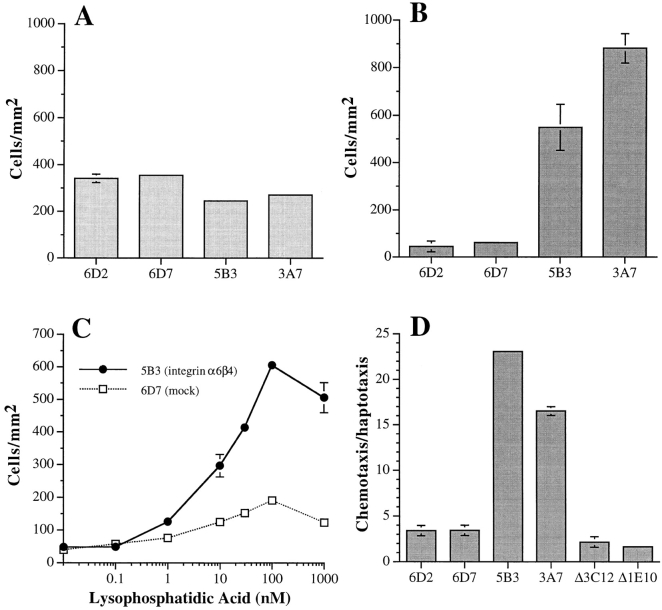
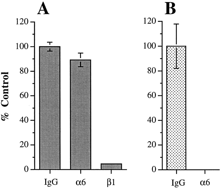
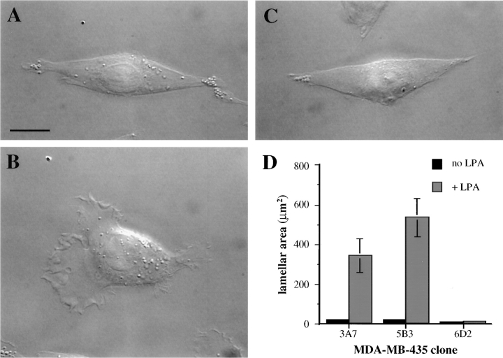
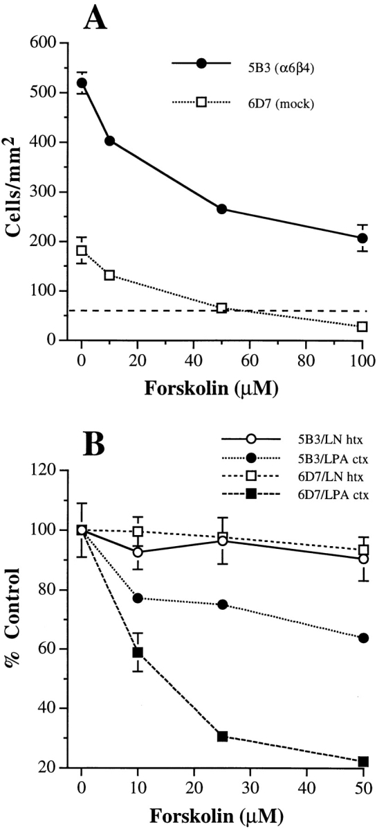
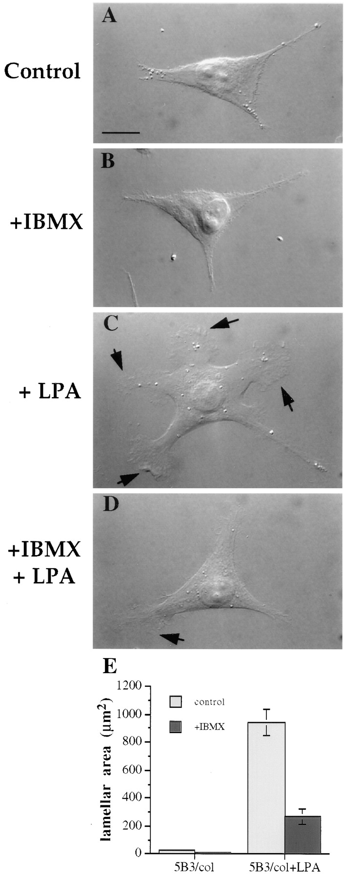
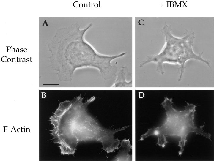
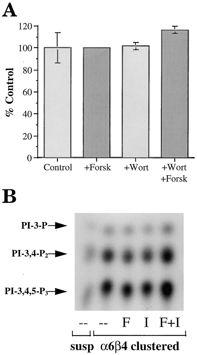
The alpha6beta4 integrin promotes carcinoma in-vasion by its activation of a phosphoinositide 3-OH (PI3-K) signaling pathway (Shaw, L.M., I. Rabinovitz, H.H.-F. Wang, A. Toker, and A.M. Mercurio. Cell. 91: 949-960). We demonstrate here using MDA-MB-435 breast carcinoma cells that alpha6beta4 stimulates chemotactic migration, a key component of invasion, but that it has no influence on haptotaxis. Stimulation of chemotaxis by alpha6beta4 expression was observed in response to either lysophosphatidic acid (LPA) or fibroblast conditioned medium. Moreover, the LPA-dependent formation of lamellae in these cells is dependent upon alpha6beta4 expression. Both lamellae formation and chemotactic migration are inhibited or "gated" by cAMP and our results reveal that a critical function of alpha6beta4 is to suppress the intracellular cAMP concentration by increasing the activity of a rolipram-sensitive, cAMP-specific phosphodiesterase (PDE). This PDE activity is essential for lamellae formation, chemotactic migration and invasion based on data obtained with PDE inhibitors. Although PI3-K and cAMP-specific PDE activities are both required to promote lamellae formation and chemotactic migration, our data indicate that they are components of distinct signaling pathways. The essence of our findings is that alpha6beta4 stimulates the chemotactic migration of carcinoma cells through its ability to influence key signaling events that underlie this critical component of carcinoma invasion.


Figures

Figure 1
Figure 1
Expression of the α6β4 integrin in MDA-MB-435 carcinoma cells stimulates chemotaxis but not haptotaxis. The migration of the MDA/β4 (5B3 and 3A7) MDA/β4-ΔCYT (Δ3C12, Δ1E10), and MDA/mock (6D2 and 6D7) transfectants toward laminin-1 (haptotaxis; A), 3T3 conditioned medium (chemotaxis; B), or LPA (chemotaxis; C and D) was assessed using a modified Boyden chamber. The lower surfaces of Transwell membranes were coated with either laminin-1 (A), conditioned medium (B), or collagen I (C and D), and then either BSA (A) 3T3 conditioned medium (B) or LPA (C and D) was added to the lower chambers. Cells (105 [A and B] or 5 × 10⁴ [C and D]) were placed in the upper chambers. After 4 h at 37°C, cells that did not migrate were removed from the upper chamber with a cotton swab and cells on the opposite side of the membrane were fixed, stained, and quantified manually as described in the Materials and Methods. (A) Haptotaxis toward laminin-1; (B) chemotaxis toward NIH-3T3 conditioned medium; (C) dose response of MDA-MB-435 subclones 5B3 (β4 transfected; solid circles) and 6D7 (mock transfected; open squares) chemotaxis toward LPA; (D) Chemotaxis toward 100 nM LPA. Data are reported as fold increases over haptotactic migration on collagen I in the absence of LPA. Data (all panels) are shown as mean ± standard deviation from triplicate determinations.
Figure 2
Figure 2
Inhibition of α6β4-stimulated migration by integrin-specific antibodies. MDA/β4 (5B3; A, gray bars) or mock transfectants (6D7; B, stippled bars) were incubated with the indicated function blocking mAbs for 30 min before their use in a chemotaxis assay using 100 nM LPA on collagen I (A) or a haptotaxis assay on laminin-1–coated wells (B) as described in Fig. 1. Nonspecific mouse IgG was used as a negative control. Data are reported as the percentage of migration observed for the IgG control ± standard deviation from triplicate determinations.
Figure 3
Figure 3
The α6β4 integrin is required for the LPA-dependent formation of lamellae in MDA-MB-435 cells. MDA/β4 (A and B) and mock transfectants (C) were plated onto coverslips that had been coated with 20 μg/ ml collagen I. Cells were allowed to adhere for 2 h at 37°C and then treated with LPA for 5 min. (B and C) or left untreated (A). The cells were visualized using Nomarski DIC optics. Note the large lamellae that are formed in response to LPA stimulation of the MDA/β4 transfectants. (D) The effect of LPA on lamellar area was quantified using IPLab Spectrum imaging software. Data are shown as mean lamellar area ± standard error in which n > 20. Bar, 10 μm.
Figure 4
Figure 4
Forskolin stimulation of adenyl cyclase inhibits LPA-mediated chemotaxis differentially in the MDA/β4 and mock transfectants. (A) MDA/β4 transfectants (5B3; solid circles) or mock transfectants (6D7; open squares) were treated with the indicated concentration of forskolin for 30 min before their addition to the upper wells of the Transwell chambers. Cells were assayed for LPA-mediated chemotaxis on collagen I as described in Fig. 1. The dashed line depicts the basal level of migration of both subclones in the absence of LPA. (B) In a separate experiment, the same cells were treated with forskolin for 30 min before assaying for LPA chemotaxis (solid symbols) or laminin haptotaxis (open symbols). Data are reported as the percent migration of cells not treated with forskolin ± standard deviation of triplicate determinations.
Figure 5
Figure 5
Intracellular cAMP content of the MDA-MB-435 transfectants. The MDA/β4 (3A7, 5B3), MDA/β4-ΔCYT (Δ3C12, Δ1E10) and MDA/mock transfectants (6D2, 6D7) were plated in DME containing 10% FCS. After 18 h, cells were harvested and cAMP content was measured using a cAMP EIA protocol as described in Materials and Methods. Data shown represent the mean of 10 sample determinations ± standard error. The difference in the [cAMP]i between the MDA/β4 and the mock transfectants is significant (P < 0.001; asterisk), but the difference between the mock and the β4-ΔCYT transfectants is not significant (P = 0.2). (B and C) Differential effects of forskolin stimulation on the [cAMP]i in the MDA/β4 and mock transfectants. The [cAMP]i was assayed in the 5B3 (solid bars) and 6D7 (stippled bars) clones plated on collagen I and treated for 15 min with either 50 μM forskolin (B) or forskolin and 1 mM IBMX (C). Note that the MDA/β4 transfectants (5B3) are more resistant to a forskolin-stimulated increase in [cAMP]i than the mock transfectants (6D7). The inhibition of PDE activity with IBMX shown in C reveals that α6β4 expression results in an increase in PDE activity and not a decrease in cAMP synthesis. Data shown are the mean values ± standard error obtained from multiple experiments.
Figure 6
Figure 6
Assay of cAMP-specific PDE activity. (A) MDA/β4 (3A7 and 5B3) or mock transfectants (6D7) plated on collagen I were treated with 50 μM forskolin or 100 nM LPA as noted. Cells were harvested and the cytosolic fraction was assayed for PDE activity as described in Materials and Methods. The PDE activity of the MDA/β4 transfectants was compared with the MDA/mock transfectants for statistical significance: *, P < 0.002; , P < 0.01. (B) Extracts from cells treated as in A were incubated with 100 μM rolipram before assaying for PDE activity to determine how much of the activity in A constitutes cAMP-specific PDE (PDE 4). Data shown are mean ± standard error of four separate determinations (A and B). ns, not significant; ⋄, P = 0.02. (C) Relative expression of PDE 4B in the MDA-MB-435 transfectants. Extracts (40 μg protein) obtained from the MDA/β4 (3A7 and 5B3) and mock (6D2 and 6D7) transfectants, as well as purified PDE 4 proteins (short form of variants A, B, and D; 10 ng each; provided by M. Conti) were resolved by SDS-PAGE and immunoblotted with a PDE 4B-specific Ab. Arrows, long and short forms of PDE 4B.
Figure 6
Figure 6
Assay of cAMP-specific PDE activity. (A) MDA/β4 (3A7 and 5B3) or mock transfectants (6D7) plated on collagen I were treated with 50 μM forskolin or 100 nM LPA as noted. Cells were harvested and the cytosolic fraction was assayed for PDE activity as described in Materials and Methods. The PDE activity of the MDA/β4 transfectants was compared with the MDA/mock transfectants for statistical significance: *, P < 0.002; , P < 0.01. (B) Extracts from cells treated as in A were incubated with 100 μM rolipram before assaying for PDE activity to determine how much of the activity in A constitutes cAMP-specific PDE (PDE 4). Data shown are mean ± standard error of four separate determinations (A and B). ns, not significant; ⋄, P = 0.02. (C) Relative expression of PDE 4B in the MDA-MB-435 transfectants. Extracts (40 μg protein) obtained from the MDA/β4 (3A7 and 5B3) and mock (6D2 and 6D7) transfectants, as well as purified PDE 4 proteins (short form of variants A, B, and D; 10 ng each; provided by M. Conti) were resolved by SDS-PAGE and immunoblotted with a PDE 4B-specific Ab. Arrows, long and short forms of PDE 4B.
Figure 7
Figure 7
cAMP specific-PDE activity is required for the chemotactic migration and invasion of MDA-MB-435 cells. The MDA/ β4 (5B3; squares) or mock transfectants (6D7; circles) were treated with varying concentrations (A) or 1 mM (B) IBMX for 30 min before their use in either an LPA chemotaxis assay (A) or a Matrigel chemoinvasion assay (B). Data shown represent mean values ± standard deviation of triplicate determinations.
Figure 8
Figure 8
cAMP specific-PDE activity is required for LPA-dependent formation of lamellae in the MDA/β4 transfectants. The MDA/β4 transfectants (5B3) were plated on collagen I–coated coverslips. After 2 h, the cells were either left untreated (A and C) or treated with 1mM IBMX (B and D) for 30 min. Subsequently, the cells were either left untreated (A and B) or treated with 100 nM LPA for 5 min (C and D). The cells were then fixed and visualized using Nomarski DIC optics. (E) The effect of LPA and IBMX on lamellar area was quantified using IPLab Spectrum imaging software. Bars represent mean lamellar area ± standard error in which n > 20. Of note, IBMX inhibited the LPA-dependent formation of lamellae by 70%.
Figure 9
Figure 9
Lamellae formation in clone A colon carcinoma cells requires PDE activity. Clone A colon carcinoma cells were either treated with solvent alone (A and B) or 1 mM IBMX in solvent (C and D) and then plated on laminin-1–coated coverslips. After 45 min the cells were fixed and stained for F-actin using TRITC-phalloidin. (A and C) Phase–contrast images; (B and D) fluorescence images. Bar, 10 μm.
Figure 10
Figure 10
(A) Evaluation of PI3-K involvement in PDE activity. The MDA/β4 transfectants were incubated for 30 min in the presence of either forskolin or wortmannin, or both in combination, before assay of PDE activity as described in Materials and Methods. (B) Evaluation of the cAMP regulation of PI3-K activity. The MDA/β4 and mock transfectants were incubated in suspension with either forskolin or IBMX or both for 10 min. Subsequently, these cells were either maintained in suspension or incubated with a β4 integrin-specific antibody and allowed to adhere to anti-mouse IgG-coated plates or laminin-1–coated plates for 30 min. Aliquots of cell extracts that contained equivalent amounts of protein were incubated with the anti-phosphotyrosine mAb 4G10 and protein A–Sepharose for 3 h. After washing, the beads were resuspended in kinase buffer and incubated for 10 min at room temperature. The phosphorylated lipids were resolved by thin layer chromatography. Arrows, position of the D3-phosphoinositides.

Similar articles

Cited by

References

    1. Aznavoorian S, Stracke ML, Parsons J, McClanahan J, Liotta LA. Integrin αvβ3 mediates chemotactic and haptotactic motility in human melanoma cells through different signaling pathways. J Biol Chem. 1996;271:3247–3254. - PubMed
    1. Borradori L, Sonnenberg A. Hemidesmosomes—roles in adhesion, signaling and human disease. Curr Opin Cell Biol. 1996;8:647–656. - PubMed
    1. Chao C, Lotz MM, Clarke AC, Mercurio AM. A function for the integrin α6β4 in the invasive properties of colorectal carcinoma cells. Cancer Res. 1996;56:4811–4819. - PubMed
    1. Clark EA, Brugge JS. Integrins and signal transduction pathways: The road taken. Science. 1995;268:233–239. - PubMed
    1. Clarke AS, Lotz MM, Chao C, Mercurio AM. Activation of the p21 pathway of growth arrest and apoptosis by the β4integrin cytoplasmic domain. J Biol Chem. 1995;270:22672–22676. - PubMed

Publication types

MeSH terms