. 1998 Dec 15;18(24):10375-88.
doi: 10.1523/JNEUROSCI.18-24-10375.1998.

Nitric oxide-producing islet cells modulate the release of sensory neuropeptides in the rat substantia gelatinosa

Affiliations

Nitric oxide-producing islet cells modulate the release of sensory neuropeptides in the rat substantia gelatinosa

P Aimar et al. J Neurosci. .

Abstract

The substantia gelatinosa of the spinal cord (lamina II) is the major site of integration for nociceptive information. Activation of NMDA glutamate receptor, production of nitric oxide (NO), and enhanced release of substance P and calcitonin gene-related peptide (CGRP) from primary afferents are key events in pain perception and central hyperexcitability. By combining reduced nicotinamide adenine dinucleotide phosphate (NADPH) diaphorase histochemistry for NO-producing neurons with immunogold labeling for substance P, CGRP, and glutamate, we show that (1) NO-producing neurons in lamina IIi are islet cells; (2) these neurons rarely form synapses onto peptide-immunoreactive profiles; and (3) NADPH diaphorase-positive dendrites are often in close spatial relationship with peptide-containing terminals and are observed at the periphery of type II glomeruli showing glutamate-immunoreactive central endings. By means of confocal fluorescent microscopy in acute spinal cord slices loaded with the Ca2+ indicator Indo-1, we also demonstrate that (1) NMDA evokes a substantial [Ca2+]i increase in a subpopulation of neurons in laminae I-II, with morphological features similar to those of islet cells; (2) a different neuronal population in laminae I-IIo, unresponsive to NMDA, displays a significant [Ca2+]i increase after slice perfusion with either substance P and the NO donor 3morpholinosydnonimine (SIN-1); and (3) the responses to both substance P and SIN-1 are either abolished or significantly inhibited by the NK1 receptor antagonist sendide. These results provide compelling evidence that glutamate released at type II glomeruli triggers the production of NO in islet cells within lamina IIi after NMDA receptor activation. The release of substance P from primary afferents triggered by newly synthesized NO may play a crucial role in the cellular mechanism leading to spinal hyperexcitability and increased pain perception.


Figures
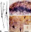
Fig. 1.
Fig. 1.
NADPH-d-stained neurons in the rat cervical dorsal horn. A, Camera lucida drawings of two NADPH-d-positive neurons from a horizontal vibratome slice cut through lamina II. The same slice was then subsequently reembedded for ultrastructural examination. Positive neurons have a fusiform shape and longitudinally oriented dendritic trees with a few long branches. Thearrows indicate axon-like processes originating from proximal dendrites. B,C, Combined enzyme histochemistry for NADPH-d (blue) and peroxidase immunocytochemistry for substance P (brown).B, NADPH-d-positive neuronal cell bodies and processes are particularly abundant in lamina IIi, whereas substance P immunoreactivity is mainly detected in lamina IIo. Lamina I shows similar densities of both labels.C, At higher magnification, NADPH-d positivity and substance P immunoreactivity appear to be present in different neuronal profiles. D, Combined enzyme histochemistry for NADPH-d (pink) and peroxidase immunocytochemistry for the NMDAR1 glutamate receptor (brownish black). A neuronal cell body in lamina IIi shows colocalization of the two labels. Scale bars: A, B, 50 μm;C, D, 25 μm.
Fig. 2.
Fig. 2.
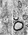
Ultrastructural visualization of NADPH-d positivity in the dorsal horn (A, B) and Lissauer’s tract (C) at the lumbar level of the rat spinal cord. The NBT-positive reaction appears in the form of coarse electrondense deposits that fill almost completely the neuronal cell body and processes without any clear association with specific cell organelles. The lamina II neuron in A has a typical bipolar shape, which is clearly appreciated in the corresponding unstained semithin section (insert) and a characteristically indented nucleus. The arrows point to a small nucleus used as an identifying landmark. Note inB and C the presence of a number of small myelinated axons containing NBT deposits within their axoplasm (arrows and insert). A positive dendrite is also indicated (long arrow) in B.a, Axon; d, dendrite; n, nucleus. Scale bars: AC, 1 μm;inserts, 0.25 μm.
Fig. 3.
Fig. 3.
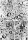
Distribution and connectivity of NADPH-d-positive nerve processes in the lumbar dorsal horn. A, An isolated labeled dendrite (arrow) in lamina I.B, A NADPH-d-positive dendrite (white d) in lamina III receives a synaptic contact (curved arrow) from an unlabeled axon (a). C, NADPH-d-positive nerve processes in lamina IIi. A large positive dendrite (white d) receives a synapse (curved arrow) from an adjacent unlabeled dendrite (black d). Other NADPH-d-positive profiles of smaller caliber (arrows) are part of the dense meshwork of neuronal processes that is observed in lamina IIi.a, Axon; d, dendrite. Scale bars, 1 μm.
Fig. 4.
Fig. 4.
Combined preembedding NADPH-d histochemistry and postembedding peptide immunogold labeling in the lumbar substantia gelatinosa. A, A NADPH-d-positive dendrite (white d, arrow) in lamina IIo is surrounded by a dense meshwork of CGRP–substance P double-labeled axonal profiles (arrowheads). B, A NADPH-d-positive dendrite (white d,arrow) is seen in proximity of a CGRP–substance P double-labeled axonal ending. Peptide immunoreactivity is restricted to the LGVs (insert). C, A NADPH-d-positive peripheral dendrite (white d, arrow) is part of a type I glomerulus showing a central bouton (C1) double-labeled for CGRP and substance P (arrowheads). d, Dendrite;LGVs, large dense-cored synaptic vesicles; CGRP, 20 nm gold; substance P, 10 nm gold. Scale bars, 0.5 μm.
Fig. 5.
Fig. 5.
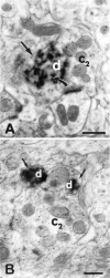
Combined preembedding NADPH-d histochemistry and postembedding glutamate immunogold labeling in the lumbar lamina IIi. A, A NADPH-d-positive dendrite (white d, arrow) is part of a type II glomerulus showing a central bouton (C2) containing 10 nm gold particles indicative of glutamate immunoreactivity. Note the axodendritic contact between the two profiles (curved arrow). B, Two NADPH-d peripheral dendrites (white d, arrow) in a glutamate-immunoreactive type II glomerulus. d, Dendrite; C2, central ending in glomerulus. Scale bars, 0.5 μm.
Fig. 6.
Fig. 6.
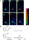
Different responses of substantia gelatinosa neurons to stimulation with substance P and NMDA. A, Time series of pseudocolor images of the [Ca2+]i changes occurring in two Indo-1-loaded cells (1 and 2) from the dorsal horn of a young rat (P8) after subsequent perfusion of the slice with 100 μm NMDA (sequence ac) and 5 μmsubstance P (sequence df). Images in af correspond to the time points af indicated inB. The R405/485 is displayed as a pseudocolor scale. Sampling rate, 2 sec. Scale bar, 10 μm. B, Kinetics of the [Ca2+]i changes in cell 1 and cell 2 after NMDA and substance P stimulation, as expressed by the ratio between Indo-1 emission wavelength at 405 and 485 nm. Calibration: 30 sec.
Fig. 7.
Fig. 7.
Kinetics of the [Ca2+]i change in one typical Indo-1-loaded neuron from the P8 rat substantia gelatinosa, after successive stimulation with the NO donor SIN-1. The NK1receptor antagonist sendide inhibited the response otherwise observed after SIN-1 application. The same neuron displayed a significant [Ca2+]i increase on substance P but not NMDA stimulation. Sampling rate, 2 sec. Calibration: 100 sec.
Fig. 8.
Fig. 8.
Hypothetical mechanism of action of NO on peptide-containing primary afferent C fibers. Stimulation of Aδ afferents evokes a release of glutamate from C2 central endings in type II glomeruli within lamina IIi. In hyperalgesic conditions, such a stimulation would be effective in activating the NMDA–NO cascade in the peripheral dendrites of glomeruli, which originate from NADPH-d-positive islet cells. NO released from the dendritic tree of islet cells diffuses throughout lamina IIo and enhances the release of substance P (and CGRP) from C fiber varicosities and terminals, which are enriched of peptide-containing LGVs. The simultaneous and prolonged release of sensory neuropeptides throughout the substantia gelatinosa represents one of the mechanisms of central sensitization.

References

    1. Abdel-Maguid TE, Bowsher D. Interneurons and proprioneurons in the adult human spinal grey matter and in the general somatic and visceral afferent cranial nerve nuclei. J Anat. 1984;139:9–19. - PMC - PubMed
    1. Aimar P, Barajon I, Merighi A. Ultrastructural features and synaptic connections of NADPH-diaphorase positive neurones in the rat spinal dorsal horn and central grey matter. Eur J Anat. 1998;2:27–33.
    1. Aimi Y, Fujimura M, Vincent SR, Kimura H. Localization of NADPH-diaphorase-containing neurons in sensory ganglia of the rat. J Comp Neurol. 1991;306:382–392. - PubMed
    1. Aley KO, McCarter G, Levine JD. Nitric oxide signaling in pain and nociceptor sensitization in the rat. J Neurosci. 1998;18:7008–7014. - PMC - PubMed
    1. Bennett GJ, Abdelmoumene M, Hayashi H, Dubner R. Physiology and morphology of substantia gelatinosa neurons intracellularly stained with horseradish peroxidase. J Comp Neurol. 1980;194:809–827. - PubMed

Publication types

MeSH terms

Grants and funding