. 1998 Dec 15;18(24):10525-40.
doi: 10.1523/JNEUROSCI.18-24-10525.1998.

The connection from cortical area V1 to V5: a light and electron microscopic study

Affiliations

The connection from cortical area V1 to V5: a light and electron microscopic study

J C Anderson et al. J Neurosci. .

Abstract

Area V5 (middle temporal) in the superior temporal sulcus of macaque receives a direct projection from the primary visual cortex (V1). By injecting anterograde tracers (biotinylated dextran and Phaseolus vulgaris lectin) into V1, we have examined the synaptic boutons that they form in V5 in the electron microscope. Nearly 80% of the target cells in V5 were spiny (excitatory). The boutons formed asymmetric (Gray's type 1) synapses with spines (54%), dendrites (33%), and somata (13%). All somatic targets and some (26%) of the target dendritic shafts showed features characteristic of smooth (inhibitory) cells. Each bouton formed, on average, 1.7 synapses. The larger boutons formed multiple synapses with the same neuron and completely enveloped the entire spine head. On most dendritic shafts and all somata the postsynaptic density en face was disk-shaped but in about half the cases the reconstructed postsynaptic densities of synapses on spines appeared as complete or partial annuli. Even in the zones of densest innervation only 3% of the asymmetric synapses were formed by the labeled boutons. Although the V1 projection forms only a small minority of synapses in V5, its affect could be considerably amplified by local circuits in V5, in a way analogous to the amplification of the small thalamic input to area V1.


Figures

Fig. 1.
Fig. 1.
Summary schematic of macaque brain showing position of three injections of neuronal tracers (2 of BDA and 1 of PHA-L) into primary visual cortex, area V1, of two animals.
Fig. 2.
Fig. 2.
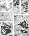
Correlated light and electron micrographs of BDA/PHA-L-labeled electron dense axon and boutons. A, Photomontage of an axon collateral located in lower layer 4 of area V5; b1, b2, b3, and b4 are selected boutons. The associated synapses of these boutons are shown in the following electron micrographs.B, Bouton b1 forms an asymmetric synapse (solid arrowhead) with a spine head (sp1) that can be traced back to the parent dendrite (d) in a single section. The dendrite produces a second spine (sp2), which receives an asymmetric synapse (small arrow) from an unidentified bouton.C, A myelinated axon collateral (m) gives rise to a bouton (b2) forming an asymmetric synapse (solid arrowhead) with a spine (sp). D, A large bouton (b3) packed with vesicles and mitochondria forms an asymmetric synapse (solid arrowheads) with a spine (sp). The spine profile has been completely embraced by the filled bouton. The postsynaptic density does not appear as a continuous structure but instead is perforated or complex.E, Another large bouton (b4) forms asymmetric synapses (solid arrowheads) with a spine (sp) and the shaft of a dendrite (d). The spine apparatus is clearly visible. The dendrite also forms an asymmetric synapse (small arrow) with an unidentified bouton. Scale bars: A, 10 μm;B, 1 μm; C–E, 0.5 μm.
Fig. 3.
Fig. 3.
Electron micrographs of BDA/PHA-L-labeled boutons found in layer 6 of area V5. A, Bouton filled with mitochondria forming two asymmetric synapses (solid arrowheads) with the same target neuron. One of the targets is clearly a spine (sp; note the spine apparatus) that can be traced back to the parent dendrite (d). The second synapse forms on a region of the dendritic shaft that projects slightly into the neuropil. Serial section reconstruction showed this projection to be a sessile or “neckless” spine. B, A spine (sp) containing spine apparatus forms an asymmetric synapse with the labeled bouton, which shows a complex postsynaptic density (solid arrowheads) within the spine. Scale bars, 0.5 μm.
Fig. 4.
Fig. 4.
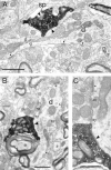
Electron micrographs of labeled synaptic boutons in contact with dendrites of smooth cells. A, Characteristically large bouton, filled with vesicles, located in lower layer 4 forms two asymmetric synapses (large, solid arrowheads), one with a dendrite (d) and the other with a spine (sp). The dendrite contains numerous mitochondria and forms many synapses of the symmetric (open arrowheads) and asymmetric (small, solid arrowheads) types with unidentified boutons. When the target dendrite was reconstructed over several serial sections, it became clear that the variations in diameter, which can be visible in the micrograph, were caused by a varicose or beaded morphology. These features are consistent with those of neurons with smooth dendrites, which contain GABA. In subsequent sections of the series, the same labeled bouton formed another two synapses with the dendrite. The spine target is almost completely enveloped by the bouton. The extensive postsynaptic density within the spine indicates that the synapse has been sectioned at an oblique plane. B, Large bouton located in layer 6 with a dendritic target similar to the one described above (Fig.3A). The labeled bouton forms an asymmetric synapse (large, solid arrowhead) with the dendrite, which also forms symmetric synapses (open arrowheads), and other asymmetric synapses (small, solid arrowheads) from unidentified boutons. C, A higher-power micrograph of the adjacent section in the series of the bouton shown in Figure3B. Detail of the unlabeled symmetric synapse (open arrowhead) and the labeled and unlabeled asymmetric synapses (large, solid and small, solid arrowheads, respectively) can be compared. Scale bars:A, B, 1 μm: C, 0.5 μm.
Fig. 5.
Fig. 5.
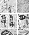
Correlated light and electron micrographs of BDA/PHA-L-filled boutons in synaptic contact with a soma (S) located in layer 4 of area V5.A, Low-power light micrograph of an identified axon collateral (large, solid arrow) rising through lower layer 4. The soma (S) and some of the boutons (small arrows) appear at higher magnification inB. B, Numbered boutons (b1, b2, b3, b4) all form contacts with the soma (S). A large-diameter myelinated axon (m) is indicated for reference. C, Low-power electron micrograph of the soma (S) seen in B. The myelinated axon (m) referred to in B can be seen in close contact with the soma. Some of the mitochondria appear to be lightly stained (small arrows) , thus enabling the soma to be seen at the light microscopic level. The large filled axon (ax) gives rise to the boutons (b2, b3) after losing its myelin sheath (not illustrated). D, High-power electron micrograph of boutons b2 and b3. The bouton b2 forms an asymmetric synapse (solid arrowhead) with the soma. Electron dense mitochondria (mit) can be seen within the cytoplasm of the soma. E, F, In sections after that shown inD, boutons b3 and b1 can be seen to form asymmetric synapses (solid arrowheads) with the soma.G, The asymmetric synapse (solid arrowhead) of the fourth bouton (b4) in contact with the soma can be compared with the asymmetric synapses (small arrows) formed by an unlabeled bouton also in contact with the soma. H, High-power micrograph of a crystalline inclusion (i) within the body of a mitochondria found in the soma contacted by the above boutons. Scale bars: A, B, 10 μm; C, 5 μm;D–H, 0.5 μm.
Fig. 6.
Fig. 6.
Three-dimensional reconstructions from serial ultrathin sections of filled boutons in area V5 showing how targets are enveloped by the bouton. A, B, Two views of a bouton (blue) found in layer 6. In A a clear depression or pocket can be seen at the top (uppermost pole) of the bouton. The edge of the postsynaptic density (yellow) can be seen at the lip of the depression. In B the bouton was rotated around a vertical axis (∼180°) to provide a better view of the postsynaptic density. C, D, A large layer 6 bouton (blue) that forms synapses with three spines (brown). In C the bouton and spines have an opaque skin, and in D the skin is transparent. The two spines on the right are deeply embedded within the bouton. The postsynaptic surface (yellow) is shown in apposition with the spines. Axes, 0.5 μm.
Fig. 7.
Fig. 7.
Reconstruction of a large bouton (blue) in layer 4 of area V5 that forms synapses with four spines and a dendrite. A, When the bouton is rendered with an opaque skin it can be seen to wrap around the dendrite (d in B). This dendrite forms three synapses with the identified bouton. The spines (s inB) are all deeply embedded in the bouton.B, Both bouton and targets are rendered transparent, showing the postsynaptic density. The asymmetric synapses areyellow. One of the spines also forms a symmetric synapse (red) with an unidentified bouton. Axes, 0.5 μm.
Fig. 8.
Fig. 8.
Two-dimensional projection of the reconstructed postsynaptic densities found on spines, soma, and dendrites postsynaptic to labeled boutons in area V5. The densities are from individual synapses and are ordered by increasing surface area calculated from the 3-D reconstructions. Scale bar, 1 μm.
Fig. 9.
Fig. 9.
Histogram of the distribution of postsynaptic areas (μm2) formed by labeled boutons in area V5.
Fig. 10.
Fig. 10.
Two different views of two boutons reconstructed and rendered with transparent skins to show solid mitochondria and postsynaptic specializations (yellow). The mitochondria are color-coded for identification of individual structures. For both boutons the right-hand image (B, D) is rotated (∼180°) about the horizontal axis of the left-hand image. A, B, Bouton found in layer 6 that formed synapses with two spines and contained four mitochondria (pink, blue, orange, and green). The longest mitochondrion (pink) was branched and formed three loops. Both synaptic surfaces (yellow) are presented at oblique, nonoptimal elevations but appear as incomplete annuli. C, D, Layer 4 bouton formed synapses with two spines and contained nine mitochondria. One mitochondrion (pink) is branched and forms loops. The synaptic specializations (yellow) become superimposed on each other in these views. One synapse is horseshoe-shaped and the second is composed of two small patches. Axes, 0.5 μm.
Fig. 11.
Fig. 11.
Histogram of synaptic targets of boutons in area V5 originating from neurons labeled in area V1.
Fig. 12.
Fig. 12.
Histogram of the number of synapses (1, 2, 3, 4, 5 or more per bouton) formed by individual labeled boutons in layers 4 and 6 of area V5.
Fig. 13.
Fig. 13.
Histogram showing the distribution of area (μm2) of labeled (black bars) and nonlabeled (white bars) synaptic boutons in area V5. Only those boutons with disappearing synapses were measured.

References

    1. Adelson EH, Movshon JA. Phenomenal coherence of moving visual patterns. Nature. 1982;300:523–525. - PubMed
    1. Ahmed B, Anderson JC, Martin KAC, Nelson JC. Polyneuronal innervation of spiny stellate neurons in cat visual cortex. J Comp Neurol. 1994;341:39–49. - PubMed
    1. Ahmed B, Anderson JC, Martin KAC, Nelson JC. Map of the synapses onto layer 4 basket cells of the primary visual cortex of the cat. J Comp Neurol. 1997;380:230–242. - PubMed
    1. Allman JM, Kaas JH. A representation of the visual field in the caudal third of the middle temporal gyrus of the owl monkey (Aotus trivirgatus). Brain Res. 1971;31:85–105. - PubMed
    1. Baude A, Nusser Z, Molnár E, McIlhenny RAJ, Somogyi P. High-resolution immunogold localization of AMPA type glutamate receptor subunits at synaptic and non-synaptic sites in rat hippocampus. Neuroscience. 1995;69:1031–1055. - PubMed

Publication types

MeSH terms