

Psychostimulant-induced Fos protein expression in the thalamic paraventricular nucleus
- PMID: 9852603
- PMCID: PMC6793371
- DOI: 10.1523/JNEUROSCI.18-24-10680.1998
Psychostimulant-induced Fos protein expression in the thalamic paraventricular nucleus
Abstract
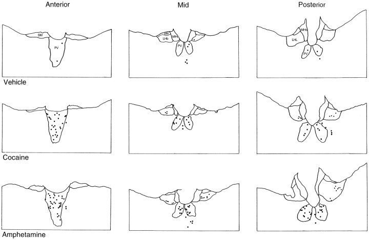
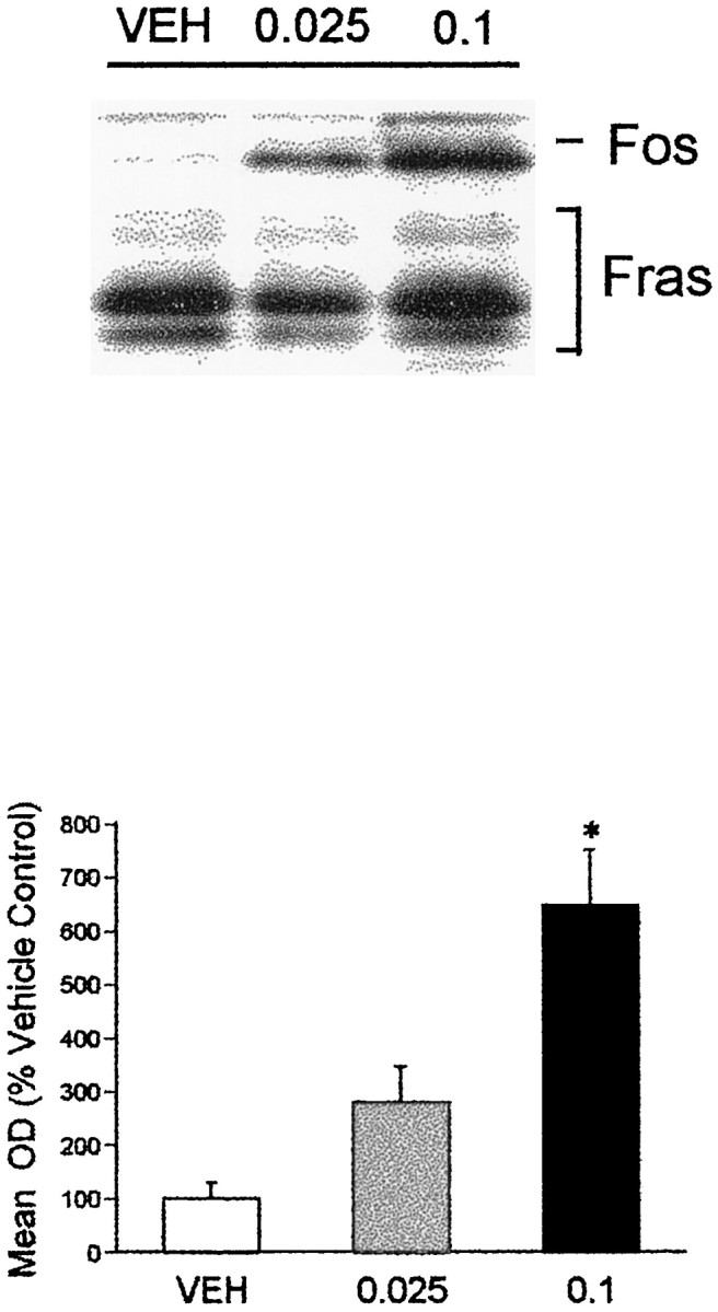
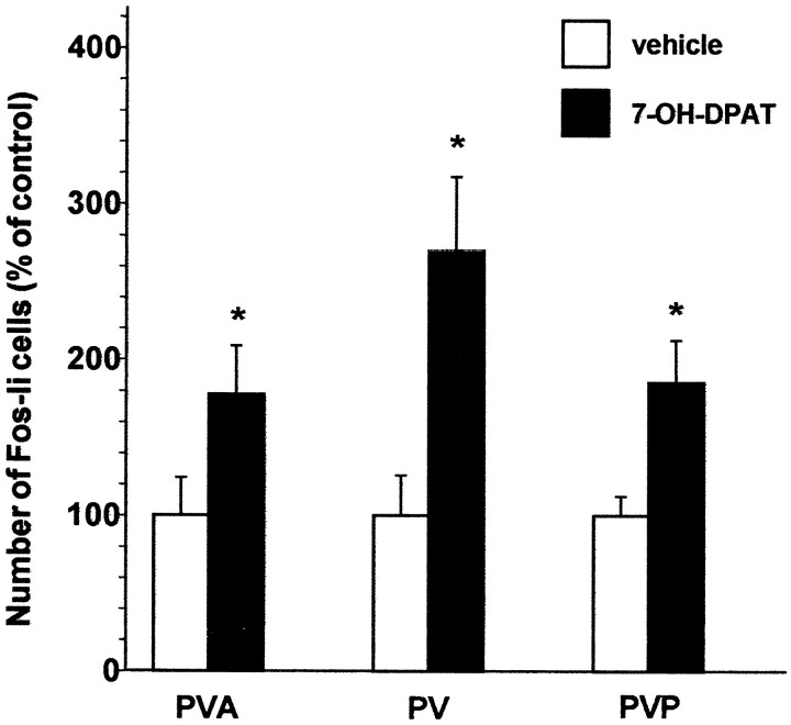
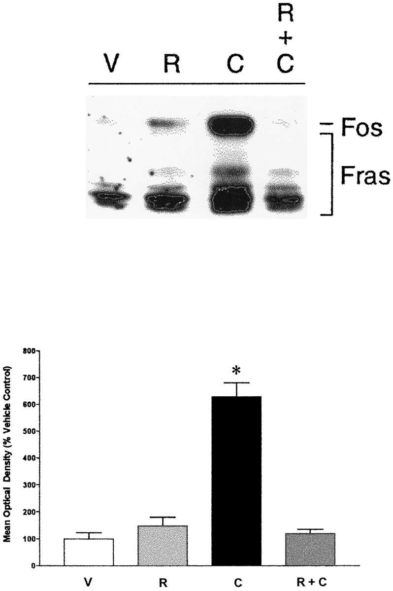
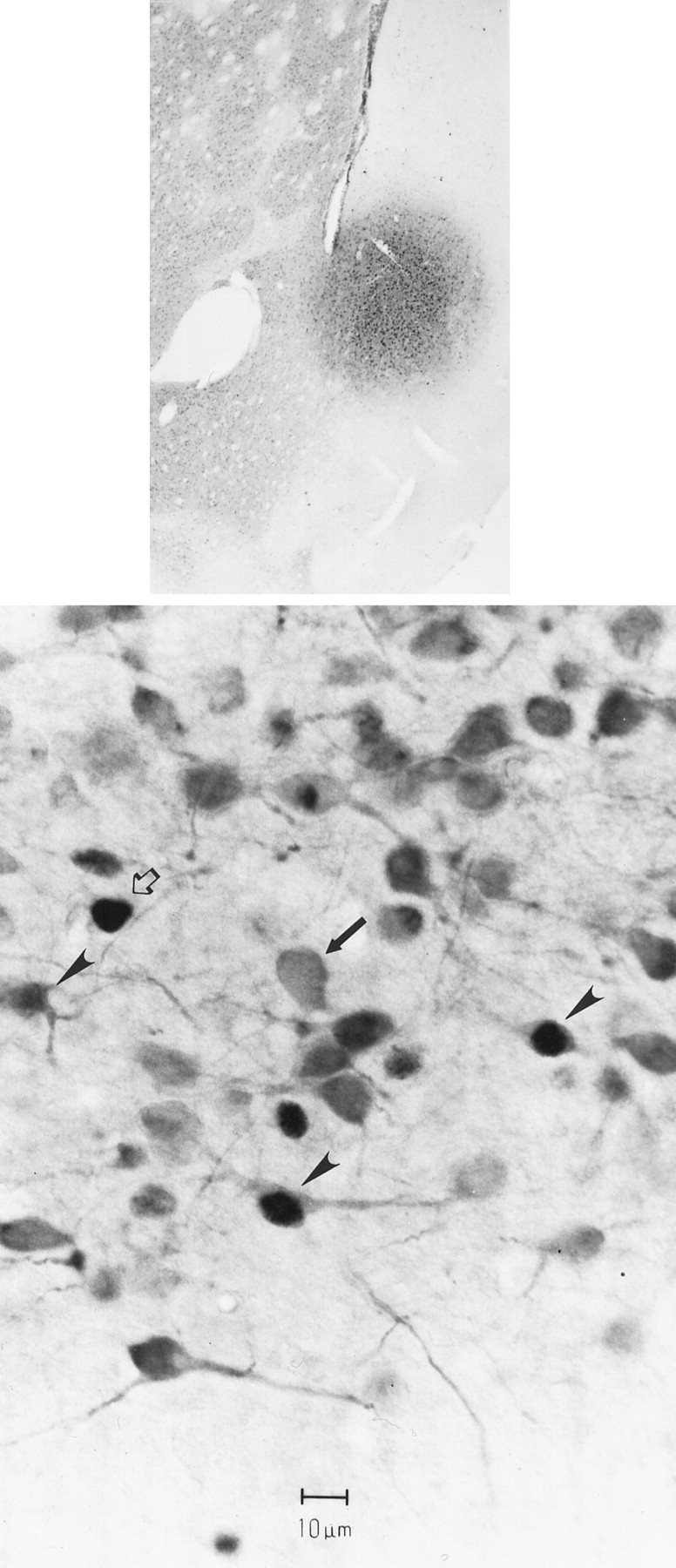
Lesions of glutamatergic afferents to the nucleus accumbens have been reported to block psychostimulant-induced behavioral sensitization. However, thalamic glutamatergic projections to the nucleus accumbens have received little attention in the context of psychostimulant actions. We examined the effects of acute amphetamine and cocaine administration on expression of Fos protein in the thalamic paraventricular nucleus (PVT), which provides glutamatergic inputs to the nucleus accumbens and also receives dopaminergic afferents. Immunoblot and immunohistochemical studies revealed that both psychostimulants dose-dependently increased PVT Fos expression. PVT neurons retrogradely labeled from the nucleus accumbens were among the PVT cells that showed a Fos response to amphetamine. D2 family dopamine agonists, including low doses of the D3-preferring agonist 7-OH-DPAT, increased the numbers of Fos-like-immunoreactive neurons in the PVT. Conversely, the effects of cocaine and amphetamine on PVT Fos expression were blocked by pretreatment with the dopamine D2/3 antagonist raclopride. Because PVT neurons express D3 but not other dopamine receptor transcripts, it appears that psychostimulants induce Fos in PVT neurons through a D3 dopamine receptor. We suggest that the PVT may be an important part of an extended circuit subserving both the arousing properties and reinforcing aspects of psychostimulants.
Figures


Similar articles
-
CART neurons in the lateral hypothalamus communicate with the nucleus accumbens shell via glutamatergic neurons in paraventricular thalamic nucleus to modulate reward behavior.Brain Struct Funct. 2018 Apr;223(3):1313-1328. doi: 10.1007/s00429-017-1544-6. Epub 2017 Nov 7. Brain Struct Funct. 2018. PMID: 29116427
-
Stress induces Fos expression in neurons of the thalamic paraventricular nucleus that innervate limbic forebrain sites.Synapse. 1999 Apr;32(1):13-22. doi: 10.1002/(SICI)1098-2396(199904)32:1<13::AID-SYN2>3.0.CO;2-R. Synapse. 1999. PMID: 10188633
-
Neurochemical evidence that postsynaptic nucleus accumbens D3 receptor stimulation enhances cocaine reinforcement.J Neurochem. 1996 Sep;67(3):1078-89. doi: 10.1046/j.1471-4159.1996.67031078.x. J Neurochem. 1996. PMID: 8752115
-
Mediation of the discriminative stimulus properties of cocaine by mesocorticolimbic dopamine systems.Pharmacol Biochem Behav. 1997 Jul;57(3):601-7. doi: 10.1016/s0091-3057(96)00434-0. Pharmacol Biochem Behav. 1997. PMID: 9218282 Review.
-
A circuitry model of the expression of behavioral sensitization to amphetamine-like psychostimulants.Brain Res Brain Res Rev. 1997 Oct;25(2):192-216. doi: 10.1016/s0165-0173(97)00021-0. Brain Res Brain Res Rev. 1997. PMID: 9403138 Review.
Cited by
-
Relative contributions of the thalamus and the paraventricular nucleus of the hypothalamus to the cardiac sympathetic afferent reflex.Am J Physiol Regul Integr Comp Physiol. 2013 Jul 1;305(1):R50-9. doi: 10.1152/ajpregu.00004.2013. Epub 2013 Apr 24. Am J Physiol Regul Integr Comp Physiol. 2013. PMID: 23616108 Free PMC article.
-
Transient inactivation of the paraventricular nucleus of the thalamus enhances cue-induced reinstatement in goal-trackers, but not sign-trackers.Psychopharmacology (Berl). 2018 Apr;235(4):999-1014. doi: 10.1007/s00213-017-4816-1. Epub 2017 Dec 28. Psychopharmacology (Berl). 2018. PMID: 29285634 Free PMC article.
-
Influences of Stress and Sex on the Paraventricular Thalamus: Implications for Motivated Behavior.Front Behav Neurosci. 2021 Feb 26;15:636203. doi: 10.3389/fnbeh.2021.636203. eCollection 2021. Front Behav Neurosci. 2021. PMID: 33716683 Free PMC article.
-
Heterogeneity in the Paraventricular Thalamus: The Traffic Light of Motivated Behaviors.Front Behav Neurosci. 2020 Oct 16;14:590528. doi: 10.3389/fnbeh.2020.590528. eCollection 2020. Front Behav Neurosci. 2020. PMID: 33177999 Free PMC article.
-
Glutamatergic pathways from medial prefrontal cortex to paraventricular nucleus of thalamus contribute to the methamphetamine-induced conditioned place preference without affecting wakefulness.Theranostics. 2025 Jan 2;15(5):1822-1841. doi: 10.7150/thno.100688. eCollection 2025. Theranostics. 2025. PMID: 39897554 Free PMC article.
References
-
- Acri JB, Carter SR, Alling K, Geter-Douglass B, Dijkstra D, Wikstrom H, Katz JL, Witkin JM. Assessment of cocaine-like discriminative stimulus effects of dopamine D3 receptor ligands. Eur J Pharmacol. 1995;260:177–181. - PubMed
-
- Berendse HW, Groenewegen HJ. Organization of the thalamostriatal projections in the rat, with special emphasis on the ventral striatum. J Comp Neurol. 1990;299:187–228. - PubMed
-
- Berendse HW, Groenewegen HJ. Restricted cortical termination fields of the midline and intralaminar nuclei in the rat. Neurosci. 1991;42:73–102. - PubMed
-
- Bevins RA, Klebaur JE, Bardo MT. 7-OH-DPAT has d-amphetamine-like discriminative stimulus properties. Pharmacol Biochem Behav. 1997;58:485–490. - PubMed
-
- Bolton RF, Cornwall J, Phillipson OT. Collateral axons of cholinergic pontine neurones projecting to midline, mediodorsal and parafascicular thalamic nuclei in the rat. J Chem Neuroanat. 1993;6:101–114. - PubMed
Publication types
MeSH terms
Substances
Grants and funding
LinkOut - more resources
Full Text Sources