. 1998 Dec 15;18(24):10749-61.
doi: 10.1523/JNEUROSCI.18-24-10749.1998.

Glutamate inhibits GABA excitatory activity in developing neurons

Affiliations

Glutamate inhibits GABA excitatory activity in developing neurons

A N van den Pol et al. J Neurosci. .

Abstract

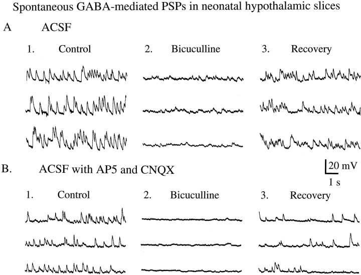
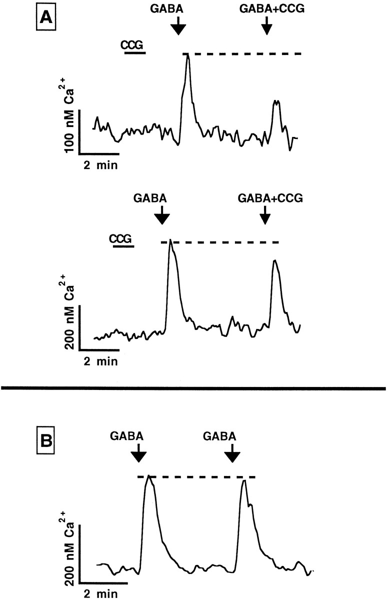
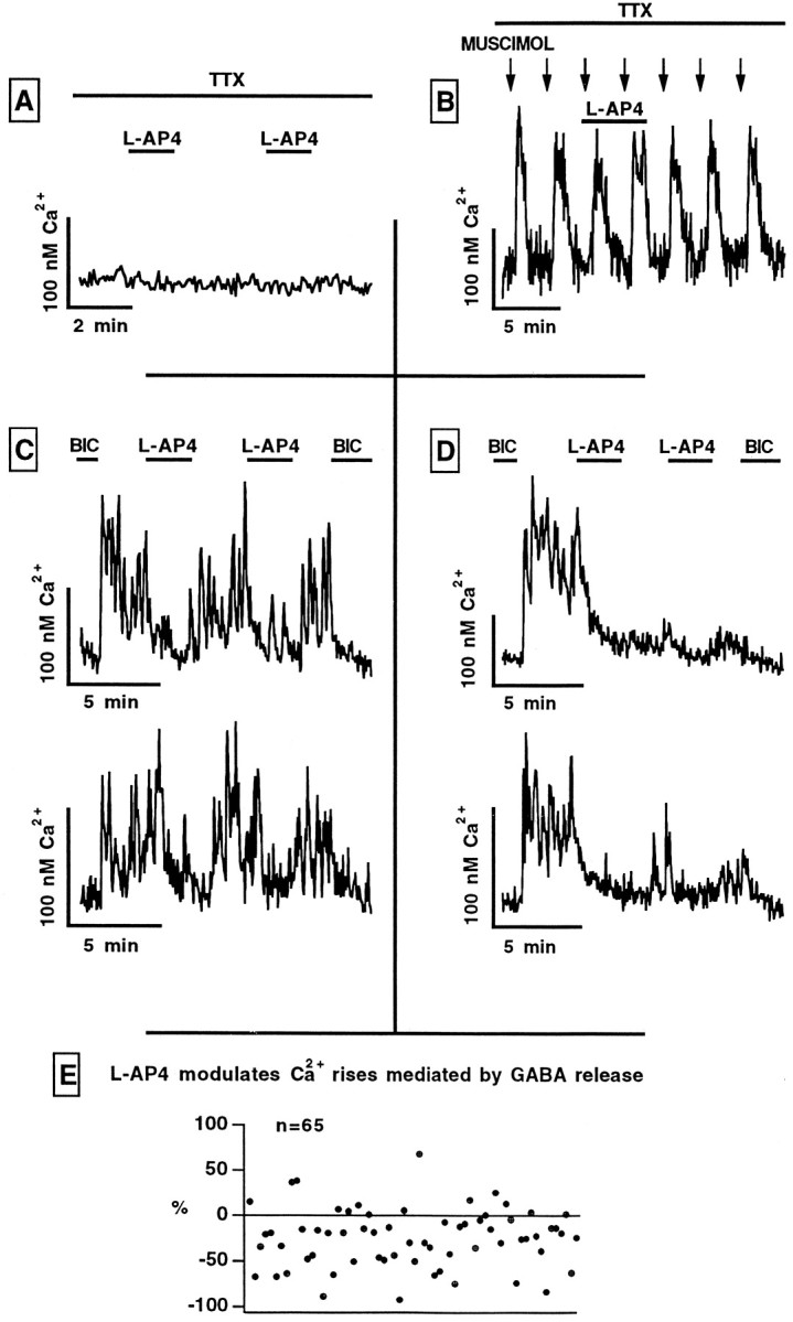
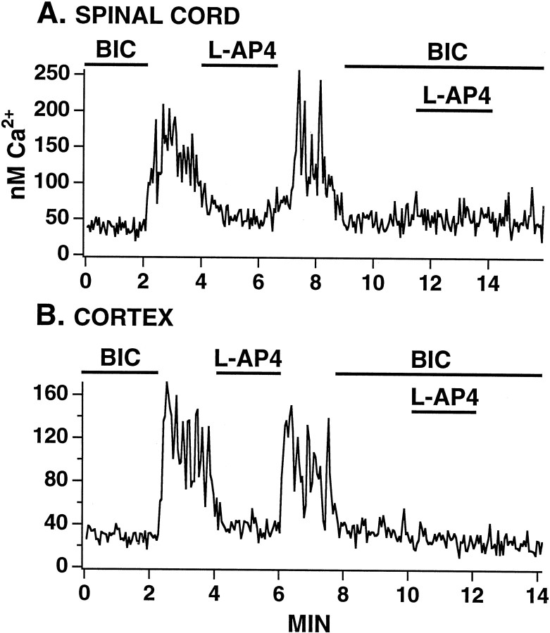
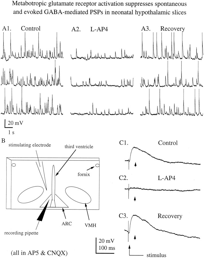
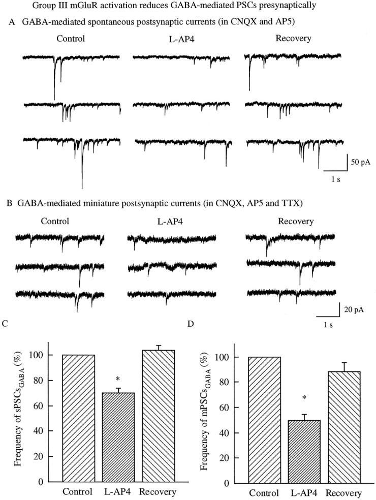
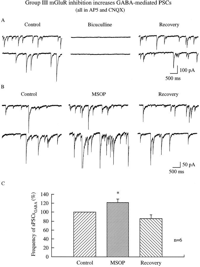
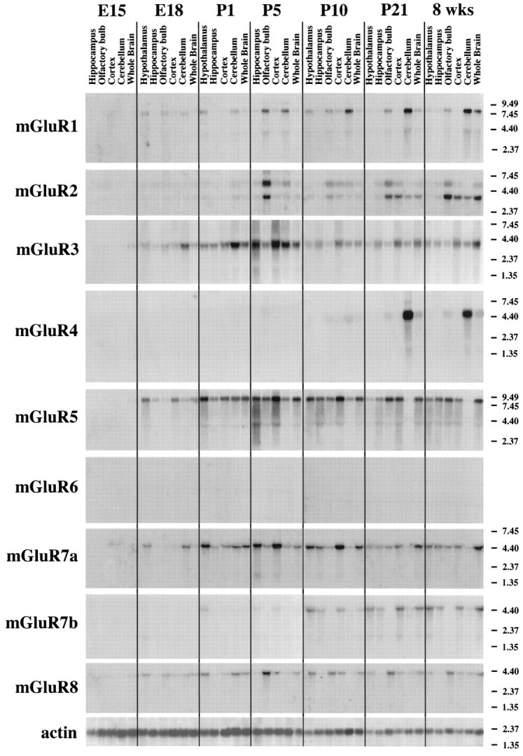
In contrast to the mature brain, in which GABA is the major inhibitory neurotransmitter, in the developing brain GABA can be excitatory, leading to depolarization, increased cytoplasmic calcium, and action potentials. We find in developing hypothalamic neurons that glutamate can inhibit the excitatory actions of GABA, as revealed with fura-2 digital imaging and whole-cell recording in cultures and brain slices. Several mechanisms for the inhibitory role of glutamate were identified. Glutamate reduced the amplitude of the cytoplasmic calcium rise evoked by GABA, in part by activation of group II metabotropic glutamate receptors (mGluRs). Presynaptically, activation of the group III mGluRs caused a striking inhibition of GABA release in early stages of synapse formation. Similar inhibitory actions of the group III mGluR agonist L-AP4 on depolarizing GABA activity were found in developing hypothalamic, cortical, and spinal cord neurons in vitro, suggesting this may be a widespread mechanism of inhibition in neurons throughout the developing brain. Antagonists of group III mGluRs increased GABA activity, suggesting an ongoing spontaneous glutamate-mediated inhibition of excitatory GABA actions in developing neurons. Northern blots revealed that many mGluRs were expressed early in brain development, including times of synaptogenesis. Together these data suggest that in developing neurons glutamate can inhibit the excitatory actions of GABA at both presynaptic and postsynaptic sites, and this may be one set of mechanisms whereby the actions of two excitatory transmitters, GABA and glutamate, do not lead to runaway excitation in the developing brain. In addition to its independent excitatory role that has been the subject of much attention, our data suggest that glutamate may also play an inhibitory role in modulating the calcium-elevating actions of GABA that may affect neuronal migration, synapse formation, neurite outgrowth, and growth cone guidance during early brain development.


Figures

Fig. 1.
Fig. 1.
Spontaneous GABA-mediated postsynaptic potentials in neonatal hypothalamic slices. A, Spontaneous depolarizing PSPs detected with whole-cell recording in a P1 arcuate–ventromedial nucleus (ARC–VMH) neuron in normal ACSF were reversibly blocked by the addition of the GABAAreceptor antagonist bicuculline (30 μm). After bicuculline washout, PSPs recover. B, Large depolarizing PSPs were also observed in neonatal ARC–VMH neurons in the presence of AP5 (50 μm) and CNQX (25 μm) and were reversibly blocked by the addition of bicuculline (30 μm). This observation suggests that in the developing hypothalamus GABA-mediated activity is not dependent on ionotropic glutamate receptor activation. The traces inB were obtained in a hypothalamic slice from a P2 rat. Recordings in A and B were performed using KCl electrodes, and the membrane potential of these neurons was held at ∼90 mV throughout the respective experiments.
Fig. 2.
Fig. 2.
Glutamate reduces the amplitude of GABA-mediated calcium rises. In three hypothalamic neurons recorded simultaneously with digital fura-2 imaging after 3 d in vitro, both GABA (5 μm) and glutamate (5 μm) evoked Ca2+ rises. In these neurons the rise evoked by GABA is of greater amplitude than that evoked by glutamate. The addition of glutamate to GABA reduced the amplitude of the GABA-evoked Ca2+ rise. Each transmitter was applied for 30 sec and allowed a 5 min recovery time before the next transmitter application.
Fig. 3.
Fig. 3.
Group II metabotropic glutamate receptor agonist reduces Ca2+ response to GABA. A, Two neurons from different hypothalamic cultures show no direct Ca2+ response to the group II metabotropic glutamate receptor agonist CCG alone, but when CCG (50 μm) is added together with GABA (5 μm), a substantial decrease in the GABA-evoked Ca2+ rise is found. B, In a control experiment, a typical cell showed similar amplitude Ca2+ rises in response to GABA (5 μm).
Fig. 4.
Fig. 4.
Group III metabotropic receptor activation modulates GABA-elevating actions presynaptically but not postsynaptically. A, l-AP4 (100 μm), a group III metabotropic glutamate receptor agonist, elicited no Ca2+ change in the presence of tetrodotoxin (TTX; 1 μm) in cultured hypothalamic neurons. B, l-AP4 (100 μm) did not modulate Ca2+ rises in response to the GABAA receptor agonist muscimol (5 μm), depicted in a typical neuron that showed a Ca2+ rise in response to muscimol in the presence of TTX (1 μm). These data suggest l-AP4 has little detectable postsynaptic effect on modulating GABA actions.C, In the presence of glutamate receptor antagonists AP5 (100 μm) and CNQX (10 μm), synaptically released GABA-generated Ca2+ rises were blocked by the GABAA receptor antagonist bicuculline (BIC; 30 μm). l-AP4 reduced the Ca2+ activity in both cells recorded at the same time. Because we found no postsynaptic modulation of GABA byl-AP4, this suggests an inhibition of GABA release from presynaptic axonal terminals. D, Some neurons showed an extended l-AP4 depression of GABA release as evidenced by the relative lack of recovery after l-AP4 washout.E, This scatterplot shows the effect ofl-AP4 on Ca2+ raised by synaptically released GABA. The values were determined by the ratio Ca2+ in control (pre-l-AP4) to the Ca2+ value during l-AP4, and are represented by the percent change. Values below zero show the percent inhibition of l-AP4 on Ca2+ levels mediated by synaptically released GABA. Each point represents the percent change in Ca2+ for a single neuron. The zero (0) baseline represents the pre-l-AP4 level for a particular cell.
Fig. 5.
Fig. 5.
Group III metabotropic receptor agonist depresses excitatory GABA actions in cortex and spinal cord neurons. In spinal cord (A) and cortical (B) neurons cultured for 4–6 d, and in the presence of AP5 (100 μm) and CNQX (10 μm), bicuculline (20 μm) depressed calcium levels, indicating a dependence on synaptic GABA activity. l-AP4 (50 μm) caused a strong decrease in calcium levels that recovered afterl-AP4 washout. In the presence of bicuculline (30 μm), l-AP4 had no effect on cytosolic calcium, suggesting that its effect was dependent on the excitatory actions of GABA.
Fig. 6.
Fig. 6.
Metabotropic glutamate receptor activation suppresses spontaneous and evoked GABA-mediated PSPs in the neonatal hypothalamic slice. A, The frequency of spontaneous GABAergic PSPs recorded from a postnatal day 4 ARC–VMH neuron was reversibly suppressed by the addition of l-AP4 (100 μm). The membrane potential of this neuron was held at approximately −95 mV throughout the experiment, and the spikes inA1 and A3 are clipped. B, Schematic diagram demonstrating the approximate configuration of stimulating and recording pipettes used in the experiments examining the effect of metabotropic glutamate receptor activation on evoked GABA-mediated PSPs. C, Application of l-AP4 (100 μm) reversibly suppressed monosynaptically evoked GABA-mediated PSPs in a P5 ARC–VMH neuron. In this experiment monosynaptic PSPs (small arrow) were examined by delivering an electrical stimulus at the junction of the ventromedial and arcuate nuclei; l-AP4 showed a substantial reduction in PSP amplitude. The baseline membrane potential of this neuron was held at approximately −70 mV in the examples shown here.
Fig. 7.
Fig. 7.
Group III metabotropic glutamate receptor activation reduces the frequency of spontaneous and miniature GABA-mediated EPSCs. A, In this typical hypothalamic neuron in culture, the addition of l-AP4 (100 μm) reduced the frequency of spontaneous EPSCs in the presence of glutamate receptor antagonists AP5 (100 μm) and CNQX (10 μm). After l-AP4 washout, the frequency of EPSCs increased again (Recovery).B, An example of mEPSCs in the presence of AP5 (100 μm), CNQX (10 μm), and TTX (1 μm). The frequency of mEPSCs is reduced byl-AP4. C, The bar graph shows the mean spontaneous EPSC frequency before, during, and afterl-AP4 administration in five of six neurons that responded to l-AP4. l-AP4 caused a statistically significant (t test; p < 0.05) decrease in GABA-mediated EPSC frequency. D, Thebar graph shows the mean frequency shift in GABA-mediated mEPSC frequency before, during, and afterl-AP4 administration. The star denotes a statistically significant (t test; p< 0.05) inhibition of mEPSC frequency in the presence ofl-AP4 in 8 of 11 neurons.
Fig. 8.
Fig. 8.
Metabotropic glutamate receptor antagonist potentiates GABA activity. A, In the presence of AP5 (100 μm) and CNQX (10 μm), PSCs are completely blocked by bicuculline (30 μm) in synaptically coupled hypothalamic neurons in culture. After washout, recovery is found. These data indicate that PSCs in these conditions are solely caused by synaptic release of GABA. In these experiments, KCl was used in the recording pipette. B, The group III antagonist MSOP (200 μm) causes an increase in the frequency of GABA-mediated PSCs that recovers after MSOP washout. C, In this bar graph, the data from all six neurons are combined. Blocking of the group III mGluR by MSOP causes a significant (p < 0.05; paired t test) increase in the frequency of GABA-mediated PSCs. After MSOP washout, the frequency of PSCs decreases. These data support the view that there is an ongoing inhibition of GABA activity in developing hypothalamic neurons by activated mGluR receptors.
Fig. 9.
Fig. 9.
Metabotropic glutamate receptors: developmental expression examined with Northern blot. Ten micrograms of RNA samples were loaded from hippocampus, hypothalamus, cortex, cerebellum, olfactory bulb, and whole brain at different development time points from E15 to adult (8 weeks). Each lane is based in a mixture of tissue from three rats. Even in the embryonic brain at E18, expression can be detected for some of the mGluRs, suggesting early expression. At the bottom are control lanes showing actin RNA. The slightly higher level of expression of actin in developing brain can be interpreted as a greater level of actin synthesis rather than differential loading.

Similar articles

Cited by

References

    1. Barbin G, Pollard H, Gaiarsa JL, Ben-Ari Y. Involvement of GABA-A receptors in the outgrowth of cultured hippocampal neurones. Neurosci Lett. 1993;152:150–154. - PubMed
    1. Belousov AB, van den Pol AN. Local synaptic release of glutamate from neurons in the rat hypothalamic arcuate nucleus. J Physiol (Lond) 1997;499:747–761. - PMC - PubMed
    1. Ben-Ari Y, Tseeb V, Raggozzino D, Khazipov R, Gaiarsa JL. Gamma-aminobutyric acid (GABA): a fast excitatory transmitter which may regulate the development of hippocampal neurones in early postnatal life. Prog Brain Res. 1994;102:261–273. - PubMed
    1. Bonci A, Grillner P, Siniscalchi A, Mercuri NB, Bernardi G. Glutamate metabotropic receptor agonists depress excitatory and inhibitory transmission on rat mesencephalic principal neurons. Eur J Neurosci. 1997;9:2359–2369. - PubMed
    1. Chen G, van den Pol AN. Coexpression of multiple metabotropic glutamate receptors in axon terminals of single suprachiasmatic nucleus neurons. J Neurophysiol. 1998;80:1932–1938. - PubMed

Publication types

MeSH terms

LinkOut - more resources