Review
. 1998;42(7):1051-65.

A systematic molecular genetic approach to study mammalian germline development

Affiliations
Review

A systematic molecular genetic approach to study mammalian germline development

K Abe et al. Int J Dev Biol. 1998.

Abstract

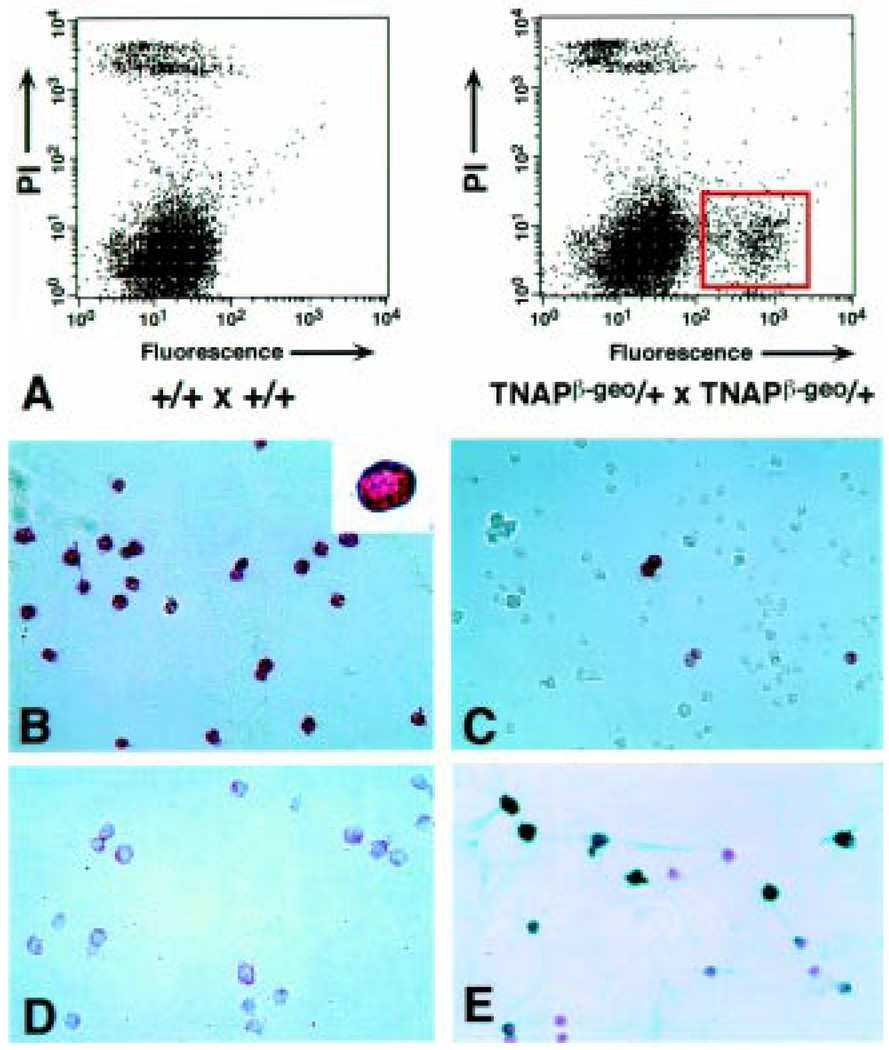
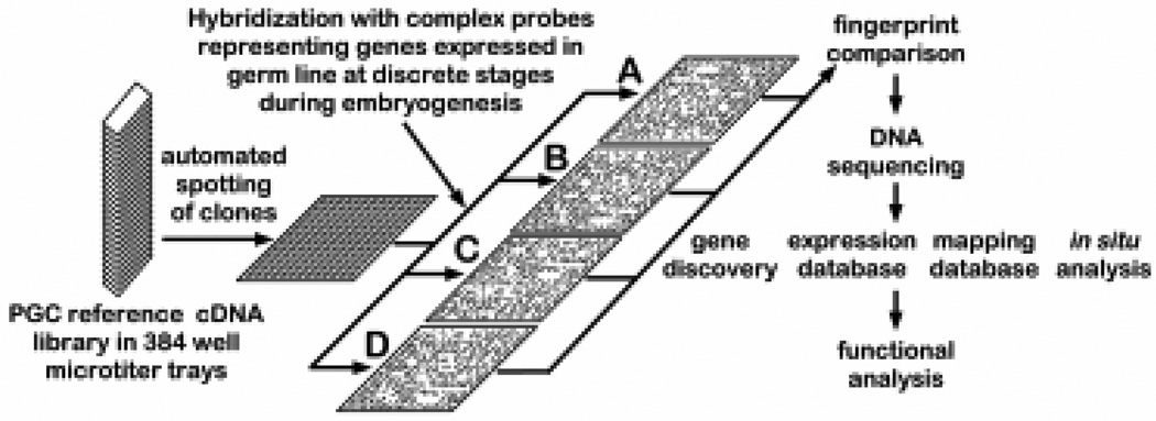
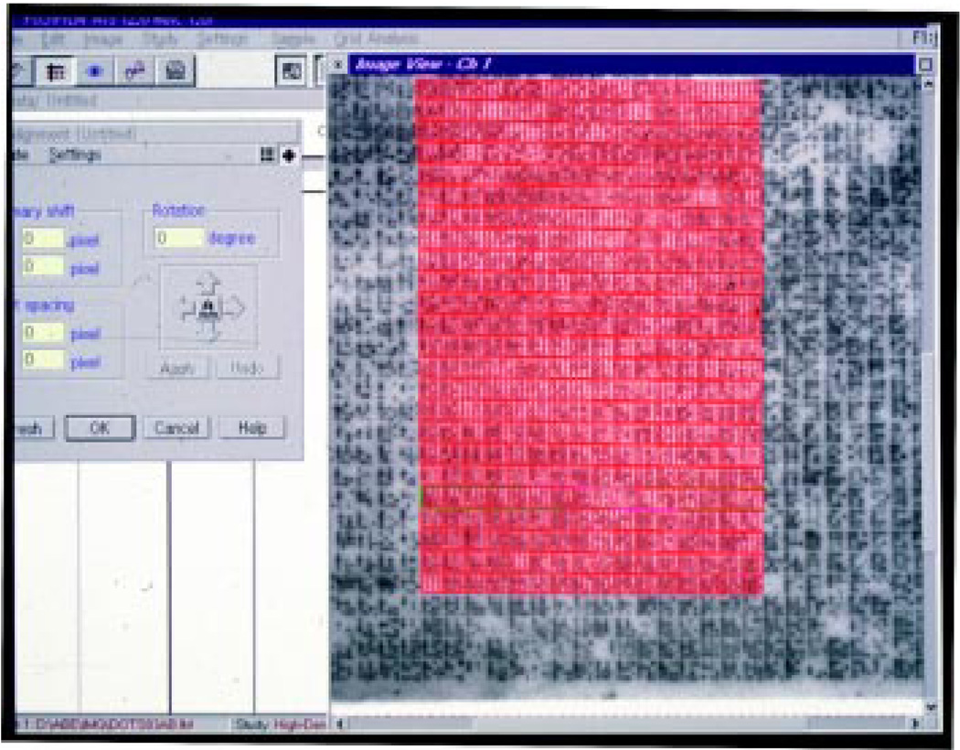
It is difficult to study gene expression in mammalian embryonic germ cells as PGCs constitute only a minor proportion of the mouse embryo. We have overcome this problem by using a novel combination of established molecular and transgenic approaches. A line of mice has been generated in which the cells of the germ lineage express the beta-galactosidase reporter gene during embryogenesis. Using this line, germ cells have been purified to near homogeneity from embryos at discrete stages during germline development by use of a stain for beta-gal activity and a fluorescence activated cell sorter. Subsequently, cDNA libraries have been constructed from each germ cell population using a modified lone-linker PCR strategy. These combined cDNA libraries represent genes expressed in PGCs during mammalian germline development. To facilitate a molecular genetic approach to studying mammalian germline development, these cDNA libraries will be pooled to form an arrayed, addressed reference embryonic germ cell cDNA library. In parallel with large-scale cDNA sequencing efforts; genes that are differentially expressed in germ cells will be identified by screening the reference library with probes generated by subtractive hybridization. Complementary DNAs identified using this approach will be analyzed by sequencing, database comparison, genomic mapping and in situ hybridization to ascertain the potential functional importance of each gene to germline development. In addition to providing a wealth of novel information regarding patterns of gene expression during mammalian germline development, these results will form the basis for future experiments to determine the function of these genes in this process.


Figures

Fig. 1
Fig. 1. Marking of the embryonic germ lineage with lacZ expression in TNAPβ-geo mouse embryos
(A) Expression of TNAPβ-geo in PGCs at the base of the allantois (al) in e7.5 mouse embryos as indicated by X-gal staining. The left embryo is heterozygous for TNAPβ-geo, right embryo is wild-type (negative control). (B) Heterozygous TNAPβ-geo embryo at e8.5. At this stage, the PGCs are located within the hindgut endoderm [see transverse section indicated by white line, shown in (C)]. (C) Transverse section of embryo in Figure 1B. The PGCs (arrowheads) at this stage are masked by expression of TNAPβ-geo in overlying neurectoderm (n) and mesoderm (m), (h) hindgut endoderm, (v) vitelline artery. (D) PGCs at e9.5 are located in developing hindgut (arrowhead). (E) By e10.5, PGCs have arrived in genital ridge (arrowhead). (F) At e14, gonocytes continue to express TNAPβ-geo and are located in the differentiating gonad (g); (r), ribs. The embryo has been cut transversely, eviscerated and the caudal half stood upright to permit observation of the gonads.
Fig. 2
Fig. 2
(A). Purification of PECs from TNAPβ-geo embryos. FACS profile of disaggregated gonads isolated from e12.5 wild-type embryos (left) or embryos obtained from a cross involving a TNAPβ-geo parent (right). The cells were stained with FDG to detect lacZ activity. X-axis indicates fluorescence, Y-axis indicates viability (propidium iodide exclusion). The F(+) population is boxed in red (right panel). (B) Alkaline phosphatase (AP) staining of the F(+) sorted cells from the e12.5 embryonic gonads. Inset – higher magnification of a single AP(+) cell. (C) AP staining of the F(−) sorted cells from e12.5 gonads. (D) The F(+) sorted cells of a e12.5 embryonic gonad were stained with the ACK2 antibody that defines c-kit expression using streptavidin-biotin peroxidase conjugate (Histostain SP kit, Zymed, CA). The red color indicates a positive reaction. Inset –higher magnification of c-kit positive cell. (E) Immunocytochemical staining of F(+) cells sorted from an e13.5 TNAPβ-geo gonad using the EMA-1 antibody. Cells were stained as in (D) except tetramethyl benzidine (blue color) was used to detect a positive immunoreactivity. Cells were counter-stained with nuclear fast red to reveal nuclei. Figure from Abe et al. (1996), used with permission.
Fig. 3
Fig. 3. Hybridization signature approach to analyze gene expression
See main text for description of details.
Fig. 4
Fig. 4. Use of AIS software to quantitate relative levels of gene expression
A 22×22cm hybridization filter containing approximately 36864 (4×4×384) individual mouse genomic DNA BAC clones spotted in 4×4 arrays was hybridized with a complex probe, washed and exposed to a phospho-storage screen. The plate was read by a phospho-imager (Fuji BAS 2000) and the image directly imported by the AIS software program. The screen-shot illustrates a grid used by the software to define individual areas that are subjected to relative quantitation. The grid represents one sixth of the entire filter. The software locates spots, quantitates the signals and converts them into numerical data that can be imported to any spread-sheet application. Signals are normalized to a standard reference set of target sequences on the filter. The AIS software can also compare one hybridization ‘signature’ with another and present the results in either a numerical or graphic format. More recently, EXPANEL software has been developed to perform relative quantitation of hybridization signatures (M.S.H.K., unpublished). The EXPANEL application provides a 2-dimensional color-graded graphical representation of differential expression of addressed cDNAs.
Fig. 5
Fig. 5. PCR based analysis of gene expression in PGCs and somatic tissues from mouse embryos
(A) Expression of M 96, Dlg2-like, fsh-related and Hprt (control) was determined by PCR analysis of the following cDNA samples-(M)-marker; (1) e6.5 epiblast; (2) e11.5 PGC; (3) e12.5 PGC; (4) e13.5 PGC; (5) e13.5 male PGC; (6) e13.5 female PGC; (7) e12.5 F(−) somatic cells. (B) Expression of fsh-related gene (H0503B12) during embryonic development and in adult mouse tissues. Northern blots containing adult (upper panel) or embryonic mouse total RNA (lower panel) were hybridized with a fsh-related cDNA. (1) heart; (2) brain; (3) spleen; (4) lung; (5) liver; (6) muscle; (7) kidney; (8) testis; (9) e7.5; (10) e11.5; (11) e15.5; (12) e17.5. Molecular weight sizes are in kb. (data courtesy of Rhonda H. Nicholson). (C) H0503B12 (fsh-related) maps to mouse chromosome 17 within the t-complex. fsh-related was mapped using an inter-specific species backcross panel (Rowe et al., 1994). (Data courtesy of Xueqian Wang).

Similar articles

Cited by

References

    1. Aaronson JS, Eckman B, Blevins RA, Borkowski JA, Myerson J, Imran S, Elliston KO. Toward the development of a gene index to the human genome: an assessment of the nature of high-throughput EST sequence data. Genome Res. 1996;6:829–845. - PubMed
    1. Abe K. Rapid isolation of desired sequences from lone linker PCR amplified cDNA mixtures: Application to identification and recovery of expressed sequences in cloned genomic DNA. Mammal. Genome. 1992;2:252–259. - PubMed
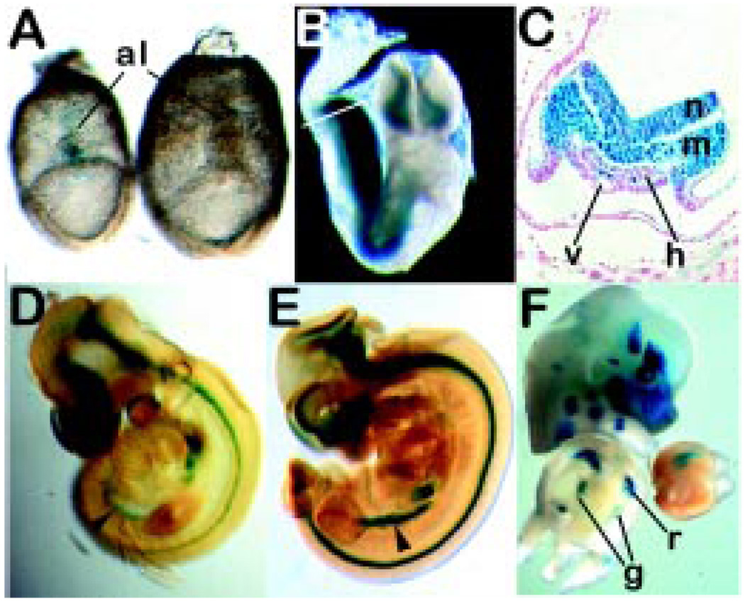
    1. Abe K, Hashiyama M, MacGregor G, Yamamura K-I, Abe K. Purification of primordial germ cells from TNAPβ-geo mouse embryos using FACS-gal. Dev. Biol. 1996;180:468–472. - PubMed
    1. Adams MD, Kelley JM, Gocayne JD, Dubnick M, Polymeropoulos MH, Xiao H, Merril CR, Wu A, Olde B, Moreno RF, et al. Complementary DNA sequencing: expressed sequence tags and human genome project. Science. 1991;252:1651–1656. - PubMed
    1. Alvarez-Buylla A, Merchant-Larios H. Mouse primordial germ cells use fibronectin as a substrate for migration. Exp. Cell Res. 1986;165:362–368. - PubMed

Publication types

LinkOut - more resources