. 1998 Dec 21;188(12):2301-11.
doi: 10.1084/jem.188.12.2301.

CD5 expression is developmentally regulated by T cell receptor (TCR) signals and TCR avidity

Affiliations

CD5 expression is developmentally regulated by T cell receptor (TCR) signals and TCR avidity

H S Azzam et al. J Exp Med. .

Abstract

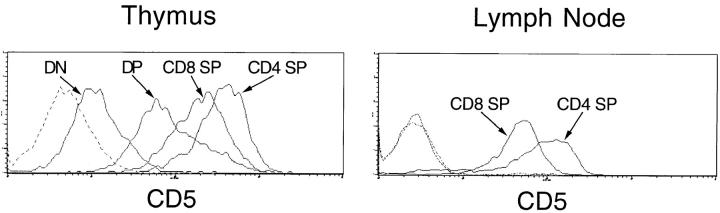
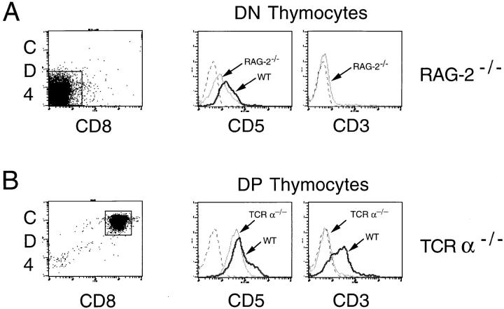
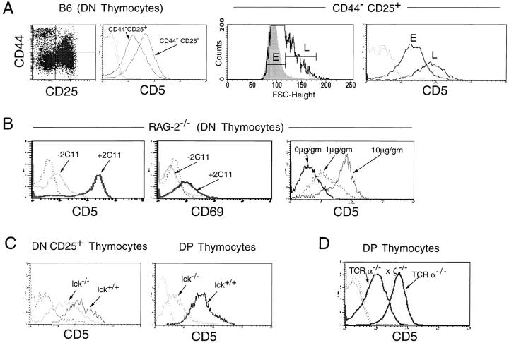
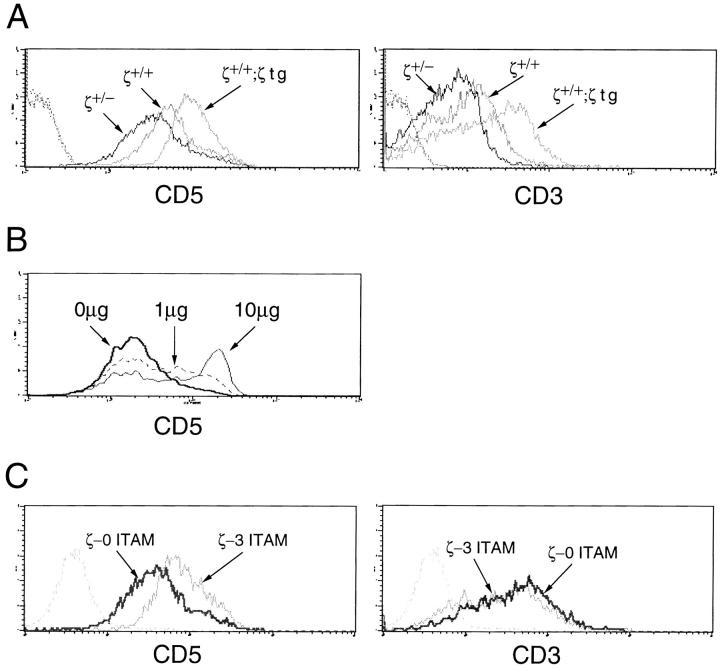
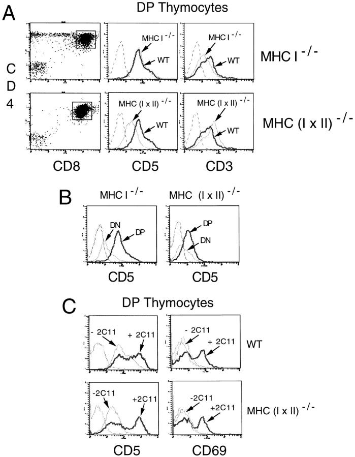
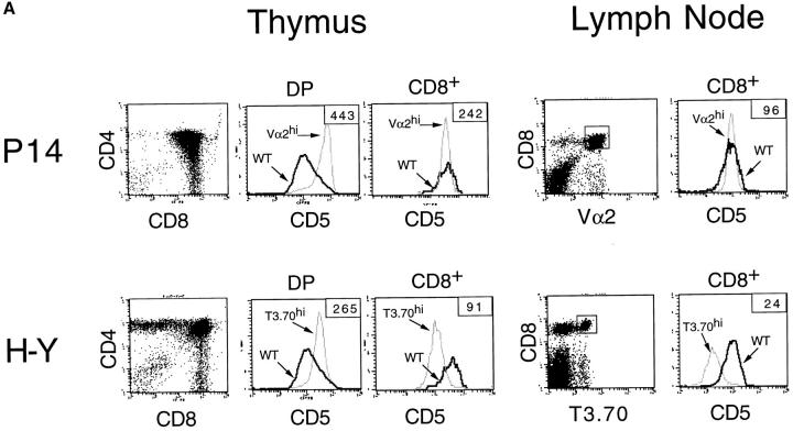
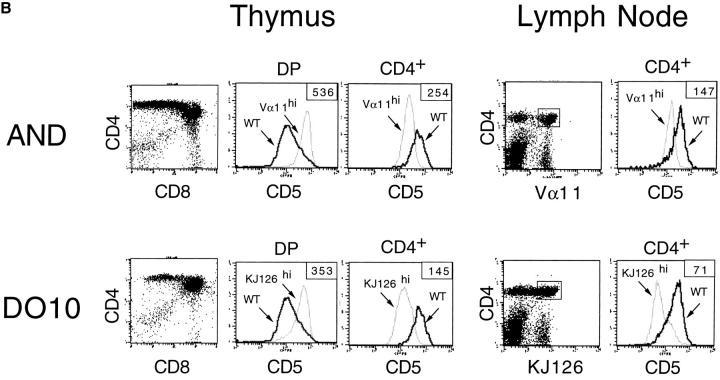
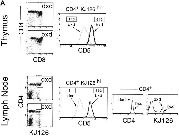
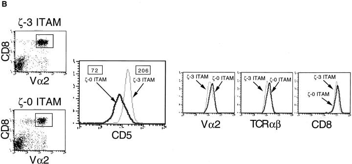
Recent data indicate that the cell surface glycoprotein CD5 functions as a negative regulator of T cell receptor (TCR)-mediated signaling. In this study, we examined the regulation of CD5 surface expression during normal thymocyte ontogeny and in mice with developmental and/or signal transduction defects. The results demonstrate that low level expression of CD5 on CD4(-)CD8(-) (double negative, DN) thymocytes is independent of TCR gene rearrangement; however, induction of CD5 surface expression on DN thymocytes requires engagement of the pre-TCR and is dependent upon the activity of p56(lck). At the CD4(+)CD8(+) (double positive, DP) stage, intermediate CD5 levels are maintained by low affinity TCR-major histocompatibility complex (MHC) interactions, and CD5 surface expression is proportional to both the surface level and signaling capacity of the TCR. High-level expression of CD5 on DP and CD4(+) or CD8(+) (single positive, SP) thymocytes is induced by engagement of the alpha/beta-TCR by (positively or negatively) selecting ligands. Significantly, CD5 surface expression on mature SP thymocytes and T cells was found to directly parallel the avidity or signaling intensity of the positively selecting TCR-MHC-ligand interaction. Taken together, these observations suggest that the developmental regulation of CD5 in response to TCR signaling and TCR avidity represents a mechanism for fine tuning of the TCR signaling response.


Figures

Figure 1
Figure 1
CD5 expression during normal T cell development. (A) Thymus. (B) Lymph node. Thymocytes and T cells from a C57 BL/6 mouse were stained with CD5, CD4, and CD8, and CD5 was assessed by gating on the various populations. Histograms represent CD5 expression on the indicated subpopulations. Dotted lines represent staining with control (nonspecific) antibody.
Figure 2
Figure 2
CD5 surface expression on thymocytes from Rag-2−/− and TCR-α−/− mice. Dot plots illustrate the CD4/CD8 staining profile and demonstrate the stage at which thymocyte development is blocked. Histograms depict CD5 and CD3 surface levels on gated CD4CD8 (DN) thymocytes (A), or CD4+CD8+ (DP) thymocytes (B). CD5 and CD3 surface expression on DN and DP thymocytes from control (WT) animals are included for comparison. Dotted lines represent control peaks.
Figure 3
Figure 3
(A) CD5 surface expression on CD4CD8 (DN) thymocyte subsets. DN thymocytes from B6 mice were isolated as described in Materials and Methods, and stained with CD25 and CD44 antibodies to distinguish the various DN subpopulations (dot plot). Histogram on the left shows CD5 expression levels on gated CD44 CD25+ and CD44CD25 thymocyte subpopulations. The middle histogram shows forward scatter profile of DN CD44 CD25+ thymocytes from B6 mice (white histogram) or from Rag-2−/− mice (gray histogram). Rag-2−/− thymocytes were used for placement of E and L gates as shown. Right histogram shows CD5 expression on E and L gated DN CD44CD25+ thymocytes from B6 mice. (B) Induction of CD5 and CD69 surface expression on Rag-2−/− DN thymocytes by anti-CD3ε antibody. Left and middle: 200 μg of anti-CD3ε (2C11) antibody was injected into Rag-2−/− mice (+2C11) and CD5 (left) or CD69 (middle) surface levels on DN thymocytes were determined 24 h later. Control animals were injected with PBS (−2C11). Right: Rag-2−/− mice were injected with 0, 1, or 10 μg 2C11/gram body wt and DN thymocytes were analyzed for CD5 surface expression 3 d later. (C) CD5 surface expression on DN CD25+ and CD4+CD8+ (DP) thymocytes from lck−/− mice. CD5 levels on DN CD25+ and DP thymocytes from control (lck +/+) mice are included for comparison. (D) CD5 surface expression on gated CD4+CD8+ (DP) thymocytes from TCR-α−/− and TCR-α−/− × ζ−/− mice. Dotted lines represent staining with control antibody.
Figure 4
Figure 4
TCR levels and TCR signaling intensity influence CD5 expression on CD4+CD8+ (DP) thymocytes. (A) Effect of TCR levels on CD5 expression. Histograms depict CD5 and CD3 expression on gated CD4+CD8+ (DP) thymocytes from TCR-ζ heterozygotes (TCR-ζ+/−) and TCR-ζ-transgenic mice (TCR-ζ+/+; ζ tg) relative to control mice (TCR-ζ+/+). (B) Relationship between TCR signal intensity and CD5 expression. DP thymocytes were stimulated for 18 h on plates coated with the indicated amount of cross-linking antibody (anti-CD3ε; mAb 2C11) then analyzed for CD5 surface expression. (C) Effect of TCR signaling capacity on CD5 expression. Histograms depict CD5 and CD3 surface expression on DP thymocytes from TCR-ζ−/− mice reconstituted with transgenes encoding either full length ζ chain (ζ-3 ITAM) or ζ chain lacking sequences required for signal transduction (ζ-0 ITAM) (reference , see Results). Dotted lines represent staining with control antibody.
Figure 5
Figure 5
CD5 surface expression on thymocytes from mice lacking MHC. (A) Histograms show CD5 and CD3 surface levels on gated CD4+CD8+ (DP) thymocyte populations from mice lacking expression of MHC class I (MHC I−/−) or MHC class I and class II [MHC (I × II−/−)]. CD3 and CD5 levels on DP thymocytes from control B6 (WT) mice are included for comparison. (B) CD5 levels on CD4CD8 (DN) and DP thymocytes from MHC I−/− and MHC (I × II)−/− mice. (C) Induction of CD5 by CD3 cross-linking. Thymocytes from C56 BL/6 (WT) and MHC (I × II)−/− mice were incubated in the presence (+2C11) or absence (−2C11) of plate-bound anti-CD3ε antibody (50 μg) for 18 h at 37°C and CD5 and CD69 expression on DP thymocytes was assessed by FACS®. Dotted lines represent staining with control antibodies.
Figure 6
Figure 6
CD5 expression on thymocytes and mature T cells from TCR transgenic mice. (A) Comparison of CD5 levels on thymocytes and lymph node T cells from MHC class I–restricted (P14 H-2b and H-Y H-2b) TCR transgenic mice. (B) Comparison of CD5 levels on thymocytes and lymph node T cells from MHC class II–restricted (AND H-2bxk and DO10 H-2d) TCR transgenic mice. Single-color histograms represent CD5 staining on CD4+CD8+ thymocytes, as well as on CD8+ SP thymocytes and T cells (HY and P14 TCR transgenic mice), or CD4+ SP thymocytes and T cells (AND and DO10 TCR transgenic mice). Shown are CD5 levels on transgenic TCRhi gated cells. Data obtained for similar cell populations from nontransgenic (WT) mice are shown for comparison. Numbers in boxes depict mean CD5 fluorescence for the transgenic TCRhi cells.
Figure 6
Figure 6
CD5 expression on thymocytes and mature T cells from TCR transgenic mice. (A) Comparison of CD5 levels on thymocytes and lymph node T cells from MHC class I–restricted (P14 H-2b and H-Y H-2b) TCR transgenic mice. (B) Comparison of CD5 levels on thymocytes and lymph node T cells from MHC class II–restricted (AND H-2bxk and DO10 H-2d) TCR transgenic mice. Single-color histograms represent CD5 staining on CD4+CD8+ thymocytes, as well as on CD8+ SP thymocytes and T cells (HY and P14 TCR transgenic mice), or CD4+ SP thymocytes and T cells (AND and DO10 TCR transgenic mice). Shown are CD5 levels on transgenic TCRhi gated cells. Data obtained for similar cell populations from nontransgenic (WT) mice are shown for comparison. Numbers in boxes depict mean CD5 fluorescence for the transgenic TCRhi cells.
Figure 7
Figure 7
(A) Comparison of CD5 levels on CD4+ thymocytes and T cells from DO10 TCR transgenic mice generated in the H-2dxd and H-2bxd backgrounds. Top: Two-color (CD4 versus CD8) analysis of thymocytes from DO10/H-2dxd and DO10/H-2bxd mice. Histogram depicts CD5 levels on CD4+ KJ126hi thymocytes. Bottom: Two-color (CD4 versus KJ126) analysis of lymph node cells from DO10/H-2dxd and DO10/H-2bxd mice. Large histogram depicts CD5 levels on CD4+ KJ126hi T cells. Small histograms show CD4 and KJ126-TCR levels on total CD4+ T cells. Numbers in boxes depict mean CD5 fluorescence for the transgenic TCRhi cells. (B) Comparison of CD5 levels on CD8+ T cells generated in P14 TCR transgenic mice expressing TCRs containing full-length (ζ-3 ITAM) or signaling defective (ζ-0 ITAM) ζ-chains Histograms depict Vα2, TCR-β, CD8, or CD5 staining on gated CD8+Vα2+ T cells (gates are shown in the two-color plots). Numbers in boxes depict mean CD5 fluorescence.
Figure 7
Figure 7
(A) Comparison of CD5 levels on CD4+ thymocytes and T cells from DO10 TCR transgenic mice generated in the H-2dxd and H-2bxd backgrounds. Top: Two-color (CD4 versus CD8) analysis of thymocytes from DO10/H-2dxd and DO10/H-2bxd mice. Histogram depicts CD5 levels on CD4+ KJ126hi thymocytes. Bottom: Two-color (CD4 versus KJ126) analysis of lymph node cells from DO10/H-2dxd and DO10/H-2bxd mice. Large histogram depicts CD5 levels on CD4+ KJ126hi T cells. Small histograms show CD4 and KJ126-TCR levels on total CD4+ T cells. Numbers in boxes depict mean CD5 fluorescence for the transgenic TCRhi cells. (B) Comparison of CD5 levels on CD8+ T cells generated in P14 TCR transgenic mice expressing TCRs containing full-length (ζ-3 ITAM) or signaling defective (ζ-0 ITAM) ζ-chains Histograms depict Vα2, TCR-β, CD8, or CD5 staining on gated CD8+Vα2+ T cells (gates are shown in the two-color plots). Numbers in boxes depict mean CD5 fluorescence.

References

    1. Ledbetter JA, Rouse RV, Micklem HS, Herzenberg LA. T cell subsets defined by expression of Lyt-1,2,3 and Thy-1 antigens: two parameter immunofluorescence and cytotoxicity analysis with monoclonal antibodies modifies current views. J Exp Med. 1980;152:280–294. - PMC - PubMed
    1. Hayakawa K, Hardy RR, Parks DR, Herzenberg LA. The “Ly-1 B” cell subpopulation in normal, immunodefective, and autoimmune mice. J Exp Med. 1983;157:202–218. - PMC - PubMed
    1. Fowlkes BJ, Edison L, Mathieson BJ, Chused TM. Early T lymphocytes: differentiation in vivo of adult intrathymic precursor cells. J Exp Med. 1985;162:802–822. - PMC - PubMed
    1. Huang HJ, Jones NH, Strominger JL, Hertzenberg LA. Molecular cloning of Ly-1, a membrane glycoprotein of mouse T lymphocytes and a subset of B cells: molecular homology to its human counterpart Leu-1/T1 (CD5) Proc Natl Acad Sci USA. 1987;84:204–208. - PMC - PubMed
    1. Van De Helde H, Von Hoegen I, Luo W, Parnes JR, Thielemans K. The B-cell surface protein CD72/Lyb2 is the ligand for CD5. Nature. 1991;351:662–665. - PubMed

MeSH terms

Substances