. 1998 Dec 21;188(12):2357-68.
doi: 10.1084/jem.188.12.2357.

The central role of CD4(+) T cells in the antitumor immune response

Affiliations

The central role of CD4(+) T cells in the antitumor immune response

K Hung et al. J Exp Med. .

Abstract

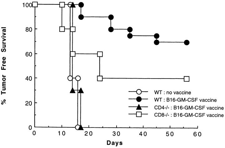
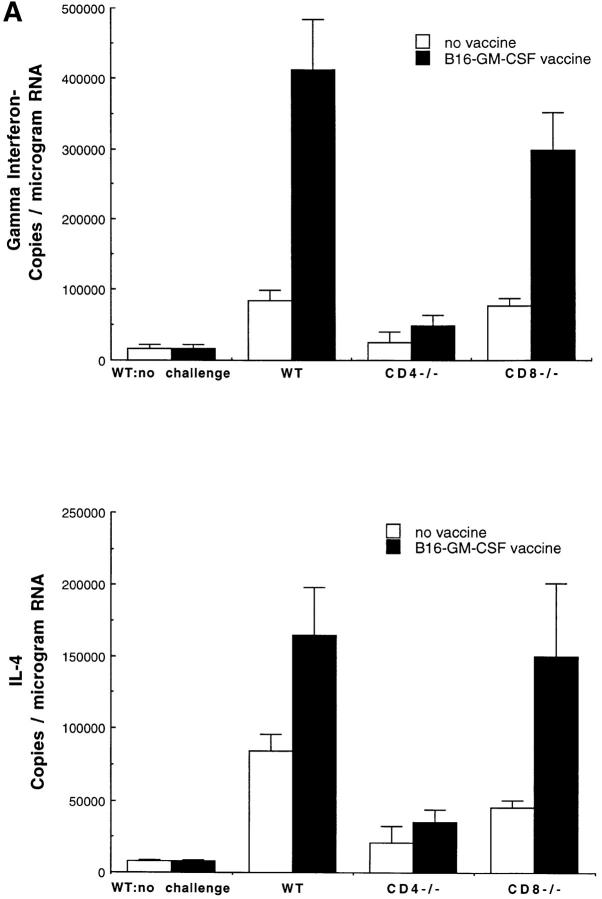
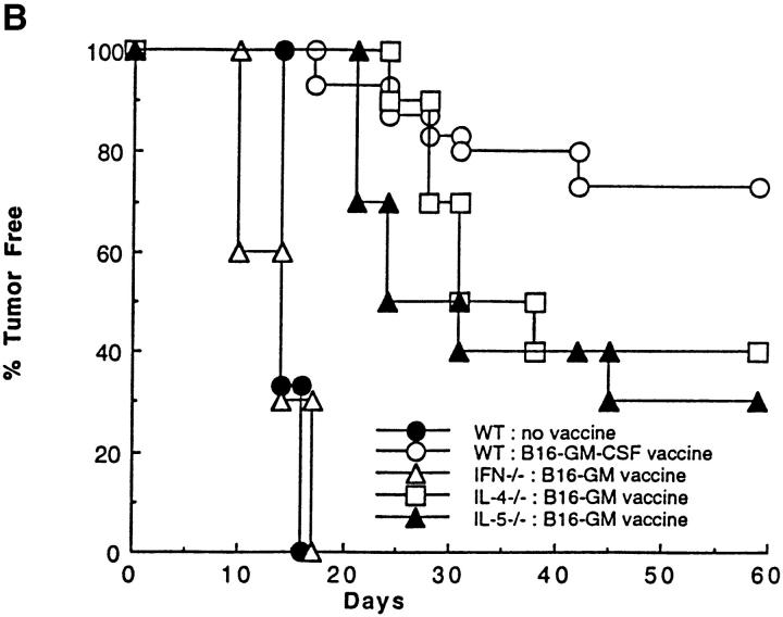
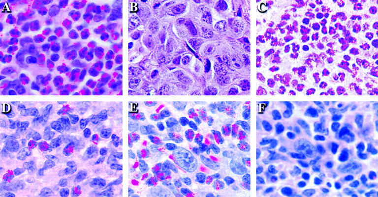
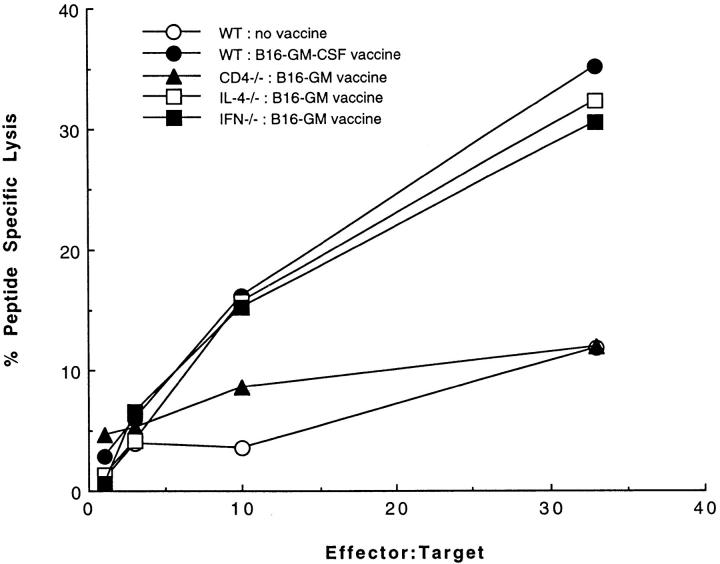
The induction of optimal systemic antitumor immunity involves the priming of both CD4(+) and CD8(+) T cells specific for tumor-associated antigens. The role of CD4(+) T helper cells (Th) in this response has been largely attributed to providing regulatory signals required for the priming of major histocompatibility complex class I restricted CD8(+) cytolytic T lymphocytes, which are thought to serve as the dominant effector cell mediating tumor killing. However, analysis of the effector phase of tumor rejection induced by vaccination with irradiated tumor cells transduced to secrete granulocyte/macrophage colony-stimulating factor indicates a far broader role for CD4(+) T cells in orchestrating the host response to tumor. This form of immunization leads to the simultaneous induction of Th1 and Th2 responses, both of which are required for maximal systemic antitumor immunity. Cytokines produced by these CD4(+) T cells activate eosinophils as well as macrophages that produce both superoxide and nitric oxide. Both of these cell types then collaborate within the site of tumor challenge to cause its destruction.


Figures

Figure 1
Figure 1
The contribution of CD4+ and CD8+ T cell subsets to the systemic response to B16-GM-CSF vaccination. Wild-type, CD4−/−, and CD8−/− mice were injected subcutaneously in the left flank with 106 irradiated (50 Gy) B16-GM-CSF cells. Mice were challenged 2 wk later in the right flank with 105 live B16-WT cells, and examined twice weekly for the development of tumor. These data represent the results of three different experiments in which each experimental group consisted of 10 mice.
Figure 2
Figure 2
The role of cytokines in the systemic immune response to vaccination with B16-GM-CSF. (A) Mice were vaccinated with 106 irradiated (50 Gy) B16-GM-CSF tumor cells and challenged 2 wk later in the opposite flank with 105 live B16-WT tumor cells. 4 d after live tumor challenge, mice were killed and total RNA was extracted from a biopsy of the tumor challenge site. At least four mice were used for each group. γ-IFN and IL-4 mRNA levels were then determined by a competitive quantitative RT-PCR assay (14). Results are shown as number of copies of γ-IFN or IL-4 message per microgram of total RNA. (B) Wild-type, γ-IFN−/−, IL-4−/−, and IL-5−/− mice were vaccinated with irradiated B16-GM-CSF cells and challenged with B16-WT cells 2 wk later in the opposite flank, as in Fig. 1. These data represent the results of three different experiments in which each experimental group consisted of 10 mice.
Figure 2
Figure 2
The role of cytokines in the systemic immune response to vaccination with B16-GM-CSF. (A) Mice were vaccinated with 106 irradiated (50 Gy) B16-GM-CSF tumor cells and challenged 2 wk later in the opposite flank with 105 live B16-WT tumor cells. 4 d after live tumor challenge, mice were killed and total RNA was extracted from a biopsy of the tumor challenge site. At least four mice were used for each group. γ-IFN and IL-4 mRNA levels were then determined by a competitive quantitative RT-PCR assay (14). Results are shown as number of copies of γ-IFN or IL-4 message per microgram of total RNA. (B) Wild-type, γ-IFN−/−, IL-4−/−, and IL-5−/− mice were vaccinated with irradiated B16-GM-CSF cells and challenged with B16-WT cells 2 wk later in the opposite flank, as in Fig. 1. These data represent the results of three different experiments in which each experimental group consisted of 10 mice.
Figure 3
Figure 3
The dependence of eosinophil recruitment to the tumor challenge site on CD4+ T cells and cytokines. Mice were vaccinated with 106 irradiated (50 Gy) B16-GM-CSF cells and challenged 2 wk later on the opposite flank with 105 live B16-WT cells. After 4 d, the site of live wild-type tumor challenge was removed and sections were stained with hematoxylin and eosin. (A) Wild-type, (B) CD4−/−, (C) CD8−/−, (D) IL-4−/−, (E) γ-IFN−/−, and (F) IL-5−/− C57BL/6 mice.
Figure 4
Figure 4
The role of CD4+ T cells and cytokines in priming tumor-specific CTL. Wild-type, CD4−/−, γ-IFN−/−, and IL-4−/− mice were vaccinated with 106 irradiated (50 Gy) B16-GM-CSF tumor cells. After 2 wk, splenocytes were isolated and cultured for 7 d with 5 μg/ml TRP-2 peptide and 10 U/ml IL-2. On day 7, live T cells were incubated with 51Cr-labeled MC57G targets at the indicated effector to target ratios in the presence of 1 μg/ml TRP-2 peptide or irrelevant peptide. The percent peptide-specific lysis is calculated as the difference between the percent specific lysis due to the TRP-2 peptide and the percent specific lysis due to the irrelevant peptide.
Figure 5
Figure 5
Macrophages and iNOS expression at the site of wild-type tumor challenge. Mice were vaccinated with 106 irradiated (50 Gy) B16-GM-CSF tumor and challenged 2 wk later with 105 live B16-WT tumor. After 4 d, mice were killed and the live wild-type tumor challenge site was sectioned and stained for macrophages (A–F) and iNOS (G–L) (24). (A) Control for macrophage staining: no primary antibody; (B) macrophages at site of tumor challenge in wild-type mice; (C) CD4−/− mice; (D) CD8−/− mice; (E) γ-IFN−/− mice; and (F) IL-4−/− mice; (G) control for iNOS staining: no primary antibody; (H) iNOS at site of tumor challenge in wild-type mice; (I) CD4−/− mice; (J) CD8−/− mice; (K) γ-IFN−/− mice; and (L) IL-4−/− mice.

References

    1. Pardoll DM. Tumour antigens. A new look for the 1990s. Nature. 1994;369:357. - PubMed
    1. Boon T, van der Bruggen P. Human tumor antigens recognized by T lymphocytes. J Exp Med. 1996;183:725–729. - PMC - PubMed
    1. Kawakami Y, Rosenberg SA. Human tumor antigens recognized by T-cells. Immunol Res. 1997;16:313–339. - PubMed
    1. Pardoll DM. Cancer vaccines. Nat Med. 1998;4:525–531. - PubMed
    1. Dranoff G, Jaffee E, Lazenby A, Golumbek P, Levitsky H, Brose K, Jackson V, Hamada H, Pardoll D, Mulligan RC. Vaccination with irradiated tumor cells engineered to secrete murine granulocyte-macrophage colony–stimulating factor stimulates potent, specific, and long-lasting anti-tumor immunity. Proc Natl Acad Sci USA. 1993;90:3539–3543. - PMC - PubMed

Publication types

MeSH terms