. 1999 Jan;19(1):57-68.
doi: 10.1128/MCB.19.1.57.

RING1 interacts with multiple Polycomb-group proteins and displays tumorigenic activity

Affiliations

RING1 interacts with multiple Polycomb-group proteins and displays tumorigenic activity

D P Satijn et al. Mol Cell Biol. 1999 Jan.

Abstract

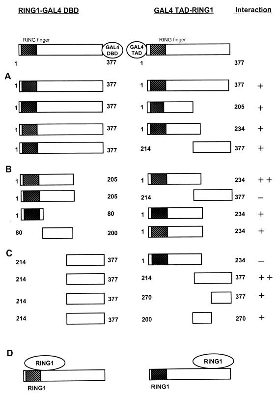
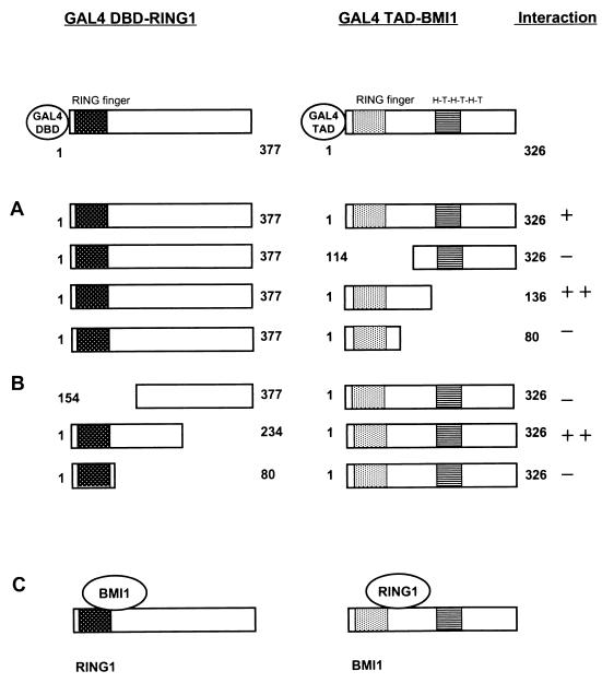
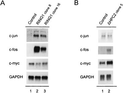
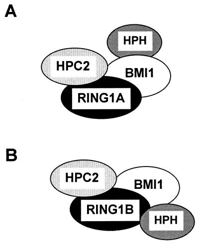
Polycomb-group (PcG) proteins form large multimeric protein complexes that are involved in maintaining the transcriptionally repressive state of genes. Previously, we reported that RING1 interacts with vertebrate Polycomb (Pc) homologs and is associated with or is part of a human PcG complex. However, very little is known about the role of RING1 as a component of the PcG complex. Here we undertake a detailed characterization of RING1 protein-protein interactions. By using directed two-hybrid and in vitro protein-protein analyses, we demonstrate that RING1, besides interacting with the human Pc homolog HPC2, can also interact with itself and with the vertebrate PcG protein BMI1. Distinct domains in the RING1 protein are involved in the self-association and in the interaction with BMI1. Further, we find that the BMI1 protein can also interact with itself. To better understand the role of RING1 in regulating gene expression, we overexpressed the protein in mammalian cells and analyzed differences in gene expression levels. This analysis shows that overexpression of RING1 strongly represses En-2, a mammalian homolog of the well-characterized Drosophila PcG target gene engrailed. Furthermore, RING1 overexpression results in enhanced expression of the proto-oncogenes c-jun and c-fos. The changes in expression levels of these proto-oncogenes are accompanied by cellular transformation, as judged by anchorage-independent growth and the induction of tumors in athymic mice. Our data demonstrate that RING1 interacts with multiple human PcG proteins, indicating an important role for RING1 in the PcG complex. Further, deregulation of RING1 expression leads to oncogenic transformation by deregulation of the expression levels of certain oncogenes.


Figures

FIG. 1
FIG. 1
Association of RING1 with itself, Bmi-1, and HPC2 and of Bmi-1 with itself in vitro. GST-RING1 fusion protein, immobilized on glutathione-Sepharose, interacted with in vitro-translated, [35S]methionine-labeled HPC2 or RING1. [35S]methionine-labeled HPC2 (lane 1) was incubated with GST-Sepharose alone (lane 2) and with GST-RING1 (lane 3). [35S]methionine-labeled RING1 (lane 4) was incubated with GST-Sepharose alone (lane 5) and with GST-RING1 (lane 6). GST-RING1 and GST–Bmi-1 fusion proteins, immobilized on glutathione-Sepharose, interacted with in vitro-translated, [35S]methionine-labeled Bmi-1. [35S]methionine-labeled Bmi-1 (lane 7) was incubated with GST-Sepharose alone (lane 8), with GST-RING1 (lane 9), and with GST–Bmi-1 (lane 10). All proteins used in the assay are full length. Molecular masses are indicated in kilodaltons. The input (lanes 1, 4, and 7) was 10% of the amount incubated with the GST fusion proteins.
FIG. 2
FIG. 2
Mapping of homodimerization domains of RING1. (A) Full-length RING1 (aa 1 to 377) was fused to the GAL4 DBD, which in all constructs shown is located at the C-terminal end of RING1. The plasmids were cotransformed with different portions of RING1, which were fused to the GAL4 TAD, which is located at the N terminus of RING1. Interactions were positive (+) when the transformants were able to grow on selective medium lacking histidine and when they were also β-galactosidase positive. Relative strength of the interactions is a qualitative indication based on the time needed for blue coloring (++, within 30 min; +, between 30 and 120 min) and the size of the colonies. (B) N-terminal portions of RING1 fused to the GAL4 DBD were tested for interactions with N- and C-terminal portions of RING1. These constructs are fused to the GAL4 TAD. (C) The C-terminal portion of RING1 fused to the GAL4 DBD was tested for interaction with C-terminal portions of RING1 fused to the GAL4 TAD. (D) Schematic representation of the two RING1-RING1 protein interaction domains. The RING finger domain of the RING1 protein is indicated as a hatched black box.
FIG. 3
FIG. 3
Mapping of interaction domains between RING1 and HPC2. (A) Indicated portions of HPC2 were fused to the GAL4 DBD, which in all constructs shown is located at the N-terminal end of HPC2. The plasmids were cotransformed with full-length RING1 (aa 1 to 377), which is fused to the GAL4 TAD. The GAL4 TAD is located at the N terminus of RING1. (B) Full-length HPC2 (aa 1 to 558) fused to the GAL4 DBD was tested for interaction with various C-terminal region of RING1 fused to the GAL4 TAD. (C) Schematic representation of the interaction domains of HPC2 and RING1. The HPC2 protein contains a chromodomain and a C box, which are indicated as grey and black dotted boxes, respectively. The RING finger domain of the RING1 protein is indicated as a hatched black box.
FIG. 4
FIG. 4
Mapping of interaction domains between RING1 and BMI1. (A) Full-length RING1 (aa 1 to 377) was fused to the GAL4 DBD, which in all constructs shown is located at the N-terminal end of RING1. The plasmid was cotransformed with the indicated portions of BMI1, which is fused to the GAL4 TAD. In all constructs, the GAL4 TAD is located at the N-terminus of BMI1. (B) The indicated portions of RING1 were fused to the GAL4 DBD and tested for interaction with full-length BMI1 fused to the GAL4 TAD. (C) Schematic representation of the interaction domains of RING1 and BMI1. The RING finger domain of the RING1 protein is indicated as a hatched black box. The BMI1 protein has a RING finger domain and a helix-turn-helix-turn-helix-turn (H-T-H-T-H-T) domain, indicated as grey and striped boxes, respectively.
FIG. 5
FIG. 5
Mapping of homodimerization domains of BMI1. (A) Full-length BMI1 (aa 1 to 326) is fused to the GAL4 DBD, which in all constructs shown is located at the C-terminal end of BMI1. The plasmid was cotransformed with different portions of BMI1, which are fused to the GAL4 TAD. In these constructs, the GAL4 TAD is located at the N terminus of BMI1. (B) An N-terminal portion of BMI1 (aa 1 to 136) fused to the GAL4 DBD was tested for interaction with full-length BMI1 (aa 1 to 326) fused to the GAL4 TAD. (C) The C-terminal portion of BMI1 (aa 1 to 136) fused to the GAL4 DBD was tested for interaction with different portions of BMI1 fused to the GAL4 TAD. (D) Schematic representation of the homodimerization domains of BMI1. The BMI1 protein has a RING finger domain and a helix-turn-helix-turn-helix-turn (H-T-H-T-H-T) domain, indicated as grey and striped boxes, respectively.
FIG. 6
FIG. 6
Western analysis of stably transfected RING1 and ΔHPC2 proteins from cell extracts of Rat1a cells. Equal amounts of proteins were Western blotted. The blots were incubated with a rabbit anti-RING1 or rabbit anti-HPC2 antibody. (A) Endogenous rat RING1 levels were detected in the untransfected cells (lane 1); elevated levels of RING1 were detected in clone 8 (lane 2) and clone 16 (lane 3). (B) Endogenous rat HPC2 levels were detected in the untransfected cells (lane 1); elevated levels of ΔHPC2 were detected in clone 5 (lane 2). Molecular masses are indicated in kilodaltons.
FIG. 7
FIG. 7
Repression of En-2 gene expression activity in RING1-transfected Rat1a cells. Poly(A)+ mRNA isolated from Rat1a control cells (lane 1) and from RING1-transfected clone 8 (lane 2) and clone 16 (lane 3) Rat1a cells was Northern blotted and probed with the En-2 gene. To verify equal RNA loading, the filter was hybridized with a GAPDH probe.
FIG. 8
FIG. 8
Expression of c-myc, c-jun, and c-fos in RING1- and ΔHPC2-transfected Rat1a cells. (A) Poly(A)+ mRNA isolated from Rat1a control cells (lane 1) and from RING1-transfected Rat1a clone 8 (lane 2) and clone 16 (lane 3) cells was Northern blotted and probed with fragments of c-jun, c-fos, and c-myc. (B) Poly(A)+ mRNA isolated from Rat1a control cells (lane 1) and from ΔHPC2-transfected Rat1a clone 5 (lane 2) cells was Northern blotted and probed with fragments of c-jun, c-fos, and c-myc. To verify equal RNA loading, the filter was hybridized with a GAPDH probe.
FIG. 9
FIG. 9
Overexpression of RING1 induces anchorage-independent growth in control Rat1a cells (A) and in Rat1a cells transformed with the c-myc oncogene (B), with the C-terminal deletion mutant of HPC2 (C), and with full-length RING1 (D). Bar = 400 μm.
FIG. 10
FIG. 10
Model of human PcG multimeric protein complexes. (A) Model of a PcG protein complex which contains RING1/Ring1A; (B) dinG/Ring1B as the central protein for the establishment of a multimeric PcG protein complex.

References

    1. Alkema M J, Bronk M, Verhoeven E, Otte A P, van’t Veer L J, Berns A, van Lohuizen M. Identification of Bmi1-interacting proteins as constituents of a multimeric mammalian Polycomb complex. Genes Dev. 1997;11:226–240. - PubMed
    1. Alkema M J, Jacobs H, Van Lohuizen M, Berns A. Perturbation of B and T cell development and predisposition to lymphomagenesis in EμBmi1 transgenic mice require the Bmi1 RING finger. Oncogene. 1997;15:899–910. - PubMed
    1. Bradley M O, Kraynak A R, Storer R D, Gibbs J B. Experimental metastasis in nude mice of NIH 3T3 cells containing various ras genes. Proc Natl Acad Sci USA. 1986;83:5277–5281. - PMC - PubMed
    1. Buchenau P, Hodgson J, Strutt H, Arndt-Jovin D J. The distribution of polycomb-group proteins during cell division and development in Drosophila embryos; impact on models for silencing. J Cell Biol. 1998;141:469–481. - PMC - PubMed
    1. Cohen K J, Hanna J S, Prescott J E, Dang C V. Transformation by the Bmi-1 oncoprotein correlates with its subnuclear localization but not its transcriptional suppression activity. Mol Cell Biol. 1996;16:5527–5535. - PMC - PubMed

MeSH terms