. 1999 Jan;19(1):136-46.
doi: 10.1128/MCB.19.1.136.

Glutamate induces phosphorylation of Elk-1 and CREB, along with c-fos activation, via an extracellular signal-regulated kinase-dependent pathway in brain slices

Affiliations

Glutamate induces phosphorylation of Elk-1 and CREB, along with c-fos activation, via an extracellular signal-regulated kinase-dependent pathway in brain slices

P Vanhoutte et al. Mol Cell Biol. 1999 Jan.

Abstract

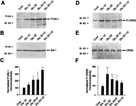
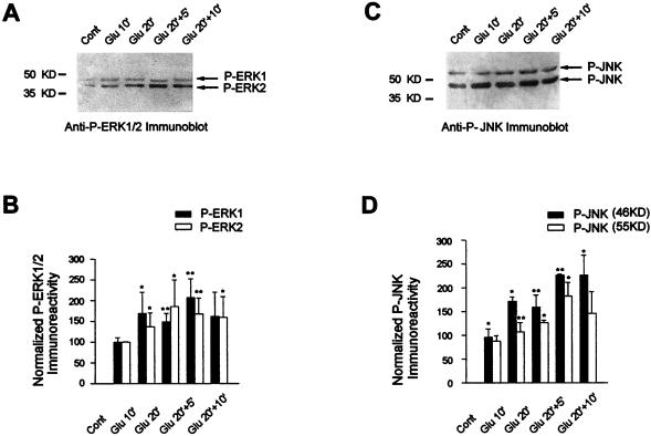
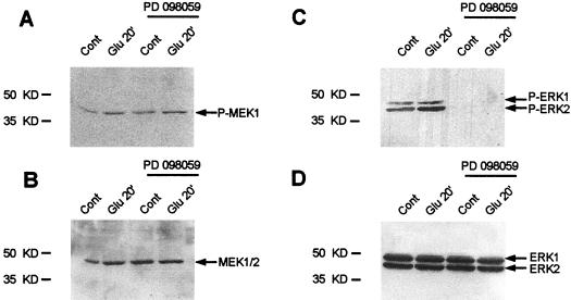
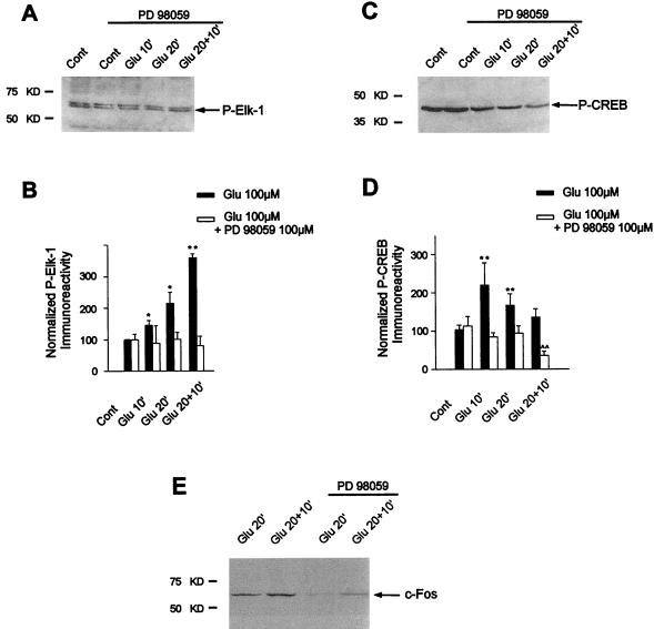
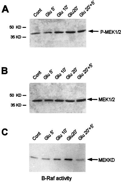
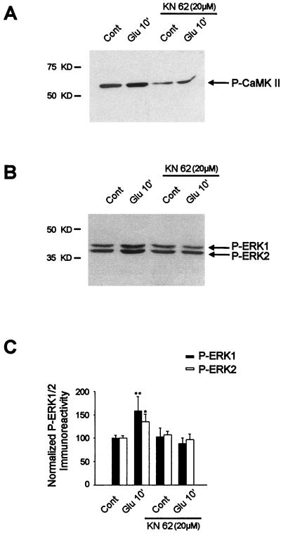
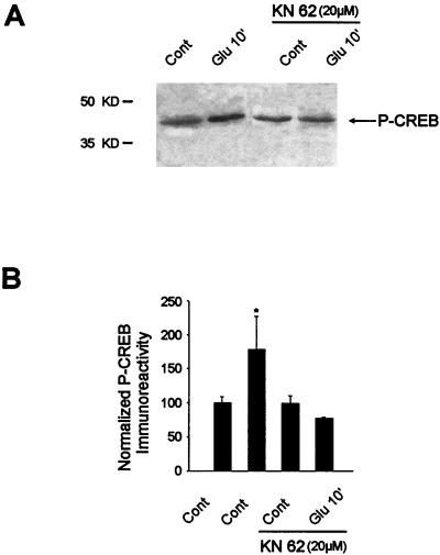
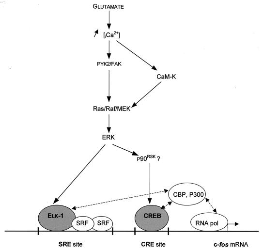
In cell culture systems, the TCF Elk-1 represents a convergence point for extracellular signal-related kinase (ERK) and c-Jun N-terminal kinase/stress-activated protein kinase (JNK/SAPK) subclasses of mitogen-activated protein kinase (MAPK) cascades. Its phosphorylation strongly potentiates its ability to activate transcription of the c-fos promoter through a ternary complex assembled on the c-fos serum response element. In rat brain postmitotic neurons, Elk-1 is strongly expressed (V. Sgambato, P. Vanhoutte, C. Pagès, M. Rogard, R. A. Hipskind, M. J. Besson, and J. Caboche, J. Neurosci. 18:214-226, 1998). However, its physiological role in these postmitotic neurons remains to be established. To investigate biochemically the signaling pathways targeting Elk-1 and c-fos in mature neurons, we used a semi-in vivo system composed of brain slices stimulated with the excitatory neurotransmitter glutamate. Glutamate treatment leads to a robust, progressive activation of the ERK and JNK/SAPK MAPK cascades. This corresponds kinetically to a significant increase in Ser383-phosphorylated Elk-1 and the appearance of c-fos mRNA. Glutamate also causes increased levels of Ser133-phosphorylated cyclic AMP-responsive element-binding protein (CREB) but only transiently relative to Elk-1 and c-fos. ERK and Elk-1 phosphorylation are blocked by the MAPK kinase inhibitor PD98059, indicating the primary role of the ERK cascade in mediating glutamate signaling to Elk-1 in the rat striatum in vivo. Glutamate-mediated CREB phosphorylation is also inhibited by PD98059 treatment. Interestingly, KN62, which interferes with calcium-calmodulin kinase (CaM-K) activity, leads to a reduction of glutamate-induced ERK activation and of CREB phosphorylation. These data indicate that ERK functions as a common component in two signaling pathways (ERK/Elk-1 and ERK/?/CREB) converging on the c-fos promoter in postmitotic neuronal cells and that CaM-Ks act as positive regulators of these pathways.


Figures

FIG. 1
FIG. 1
Glutamate-induced expression of c-Fos protein (A) and c-fos mRNA (B) in striatal slices. (A) Striatal slices were superfused with Krebs buffer alone (Cont) or in the presence of glutamate (100 μM) for 10 min (Glu 10′), 20 min (Glu 20′), or 20 min followed by 5 min (Glu 20′+5′) or 10 min (Glu 20′+10′) of Krebs buffer superfusion. At the end of the experiment, striatal slices were immediately lysed and processed for extraction of proteins. c-Fos protein expression was analyzed at the various time points by Western blotting with a specific anti c-Fos antibody. Fos protein is detectable at Glu20 and then increases at Glu20+5 and Glu20+10. (B) Total RNAs were extracted from the same striatal extracts (see Materials and Methods). c-fos and GAPDH mRNAs were detected on the same Northern blot. While GAPDH hybridization signals remain identical, c-fos mRNAs are induced at Glu10 and Glu20 and then their levels decrease slightly.
FIG. 2
FIG. 2
Kinetics of Ser383 Elk-1 and Ser133 CREB phosphorylation after glutamate application. The activation of Elk-1 and CREB was studied by Western blotting from the same striatal extracts as those used for the experiment in Fig. 1. (A) Immunolabeling obtained with an anti-active Elk-1 antibody (antiphospho- Ser383–Elk-1). (B) Detection of Elk-1 proteins on the same blot after a stripping procedure. Note the marked increase of phosphorylated 62-kDa proteins after glutamate application and the equal amount of Elk-1 proteins in all extracts. (C) Densitometric measurements of digitized images of autoradiograms were performed in four independent experiments (representing 12 striatal slices for each time point). Relative Elk-1 activation was determined by normalization of the density of images from phosphorylated Elk-1 to that of the total Elk-1 from parallel experiments in the same samples. (D) Immunolabeling obtained with an anti-active CREB antibody (antiphospho-Ser133–CREB). (E) Detection of CREB proteins on the same blot after a stripping procedure. Note the marked increase of phosphorylated 43-kDa proteins after glutamate application and the equal amount of CREB proteins in all extracts. (F) Determination of relative CREB activation. This was performed as specified for panel C. Statistical analysis: *, P < 0.05; **, P < 0.005 (unpaired Student’s t test) when comparing Glu chambers with control chambers.
FIG. 3
FIG. 3
ERK and JNK proteins are activated by glutamate. (A and C) The activation of ERK (A) and JNK (C) proteins was studied by Western blotting with specific anti-active ERK and anti-active JNK antibodies, respectively. (B) Relative ERK activation was determined by normalization of the density of images from phosphorylated ERK1 and ERK2 to that of total ERK1 and ERK2. (D) Relative JNK activation after normalization from phosphorylated JNK-p46 and JNK-p55 to that of total JNK-p46 and JNK-p55. Statistical analysis: *, P < 0.05; **, P < 0.005 (unpaired Student’s t test) when comparing Glu chambers with control chambers.
FIG. 4
FIG. 4
The MEK inhibitor PD98059 abolishes ERK activation by glutamate. Striatal slices were treated with PD98059 (100 μM) for 30 min prior to and during glutamate application (Glu10, Glu20, and Glu20+10). Shown are Western blots obtained with striatal slices (Glu 20′) with anti-active antibodies (A and C) or antibodies labeling the total proteins (B and D). Similar results were observed at the various time points (data not shown). Note that PD98059 treatment completely abolishes ERK activation in the presence or absence of glutamate (C). Glutamate-induced activation of MEK (A) remained unchanged after PD98059 treatment. Total levels of proteins remain unchanged whatever the treatment (B and D).
FIG. 5
FIG. 5
PD98059 abolishes glutamate-induced Elk-1, CREB phosphorylation and c-Fos induction by glutamate. The phosphorylation of Elk-1 and CREB and the induction of c-Fos proteins were studied by Western blotting of the same striatal extracts as used in the experiment in Fig. 4. For each time point, the efficacy of glutamate superfusion was verified (data not shown). (A) Immunolabeling obtained with antiphospho-Ser383–Elk-1 from striatal extracts activated by glutamate (Glu 10′, Glu 20′, and Glu 20+10′) in presence of PD98059 (100 μM). (B) Densitometric measurements were performed in five independent experiments (representing 15 striatal slices) in the presence or absence of PD98059. They show a complete inhibition of glutamate-induced phosphorylation of Elk-1 after PD98059 treatment. (C) Immunolabeling obtained with antiphospho-Ser133–CREB from striatal extracts activated by glutamate (Glu 10′, Glu 20′, Glu 20+10′) in the presence of PD98059 (100 μM). (D) Densitometric measurements performed in five independent experiments (representing 15 striatal slices) show the complete inhibition of glutamate-induced phosphorylation of CREB after PD98059 treatment at Glu10 and Glu20 and a decreased level of CREB phosphorylation at Glu20+10 compared to control slices. Statistical analysis: *, P < 0.05; **, P < 0.005 (unpaired Student’s t test) when comparing glutamate alone with control chambers; ∧∧, P < 0.005 when comparing glutamate plus PD98059 with control chambers. (E) c-Fos protein expression was analyzed by Western blotting at Glu 20′ and Glu 20+10′ in the presence or absence of PD98059.
FIG. 6
FIG. 6
Kinetics of MEK1 and B-Raf activation on glutamate stimulation of striatal slices. (A) Western blot analysis of MEK1 and MEK2 phosphorylation with a specific antiphospho-Ser217-Ser221 MEK1/2 antibody. This antibody stained a single band (43 kDa) on immunoblots, consistent with the molecular masses of the MEK1 and MEK2 proteins. (B) The same blot was stripped and rehybridized with the antibody corresponding to the inactive MEK1 and MEK2 proteins. This step allowed us to detect the total amounts of MEK proteins in the immunoblot. (C) B-Raf protein kinases were isolated by immunoprecipitation, and B-Raf protein kinase activity was detected in the immune complex by using [γ-32P]ATP and the MEK-kinase dead (MEKKD) fusion protein as the substrate. Note the increase of B-Raf activity at Glu 10′ and Glu 20′.
FIG. 7
FIG. 7
Role of the CaM-K inhibitor KN62 in glutamate-induced ERK activation. Striatal slices were superfused with KN62 (20 μM) for 30 min prior to and during glutamate application. (A) The efficacy of this compound was analyzed by Western blotting with an antiphospho-Thr286–CaM-KII antibody. Note that KN62 strongly decreases both basal levels and glutamate-induced phospho-Thr286-CaM-KII levels. (B) The same striatal extracts were analyzed with an anti-active ERK antibody. Note the inhibition of glutamate-induced ERK activation by KN62. (C) Densitometric measurements were performed in three independent experiments (representing nine striatal slices) in the presence or absence of KN62 (for each experiment, the inhibition of CaM-K activity by KN62 was verified as specified for panel A). Statistical analysis: *, P < 0.05; **, P < 0.005 (unpaired Student’s t test) when comparing glutamate alone with control chambers.
FIG. 8
FIG. 8
Ser133-CREB phosphorylation by glutamate is inhibited by KN62 treatment. (A) Glutamate-induced Ser133-CREB phosphorylation in the presence or absence of KN62 was analyzed by Western blotting from the same striatal extracts as used in the experiment in Fig. 7. (B) Densitometric measurements were performed in three independent experiments (representing nine striatal slices) in the presence or absence of KN62. Statistical analysis: *, P < 0.05 (unpaired Student’s t test) when comparing glutamate alone with control chambers.
FIG. 9
FIG. 9
CaM-Ks act as positive regulators of the ERK signaling cascade.

References

    1. Abate C, Luck D, Gagne E, Roeder R G, Curran T. Fos and Jun cooperate in transcriptional regulation via heterologous activation domains. Mol Cell Biol. 1990;10:5532–5535. - PMC - PubMed
    1. Ahn S, Olive M, Aggarwal S, Krylov D, Ginty D D, Vinson C. A dominant-negative inhibitor of CREB levels that it is a general mediator of stimulus-dependent transcription of c-fos. Mol Cell Biol. 1998;18:967–977. - PMC - PubMed
    1. Alessi D R, Saito Y, Campbell D G, Cohen P, Sithanandam G, Rapp U, Ashworth A, Marshall C J, Cowley S. Identification of the sites in MAP kinase kinase 1 phosphorylated by p74raf1. EMBO J. 1994;13:1610–1619. - PMC - PubMed
    1. Bading H, Ginty D D, Greenberg M E. Regulation of gene expression in hippocampal neurons by distinct signaling pathways. Science. 1993;260:181–186. - PubMed
    1. Barnier J V, Papin C, Eychène A, Lecoq O, Calothy G. The mouse B-raf gene encodes multiple protein isoforms with tissue-specific expression. J Biol Chem. 1995;270:23381–23389. - PubMed

Publication types

MeSH terms

Substances