. 1999 Jan;19(1):274-83.
doi: 10.1128/MCB.19.1.274.

Transcriptional and posttranscriptional silencing of rodent alpha1(I) collagen by a homologous transcriptionally self-silenced transgene

Affiliations

Transcriptional and posttranscriptional silencing of rodent alpha1(I) collagen by a homologous transcriptionally self-silenced transgene

M B Bahramian et al. Mol Cell Biol. 1999 Jan.

Abstract

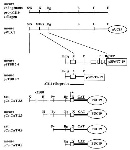
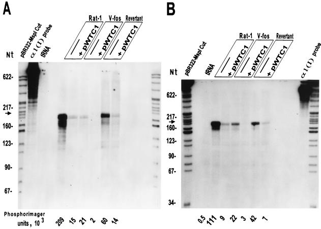
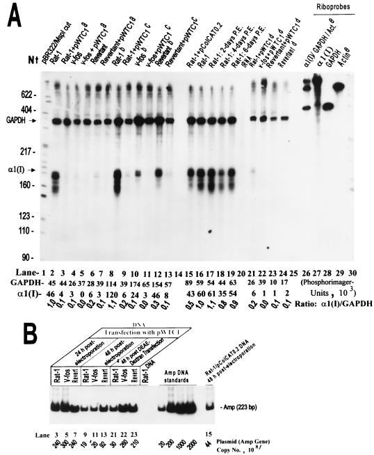
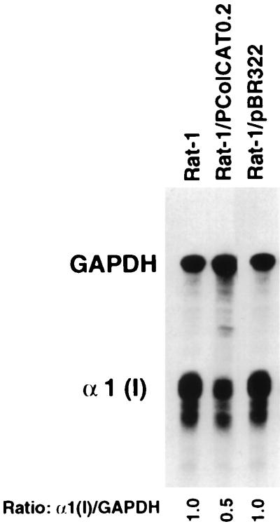
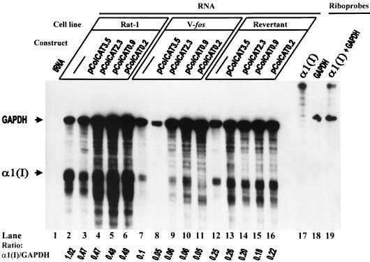
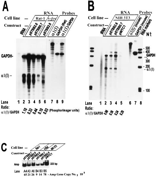
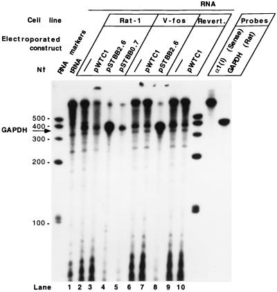
Transient transfection of rodent fibroblasts with plasmids carrying a full-size pro-alpha1(I) collagen gene (pWTC1) results in rapid reduction of the endogenous transcripts by >90%, while the transgene mRNA is undetectable. Using deletion constructs, two adjacent 5' noncoding regions of the gene are identified as causing transcriptional silencing of the endogene in normal and v-fos-transformed cells but not in nontumorigenic revertants, which show partial relief from v-fos transformation-induced alpha1(I) gene suppression. The 3' end of the transgene carries an additional element(s), causing posttranscriptional silencing of the endogene in all cells including the revertant. Data indicate that the transgenes are transcriptionally self-silenced. Genome-integrated transgenes that are transcriptionally active also allow expression of the endogene, suggesting gene activation by chromosomal factors missing in pWTC1. Silencing is not regulated by antisense RNA. Silencing of the endogenous pro-alpha1(I) collagen gene is not linked to the level of transgene expression.


Figures

FIG. 1
FIG. 1
Map of pro-α1(I) collagen plasmids and structure of RNase protection riboprobe. Positions of the first five mRNA exons are indicated by open boxes. The vertical insert marked X/B/X indicates the position of the insertion of the BamHI linker within an XbaI site. This insertion is within the 5′ untranslated portion of the mRNA. Horizontal solid and dotted lines represent the procollagen gene sequences. Arrow shows the transcription start position and direction. Relevant restriction sites for the enzymes SalI (S), XbaI (X), HindIII (H), PvuII (Pv), BglII (Bg), BamHI (B), PstI (P), and EcoRI (E) are indicated. The position of pro-α1(I) collagen gene probe transcribed in vitro by T7 RNA polymerase from pSTBB0.7 (EcoRI digested) is shown. This antisense riboprobe of about 850 nt protects the 194-nt endogenous mouse or rat α1(I) mRNA corresponding to exon 1.
FIG. 2
FIG. 2
RNase protection assays showing endogenous pro-α1(I) collagen mRNA levels in Rat-1, v-fos-transformed 1302-4-1, and revertant EMS-1-19 cells, untransfected or transiently transfected with pWTC1. The 850-nt antisense riboprobes transcribed by T7 RNA polymerase from a mouse pro-α1(I) collagen fragment (HindIII/EcoRI) of pSTBB0.7 were hybridized with total RNA extracted from equal number of cultured cells (about 106), either untransfected or transfected by pWTC1 and harvested at 24 (A) and 48 (B) h after electroporation. Endogenous rat collagen mRNA protects a 194-nt major band (shown by the arrow) and some smaller minor bands of mouse α1(I) probe after treatment with RNase 1. Mouse α1(I) transcripts from pWTC1 are expected to protect 118- and 76-nt bands. PhosphorImager units corresponding to the protected major bands of endogenous α1(I) mRNA are indicated. This RNase protection experiment is representative of four separate assays with similar results.
FIG. 3
FIG. 3
Specific suppression of pro-α1(I) collagen mRNA by transiently transfected α1(I) collagen genes. (A) RNase protection results of assays using total RNA from Rat-1, v-fos-transformed, and revertant cells, either untransfected or transfected with plasmid DNA. The α1(I) antisense riboprobe and the expected protected bands are described in the legend to Fig. 2. The 473-nt antisense riboprobe transcribed by T7 RNA polymerase from a rat GAPDH fragment (SmaI/HindIII) of pLS-1 protects a 361-nt GAPDH mRNA fragment which serves as an internal standard. PhosphorImager units determined for each set of GAPDH and α1(I) bands and the corresponding α1(I)/GAPDH ratio are indicated. Superscripts: a, cells harvested 24 h after electroporation with pWTC1; b cells electroporated without any DNA 16 h before harvesting; c cells harvested 48 h after electroporation with plasmid; d DEAE-dextran transfection (viability of cells after this transfection was less than one-fifth of that by electroporation, and therefore less RNA was available for use); e rat β-actin sense riboprobe, described in Materials and Methods, used as a negative internal control (in addition to tRNA) for RNase protection assays. Cells used for RNA preparations 17 to 19 were harvested at indicated number of days postelectroporation (P.E.) with no DNA. (B) Determination of plasmid (or β-lactamase gene) copy numbers corresponding to the transfected samples in panel A, using quantitative PCR amplification of a 223-bp fragment of the amp gene. Lane numbers in panels A and B are related. f, numbers refer to total number of plasmids in total number of transfected cells (106 to 107).
FIG. 4
FIG. 4
RNase protection assays showing endogenous pro-α1(I) collagen mRNA levels in Rat-1 cells, untransfected or transiently transfected with either ColCAT0.2 or pBR322. RNase protection assays and determination of the corresponding α1(I)/GAPDH ratio were as described in Fig. 3 at 24 h after transfection.
FIG. 5
FIG. 5
RNase 1 protection assay-based deletion mapping of pro-α1(I) collagen promoter region to identify the collagen mRNA-suppressive element(s) in Rat-1, v-fos-transformed 1302-4-1, and revertant EMS-1-19 cells. The α1(I) and rat GAPDH antisense riboprobes and the expected protected bands are described in the legends to Fig. 2 and 3. The α1(I)/GAPDH protected-band ratios, quantitated with a PhosphorImager, are shown.
FIG. 6
FIG. 6
RNase A/T1 protection assay used to identify the collagen mRNA-suppressive element(s) in the first five exon/intron regions in various rodent fibroblast cell lines. (A) RNase protection assay using total RNA from Rat-1 and v-fos-transformed 1302-4-1 cells untransfected or transiently transfected with either pSTBB2.6 or pSTBB0.7. (B) RNase protection assay using total RNA from mouse NIH 3T3 fibroblasts untransfected or transiently transfected with the plasmids indicated. The α1(I) and rat GAPDH antisense riboprobes and the expected protected bands are described in the legends to Fig. 2 and 3. The 406-nt antisense mouse GAPDH riboprobe transcribed from pTRI-GAPDH-mouse protects a 316-nt mouse GAPDH mRNA fragment. (C) Determination of the number of copies of various plasmids transfected into different rodent cell lines shown in panels A and B, using quantitative PCR amplification of the amp gene and PhosphorImager analyses.
FIG. 7
FIG. 7
RNase T1 protection assay demonstrating the absence of antisense pro-α1(I) collagen mRNA in various rat fibroblast lines which were either untransfected or transiently transfected by constructs carrying different lengths of the collagen gene. The rat GAPDH antisense riboprobe and the expected protected band are described in the legend to Fig. 3. The 850-nt α1(I) sense riboprobes transcribed in vitro by SP6 RNA polymerase from pSTBB0.7 are expected to protect 5′-end α1(I) antisense transcripts of up to 585 nt. Evaluation of transfections by different plasmids is shown in Fig. 6C.

References

    1. Bahramian M B, Zarbl H. Direct gene quantitations by PCR reveal differential accumulation of ectopic enzyme in Rat-1 cells, v-fos transformants and revertants. PCR Methods Appl. 1994;4:145–153. - PubMed
    1. Bahramian, M. B., C. D. Hoemann, and H. Zarbl. Unpublished data.
    1. Barker D D, Wu H, Hartung S, Breindl M, Jaenisch R. Retrovirus-induced insertional mutagenesis: mechanism of collagen mutation in Mov13 mice. Mol Cell Biol. 1991;11:5154–5163. - PMC - PubMed
    1. Baulcombe D C, English J J. Ectopic pairing of homologous DNA and posttranscriptional gene silencing in transgenic plants. Curr Opin Biotechnol. 1996;7:173–180.
    1. Bingham P M. Cosuppression comes to the animals. Cell. 1997;90:385–387. - PubMed

Publication types

LinkOut - more resources