. 1999 Jan;19(1):364-75.
doi: 10.1128/MCB.19.1.364.

CA- and purine-rich elements form a novel bipartite exon enhancer which governs inclusion of the minute virus of mice NS2-specific exon in both singly and doubly spliced mRNAs

Affiliations

CA- and purine-rich elements form a novel bipartite exon enhancer which governs inclusion of the minute virus of mice NS2-specific exon in both singly and doubly spliced mRNAs

A Gersappe et al. Mol Cell Biol. 1999 Jan.

Abstract

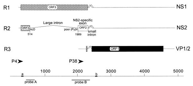
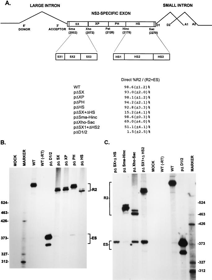
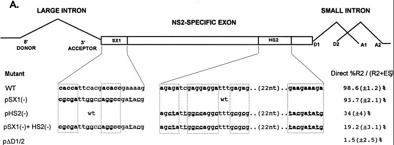
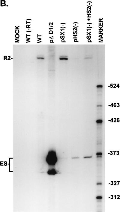
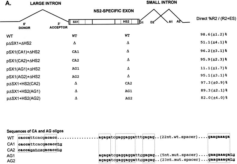
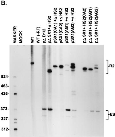
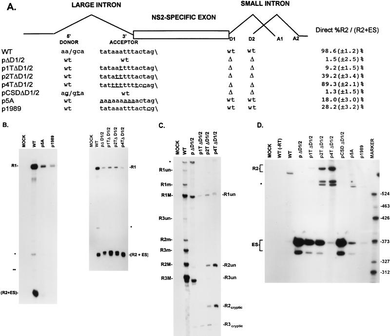
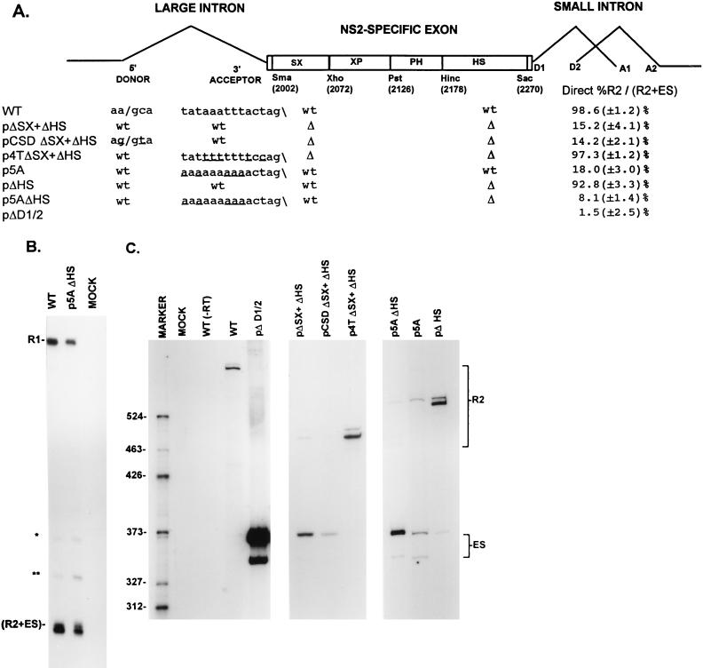
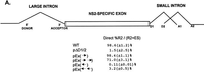
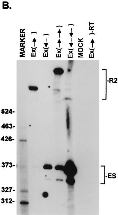
The alternatively spliced 290-nucleotide NS2-specific exon of the parvovirus minute virus of mice (MVM), which is flanked by a large intron upstream and a small intron downstream, constitutively appears both in the R1 mRNA as part of a large 5'-terminal exon (where it is translated in open reading frame 3 [ORF3]), and in the R2 mRNA as an internal exon (where it is translated in ORF2). We have identified a novel bipartite exon enhancer element, composed of CA-rich and purine-rich elements within the 5' and 3' regions of the exon, respectively, that is required to include NS2-specific exon sequences in mature spliced mRNA in vivo. These two compositionally different enhancer elements are somewhat redundant in function: either element alone can at least partially support exon inclusion. They are also interchangeable: either element can function at either position. Either a strong 3' splice site upstream (i.e., the exon 5' terminus) or a strong 5' splice site downstream (i.e., the exon 3' terminus) is sufficient to prevent skipping of the NS2-specific exon, and a functional upstream 3' splice site is required for inclusion of the NS2-specific exon as an internal exon into the mature, doubly spliced R2 mRNA. The bipartite enhancer functionally strengthens these termini: the requirement for both the CA-rich and purine-rich elements can be overcome by improvements to the polypyrimidine tract of the upstream intron 3' splice site, and the purine-rich element also supports exon inclusion mediated through the downstream 5' splice sites. In summary, a suboptimal large-intron polypyrimidine tract, sequences within the downstream small intron, and a novel bipartite exonic enhancer operate together to yield the balanced levels of R1 and R2 observed in vivo. We suggest that the unusual bipartite exonic enhancer functions to mediate proper levels of inclusion of the NS2-specific exon in both singly spliced R1 and doubly spliced R2.


Figures

FIG. 1
FIG. 1
Genetic map of MVM. The three major transcript classes and protein-encoding ORFs are shown. The two promoters (P4 and P38) are indicated by arrowheads. The large intron, small intron, and NS2-specific exon are indicated. The nonconsensus donor (ncD) and the poor polypyrimidine tract [poor (Py)n] of the large intron are also shown. The bottom diagram shows nucleotide locations, the two probes (A [nt 385 to 650] and B [nt 1854 to 2378]) used for RNase protection assays, and the two primers [a (nt 326 to 345) and b (nt 2557 to 2538)] used for RT-PCR, as described fully in Materials and Methods.
FIG. 2
FIG. 2
A bipartite enhancer within the NS2-specific exon is required for inclusion of this exon in mature spliced mRNA. (A) The restriction sites within the NS2-specific exon (SmaI, XhoI, PstI, HincII, and SacI) which divide the exon into four regions (SX, XP, PH, and HS) and were used to generate the different exon deletion mutants are indicated (ΔSX, for example, is a deletion between the SmaI and XhoI sites [see the text]). The SX1 to SX3 and HS1 to HS3 regions extend between the following nucleotide positions: SX1, nt 2002 to 2025; SX2, nt 2019 to 2052; SX3, nt 2050 to 2109; HS1, nt 2177 to 2196; HS2, nt 2196 to 2253; HS3, nt 2252 to 2270 (MVM nucleotide numbers as in reference 1). Quantitations of the direct percent R2/(R2+ES) ratio obtained by RT-PCR analysis for various mutants are also shown. All the values are the average of at least three separate experiments. Standard deviations are indicated in parentheses. ES values for pΔD1/2 are shown for comparison; the mutation is shown in Fig. 5A. (B) RT-PCR analysis of RNA generated by wild-type MVM (WT), mutants (described in the text), or mock transfected, as designated at the top of each lane. Samples were run on a 6% acrylamide—urea gel. pΔD1/2 was used as a control for amplification of the ES. WT(−RT) is a control reaction with wild-type RNA but excluding reverse transcriptase. An RNase protection analysis, with probe B, of RNA generated by the wild type was used as a marker (MARKER; the sizes of the marker bands are shown on the left) for the sizes of the RT-PCR-amplified bands. Wild-type RNA generated a 658-nt amplified R2 product, while RNA generated by the mutants showed R2 products of sizes consistent with the sizes of the deletions in these mutants. As explained in the legend to Fig. 5D, two kinds of amplified ES, using either A1 or A2, were observed. (C) RT-PCR analysis of RNA generated by wild-type MVM (WT), mutants (described in the text), or mock transfected, as designated at the top of each lane. Samples were run on a 6% acrylamide–urea gel. pΔD1/2 and WT(−RT) controls, and the marker (MARKER; the sizes of the marker bands are shown on the right for the sizes of the RT-PCR-amplified bands) are as in panel B. Wild-type RNA generated a 658-nt amplified R2 product, while RNA generated by the mutants showed R2 products of sizes consistent with the sizes of the deletions in these mutants. As explained in the legend to Fig. 5D, two kinds of amplified ES using either A1 or A2 were observed.
FIG. 3
FIG. 3
Point mutations within the bipartite enhancer result in exon skipping. (A) The sequences of the wild-type and mutant SX1 and HS2 regions are shown underneath their appropriate map positions (with deviations from the wild-type sequence underlined), together with quantitations of the direct percent R2/(R2+ES) ratio obtained by RT-PCR analysis for each mutant. The CA-rich sequences in the SX1 region and the purine-rich sequences in the HS2 region are boxed and in boldface type. All the values are the average of at least three separate experiments. Standard deviations are indicated in parentheses. ES values for pΔD1/2 are shown for comparison; the mutation is shown in Fig. 5A. wt, wild type sequence. (B) RT-PCR analysis of RNA generated by wild-type MVM (WT), mutants (described in the text), or mock transfected, as designated at the top of each lane. Samples were run on a 6% acrylamide–urea gel. pΔD1/2 and WT(−RT) controls and MARKER (the sizes of the marker bands are shown on the right) are as described in the legend to Fig. 2B. RNAs generated by either the wild type or the point mutants showed a 658-nt amplified R2 product. As explained in the legend to Fig. 5D, two kinds of amplified ES, using either A1 or A2, were observed.
FIG. 3
FIG. 3
Point mutations within the bipartite enhancer result in exon skipping. (A) The sequences of the wild-type and mutant SX1 and HS2 regions are shown underneath their appropriate map positions (with deviations from the wild-type sequence underlined), together with quantitations of the direct percent R2/(R2+ES) ratio obtained by RT-PCR analysis for each mutant. The CA-rich sequences in the SX1 region and the purine-rich sequences in the HS2 region are boxed and in boldface type. All the values are the average of at least three separate experiments. Standard deviations are indicated in parentheses. ES values for pΔD1/2 are shown for comparison; the mutation is shown in Fig. 5A. wt, wild type sequence. (B) RT-PCR analysis of RNA generated by wild-type MVM (WT), mutants (described in the text), or mock transfected, as designated at the top of each lane. Samples were run on a 6% acrylamide–urea gel. pΔD1/2 and WT(−RT) controls and MARKER (the sizes of the marker bands are shown on the right) are as described in the legend to Fig. 2B. RNAs generated by either the wild type or the point mutants showed a 658-nt amplified R2 product. As explained in the legend to Fig. 5D, two kinds of amplified ES, using either A1 or A2, were observed.
FIG. 4
FIG. 4
A CA-rich element and a purine-rich element together constitute the bipartite enhancer of the NS2-specific exon. (A) The sequences of the CA-rich and AG-rich oligonucleotides that were tested (deviations from the wild-type sequence are underlined; the sequences of the wild-type spacer in HS1 or the mutant spacers in AG1 and AG2 are described in Materials and Methods), and the positions at which they were replaced into pΔSX1+ΔHS2 are indicated (see the text for details). The CA-rich sequences in the SX1 region and the purine-rich sequences in the HS2 region are boxed and in boldface type. Quantitations of the direct percent R2/(R2+ES) ratio obtained by RT-PCR analysis for each mutant are also shown. All the values are the average of at least three separate experiments. Standard deviations are indicated in parentheses. WT, wild-type sequence. Δ, deletion. (B) RT-PCR analysis of RNA generated by wild-type MVM (WT), mutants (described in the text), or mock transfected, as designated at the top of each lane. Samples were run on a 6% acrylamide—urea gel. pΔD1/2 and WT(−RT) controls and MARKER (the sizes of the marker bands are shown on the left) are as described in the legend to Fig. 2B. RNA generated by WT showed a 658-nt amplified R2 product, while RNA generated by the mutants showed R2 products of sizes consistent with the sizes of the deletions and/or substitutions in these mutants. As explained in the legend to Fig. 5D, two kinds of amplified ES, using either A1 or A2, were observed.
FIG. 4
FIG. 4
A CA-rich element and a purine-rich element together constitute the bipartite enhancer of the NS2-specific exon. (A) The sequences of the CA-rich and AG-rich oligonucleotides that were tested (deviations from the wild-type sequence are underlined; the sequences of the wild-type spacer in HS1 or the mutant spacers in AG1 and AG2 are described in Materials and Methods), and the positions at which they were replaced into pΔSX1+ΔHS2 are indicated (see the text for details). The CA-rich sequences in the SX1 region and the purine-rich sequences in the HS2 region are boxed and in boldface type. Quantitations of the direct percent R2/(R2+ES) ratio obtained by RT-PCR analysis for each mutant are also shown. All the values are the average of at least three separate experiments. Standard deviations are indicated in parentheses. WT, wild-type sequence. Δ, deletion. (B) RT-PCR analysis of RNA generated by wild-type MVM (WT), mutants (described in the text), or mock transfected, as designated at the top of each lane. Samples were run on a 6% acrylamide—urea gel. pΔD1/2 and WT(−RT) controls and MARKER (the sizes of the marker bands are shown on the left) are as described in the legend to Fig. 2B. RNA generated by WT showed a 658-nt amplified R2 product, while RNA generated by the mutants showed R2 products of sizes consistent with the sizes of the deletions and/or substitutions in these mutants. As explained in the legend to Fig. 5D, two kinds of amplified ES, using either A1 or A2, were observed.
FIG. 5
FIG. 5
The NS2-specific exon of MVM requires only one strong exon terminus (upstream 3′ splice site or downstream 5′ splice site) to prevent exclusion from mature spliced mRNA. (A) The sequences of the large-intron 5′-splice-site and 3′-splice-site polypyrimidine tract and cleavage site in wild-type MVM and mutants are shown underneath their appropriate map positions (deviations from the wild-type sequence are underlined), with quantitations of the direct percent R2/(R2+ES) ratio obtained by quantitative RT-PCR (described fully in Materials and Methods). The presence or absence of the small-intron 5′ splice sites in these constructs is indicated by either wt (presence) or Δ (absence). All the values are the average of at least three separate experiments. Standard deviations are indicated in parentheses. wt, wild type sequence. Δ, deletion. (B) The two panels show RNase protection analyses with probe A (Fig. 1) of RNA generated by wild-type MVM (WT), mutants (as described in the text), or mock transfected, as designated at the top of each lane. The identities of the protected bands are shown on the left (in the left panel) and on the right (in the right panel). The larger species (R1) represents mRNA R1, while the major smaller species (R2+ES) represents RNA that uses the upstream large-intron 5′ splice site at nt 514 (i.e., mRNA R2 plus the ES). The identities of the bands designated ∗ and ∗∗ are unknown; however, they are probably breakdown products of the probe since they are not reproducibly seen and occasionally appear in lanes of mock-infected RNA (data not shown). (C) RNase protection analysis with probe B (Fig. 1), of RNA generated by wild-type MVM (WT), mutants (as described in the text), or mock transfected, as designated at the top of each lane. The identities of the protected bands for the wild type are shown on the left. The mutants were protected with versions of probe B homologous to the mutant region of the upstream intron 3′-splice-site polypyrimidine tract but nonhomologous in the region of D1 and D2, and therefore the RNA generated by these mutants shows fragments shorter than those generated by wild-type RNA. These mutants produce R1, R2, and R3 products that are unspliced across the small intron (designated R1un, R2un, and R3un, respectively) due to the loss of the two downstream small intron 5′ splice sites, as well as R2 and R3 products that are spliced across the downstream small intron (designated R2cryptic and R3cryptic, respectively) by using a cryptic donor within the NS2-specific exon (see Discussion). Improvements of the upstream intron 3′-splice-site polypyrimidine tract disrupted the P38 promoter, resulting in decreased production of R3cryptic as well as R3un. ∗, undigested probe B. (D) RT-PCR analysis of RNA generated by wild-type MVM (WT), mutants (as described in the text), or mock transfected, as designated at the top of each lane, with primers a and b as shown in Fig. 1 and performed as explained in Materials and Methods. Samples were run on a 6% acrylamide–urea gel. WT(−RT) is a control reaction with wild-type RNA but excluding reverse transcriptase. An RNase protection analysis with probe B of RNA generated by the wild type was used as a marker (MARKER; the sizes of the marker bands are shown on the right) for the sizes of the RT-PCR-amplified bands. WT RNA generated a 658-nt amplified R2 product. RNA generated by the mutants showed two kinds of amplified ES, both of which were considered in the quantitations: a larger (368-nt) product and a smaller (346-nt) product which represent exon skipping to 3′ splice sites A1 and A2, respectively, of the downstream small intron. RNAs generated by pΔD1/2, p1TΔD1/2, p2TΔD1/2, and p4TΔD1/2 also show two kinds of amplified R2 products, which were considered in the quantitations; the largest of these is unspliced across the small intron, and the smaller two products (designated ∗) probably utilized a donor within the NS2-specific exon (see Discussion). RNAs generated by p5A and p1989 also showed the smaller R2 product that probably utilized a donor within the exon. The ES has previously been sequenced across the splice junction to confirm its identity (56).
FIG. 6
FIG. 6
Improvement of the upstream intron 3′-splice-site polypyrimidine tract can overcome exon skipping caused by deletion of the bipartite exon enhancer. (A) The restriction sites within the NS2-specific exon (SmaI, XhoI, PstI, HincII, and SacI) which were used to generate the exon deletion mutants and the sequences of the large-intron 5′-splice-site and 3′-splice-site polypyrimidine tract and cleavage site in the wild type and mutants are shown underneath their appropriate map positions (deviations from the wild-type sequence are underlined), together with quantitations of the direct percent R2/(R2+ES) ratio obtained by RT-PCR analysis for each mutant. All the values are the averages of at least three separate experiments. Standard deviations are indicated in parentheses. ES values for pΔD1/2 are shown for comparison; the mutation is shown in Fig. 5A. wt, wild-type sequence. Δ, deletion. (B) RNase protection analyses with probe A (Fig. 1) of RNA generated by wild-type MVM (WT), mutants (as described in the text), or mock transfected, as designated at the top of each lane. The identities of the protected bands are shown on the left and explained in the legend to Fig. 5B. (C) RT-PCR analysis of RNA generated by wild-type MVM (WT), mutants (as described in the text), or mock transfected, as designated at the top of each lane. Samples were run on a 6% acrylamide–urea gel. pΔD1/2 and WT(−RT) controls and MARKER (the sizes of the marker bands are shown on the left) are as described in the legend to Fig. 2B. RNA generated by the wild type showed a 658-nt amplified R2 product, while RNA generated by the mutants showed R2 products of sizes consistent with the sizes of the deletions in these mutants. As explained in the legend to Fig. 5D, two kinds of amplified ES, using either A1 or A2, were observed.
FIG. 7
FIG. 7
Inclusion of the NS2-specific exon tolerates doubling its length. (A) Quantitations of the direct percent R2/(R2+ES) ratio obtained by RT-PCR analysis for each mutant are shown. Construction of the mutants is explained in Materials and Methods. All the values are the average of at least three separate experiments. Standard deviations are indicated in parentheses. (B) RT-PCR analysis of RNA generated by wild-type MVM (WT), mutants (as described in the text), or mock transfected, as designated at the top of each lane. Samples were run on a 6% acrylamide–urea gel. MARKER is as described in the legend to Fig. 2B (the sizes of the marker bands are shown on the left). (Ex→)-RT is a control reaction with p(Ex→)-generated RNA but excluding reverse transcriptase. RNA generated by the wild type showed a 658-nt amplified R2 product, while RNA generated by the mutants showed R2 products of sizes consistent with the sizes of the substitutions in these mutants. As explained in the legend to Fig. 5D, two kinds of amplified ES, using either A1 or A2, were observed. The parent mutant bearing only the Sma sites at nt 2005 and 2270 [pEx(→)] is indistinguishable from the wild type with respect to splicing of the upstream intron and inclusion of the NS2-specific exon (56). Inclusion of the exon that is doubled in length still required wild-type exon sequences; when either one copy or two copies of the exon in the reverse orientation were inserted between the Sma sites [pEx(←) and pEx(←←), respectively (56)], the exon was almost uniformly skipped.
FIG. 7
FIG. 7
Inclusion of the NS2-specific exon tolerates doubling its length. (A) Quantitations of the direct percent R2/(R2+ES) ratio obtained by RT-PCR analysis for each mutant are shown. Construction of the mutants is explained in Materials and Methods. All the values are the average of at least three separate experiments. Standard deviations are indicated in parentheses. (B) RT-PCR analysis of RNA generated by wild-type MVM (WT), mutants (as described in the text), or mock transfected, as designated at the top of each lane. Samples were run on a 6% acrylamide–urea gel. MARKER is as described in the legend to Fig. 2B (the sizes of the marker bands are shown on the left). (Ex→)-RT is a control reaction with p(Ex→)-generated RNA but excluding reverse transcriptase. RNA generated by the wild type showed a 658-nt amplified R2 product, while RNA generated by the mutants showed R2 products of sizes consistent with the sizes of the substitutions in these mutants. As explained in the legend to Fig. 5D, two kinds of amplified ES, using either A1 or A2, were observed. The parent mutant bearing only the Sma sites at nt 2005 and 2270 [pEx(→)] is indistinguishable from the wild type with respect to splicing of the upstream intron and inclusion of the NS2-specific exon (56). Inclusion of the exon that is doubled in length still required wild-type exon sequences; when either one copy or two copies of the exon in the reverse orientation were inserted between the Sma sites [pEx(←) and pEx(←←), respectively (56)], the exon was almost uniformly skipped.

Similar articles

Cited by

References

    1. Astell C R, Gardiner E M, Tattersall P. DNA sequence of the lymphotropic variant of minute virus of mice, MVM(i), and comparison with the DNA sequence of the fibrotropic prototype strain. J Virol. 1986;57:656–669. - PMC - PubMed
    1. Berget S M. Exon recognition in vertebrate splicing. J Biol Chem. 1995;270:2411–2444. - PubMed
    1. Black D L. Finding splice sites within a wilderness of RNA. RNA. 1995;1:763–771. - PMC - PubMed
    1. Buvoli M, Mayer S A, Patton J G. Functional crosstalk between exon enhancers, polypyrimidine tracts and branchpoint sequences. EMBO J. 1997;16:7174–7183. - PMC - PubMed
    1. Clemens K E, Cerutis D R, Burger L R, Yang G Q, Pintel D. Cloning of minute virus of mice cDNAs and preliminary analysis of individual viral proteins expressed in murine cells. J Virol. 1990;64:3967–3973. - PMC - PubMed

Publication types

LinkOut - more resources