. 1999 Jan;19(1):612-22.
doi: 10.1128/MCB.19.1.612.

Cyclin E2, a novel G1 cyclin that binds Cdk2 and is aberrantly expressed in human cancers

Affiliations

Cyclin E2, a novel G1 cyclin that binds Cdk2 and is aberrantly expressed in human cancers

J M Gudas et al. Mol Cell Biol. 1999 Jan.

Abstract

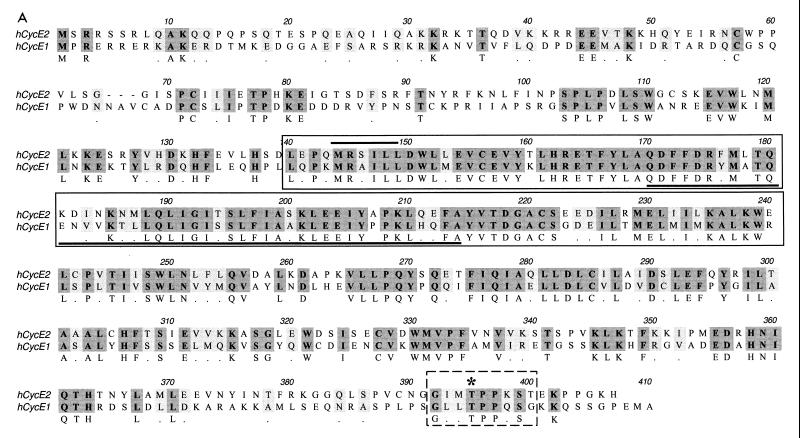
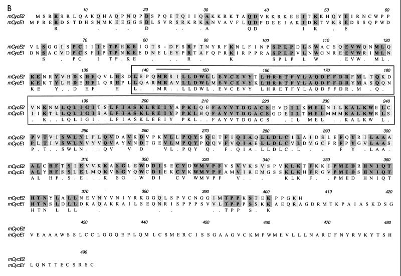
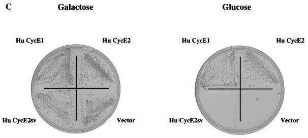
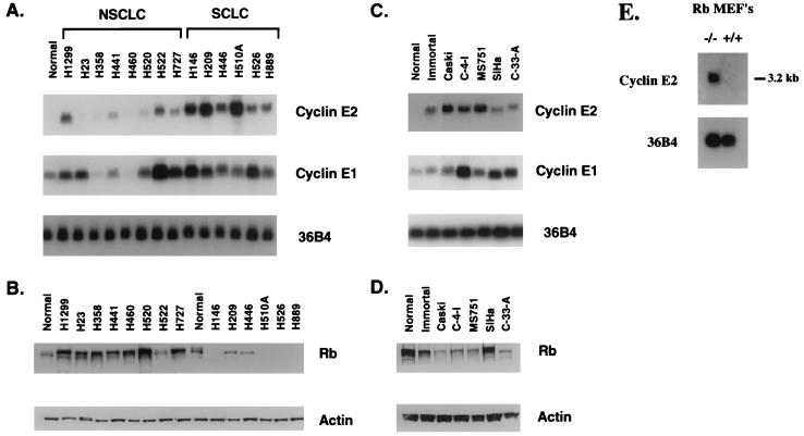
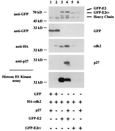
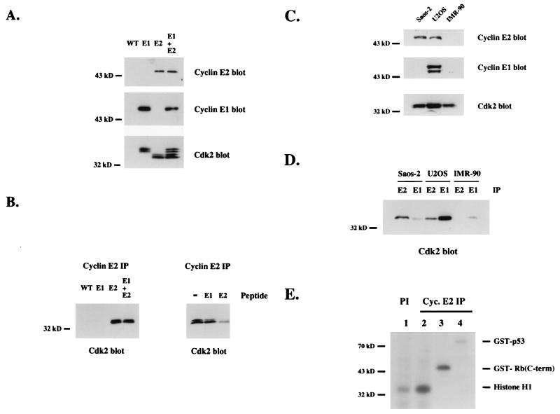
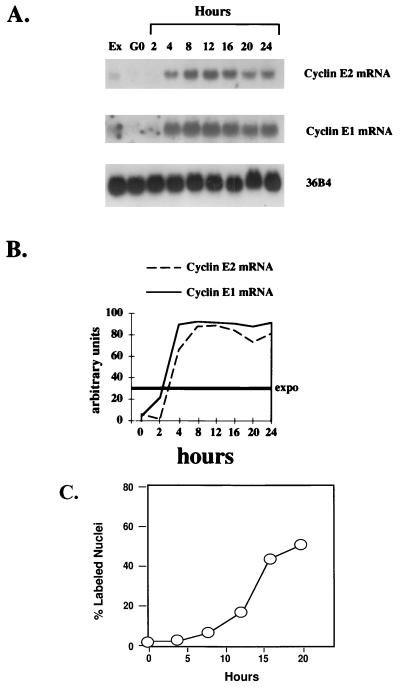
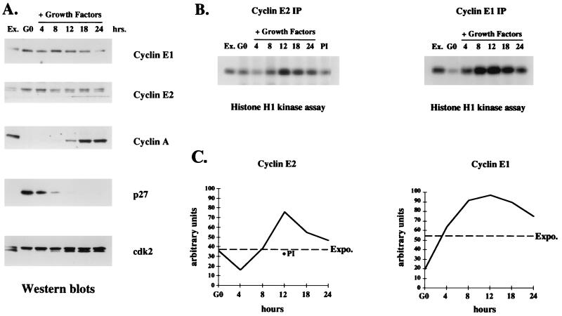
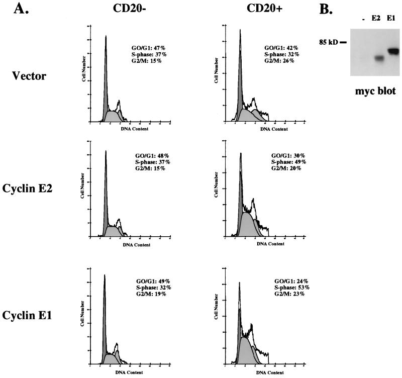
A novel cyclin gene was discovered by searching an expressed sequence tag database with a cyclin box profile. The human cyclin E2 gene encodes a 404-amino-acid protein that is most closely related to cyclin E. Cyclin E2 associates with Cdk2 in a functional kinase complex that is inhibited by both p27(Kip1) and p21(Cip1). The catalytic activity associated with cyclin E2 complexes is cell cycle regulated and peaks at the G1/S transition. Overexpression of cyclin E2 in mammalian cells accelerates G1, demonstrating that cyclin E2 may be rate limiting for G1 progression. Unlike cyclin E1, which is expressed in most proliferating normal and tumor cells, cyclin E2 levels were low to undetectable in nontransformed cells and increased significantly in tumor-derived cells. The discovery of a novel second cyclin E family member suggests that multiple unique cyclin E-CDK complexes regulate cell cycle progression.


Figures

FIG. 1
FIG. 1
Sequence alignment (using the Clustal alignment program) and functional activity of cyclin E2 and cyclin E1. (A) Alignment of full-length human cyclin E2 protein with human cyclin E1 protein. The cyclin box is boxed with an unbroken line, the MRSILL sequence is overlined, and the peptide sequence missing from cyclin E2SV is underlined. The C-terminal PEST sequence motif is outlined with a dashed line, and the CDK consensus threonine phosphorylation site is starred. (B) Clustal alignment of murine cyclin E2 and murine cyclin E1. The cyclin box motif is boxed, and the MRSILL sequence is overlined. (C) Functional complementation of yeast G1 cyclins by human cyclin E2. S. cerevisiae MTY618 has a deletion of the CLN1, CLN2, and CLN3 genes and is kept alive by an integrated GAL-CLN3 gene. On galactose medium, CLN3 is transcribed and the cells live; on glucose, the GAL promoter is shut off and the cells fail to grow. Yeast expressing either human cyclin E1 (Hu CycE1) or human cyclin E2 (Hu CycE2) are able to grow on glucose, while yeast harboring vector alone or the human cyclin E2SV (Hu CycE2sv) fail to grow on glucose.
FIG. 1
FIG. 1
Sequence alignment (using the Clustal alignment program) and functional activity of cyclin E2 and cyclin E1. (A) Alignment of full-length human cyclin E2 protein with human cyclin E1 protein. The cyclin box is boxed with an unbroken line, the MRSILL sequence is overlined, and the peptide sequence missing from cyclin E2SV is underlined. The C-terminal PEST sequence motif is outlined with a dashed line, and the CDK consensus threonine phosphorylation site is starred. (B) Clustal alignment of murine cyclin E2 and murine cyclin E1. The cyclin box motif is boxed, and the MRSILL sequence is overlined. (C) Functional complementation of yeast G1 cyclins by human cyclin E2. S. cerevisiae MTY618 has a deletion of the CLN1, CLN2, and CLN3 genes and is kept alive by an integrated GAL-CLN3 gene. On galactose medium, CLN3 is transcribed and the cells live; on glucose, the GAL promoter is shut off and the cells fail to grow. Yeast expressing either human cyclin E1 (Hu CycE1) or human cyclin E2 (Hu CycE2) are able to grow on glucose, while yeast harboring vector alone or the human cyclin E2SV (Hu CycE2sv) fail to grow on glucose.
FIG. 2
FIG. 2
Northern blot analysis of cyclin E2 and cyclin E1 in human lung and cervical epithelial cells. (A) RNAs isolated from normal bronchial epithelial cells (Normal), NSCLC cell lines H1299, H23, H358, H441, H460, H520, H522, and H727, and SCLC cell lines H146, H209, H446, H510A, H526, and H889 were electrophoresed, transferred, and hybridized with probes for cyclin E1 and cyclin E2. 36B4 was included as an internal control for loading efficiency. (B) Western blot analysis of Rb in the same panel of lung cancer cells as used for panel A. Total protein extracts (50 μg) from exponentially growing cells was electrophoresed, transferred, and subsequently probed with antibodies against Rb. The blots were also probed with actin antibody as an internal control for loading efficiency. (C) RNAs isolated from normal cervical epithelial cells (Normal), immortalized cervical epithelial cells expressing the HPV E6 and E7 genes (Immortal), and the cervical cancer cells Caski, C-4-I, MS751, SiHa, and C-33-A were prepared and treated as described for panel A. (D) Total protein extracts (50 μg) from the cervical cells used for panel C were examined for Rb and actin expression as described for panel B. (E) RNA isolated from proliferating primary Rb+/+ MEFs and Rb−/− MEFs was electrophoresed, transferred, and hybridized with a probe specific for murine cyclin E2. 36B4 was included as an internal control for loading efficiency.
FIG. 3
FIG. 3
Cyclin E2 forms a catalytically active complex with Cdk2. 293T cells were transiently transfected with empty GFP vector (lanes 1 and 2) or a vector expressing GFP-E2 (lanes 3 and 4) or GFP-E2SV (lanes 5 and 6). Where indicated, HA-Cdk2 (lanes 1 to 6) and p27Kip1 (lanes 1, 4 and 6) plasmids were cotransfected. Immunoprecipitations were performed with GFP antibodies followed by either Western blotting or kinase assays. The top four panels represent Western blot analyses, and the bottom panel represents histone H1 kinase assays performed on the GFP immunoprecipitates.
FIG. 4
FIG. 4
Endogenous cyclin E2 binds Cdk2. (A) His-tagged human cyclin E2-Cdk2 complexes and His-tagged human cyclin E1-HA-Cdk2 complexes were expressed in insect cells, purified by nickel chromatography, and analyzed by cyclin E2, cyclin E1, and Cdk2 Western blotting. Extracts made from wild-type virus (WT) were compared to cyclin E1 (E1) and cyclin E2 (E2) complexes alone or mixed at a 1:1 ratio (E1 + E2). (B) The same recombinant cyclin-Cdk2 complexes as used for panel A were immunoprecipitated with cyclin E2 polyclonal antiserum and analyzed by Cdk2 western blots (left), or recombinant cyclin E2-Cdk2 complexes were immunoprecipitated with cyclin E2 antiserum alone (−) or antiserum plus competing N-terminal cyclin E1 peptides (E1) or N-terminal cyclin E2 peptides (E2) and analyzed by Cdk2 Western blot analysis (right). (C) Western blot analysis of cyclin E2, cyclin E1, or Cdk2 in proliferating Saos-2, U2OS, and IMR-90 cells. (D) Extracts made from proliferating Saos-2, U2OS, and IMR-90 cells were immunoprecipitated (IP) with either cyclin E2 (E2) or cyclin E1 (E1) antiserum and analyzed by Cdk2 Western blot analysis. (E) Kinase assays were performed on anti-cyclin (Cyc.) E2 (lanes 2 to 4) and preimmune (lane 1) immunoprecipitates from proliferating Saos-2 cell extracts. Purified recombinant proteins comprised of histone H1 (lanes 1 and 2), GST fused to a C-terminal (C-term) fragment of Rb (lane 3), and GST fused to full-length p53 (lane 4) were used as substrates. GST protein alone was not phosphorylated by cyclin E2 immunoprecipitates (data not shown).
FIG. 5
FIG. 5
Cyclin E2 mRNA expression is cell cycle regulated. (A) Northern blot analysis of cyclin E2 and cyclin E1 in immortalized MCF10 breast epithelial cells. Quiescent normal mammary epithelial cells (G0) were restimulated to enter the cell cycle by the addition of growth factors, and RNA was harvested at the time points indicated; 20 μg of total RNA was loaded in each lane, and Northern blotting was performed with probes specific for human cyclin E2, human cyclin E1, and 36B4 (internal control for loading efficiency). Lane 1 represents cyclin expression in exponentially proliferating MCF10 cells (Ex). (B) Relative intensities of bands determined by densitometric scanning. The intensity of the bands is depicted on the y axis as arbitrary units, 0 indicates quiescent cells, and Expo. indicates the cyclin expression levels in exponentially growing cells, which averaged between 25 to 30% of the peak expression level. (C) Cumulative [3H]thymidine uptake into DNA determined in 24-well cultures that were plated and synchronized as described in Materials and Methods. At least 300 individual cells were examined to obtain data for each time point.
FIG. 6
FIG. 6
Cyclin E2-associated kinase activity is cell cycle regulated. (A) Western blot analyses with cell extracts harvested at the times indicated following growth factor stimulation of quiescent human MCF10 cells. Ex., exponentially proliferating cells; G0, quiescent cells at time zero. The membranes were probed with antibodies for cyclin E1, cyclin E2, cyclin A, p27Kip1, and Cdk2 expression. (B) Histone H1 kinase assays performed with cyclin E1 and cyclin E2 immunoprecipitates (IP) from the same extracts as used in the Western blots depicted in panel A. PI indicates kinase activity present in preimmune serum immunoprecipitates from MCF10 extracts harvested 12 h after growth factor addition. (C) Relative intensities of phosphorylated histone H1 bands determined by densitometric scanning. The intensity of each band is depicted on the y axis in arbitrary units, 0 indicates quiescent cells, and Expo. indicates the cyclin-associated kinase activity in exponentially growing cells. For the cyclin E2 analysis, the level of kinase activity in preimmune immunoprecipitates at 12 h is depicted with a filled circle.
FIG. 7
FIG. 7
Cyclin E2 overexpression decreases G1 length. (A) Proliferating Saos-2 cells were transiently transfected with CD20 plasmid plus vector alone, Myc-tagged cyclin E1, or Myc-tagged cyclin E2. Cells were harvested 40 h later, and the gated CD20 and CD20+ populations were analyzed by flow cytometry to determine their cell cycle profile. (B) Extracts made from the total population of cells transfected with vector alone (−), cyclin E2 (E2), or cyclin E1 (E1) plasmids were analyzed by Myc western blotting. All samples for flow cytometry and Western blot analysis were derived from the same experiment.

References

    1. Arends M J, Buckley C H, Wells M. Aetiology, pathogenesis and pathology of cervical neoplasias. J Clin Pathol. 1998;51:96–103. - PMC - PubMed
    1. Brugarolos J, Bronson R T, Jacks T. p21 is a critical CDK2 regulator essential for proliferation control in Rb-deficient cells. J Cell Biol. 1998;141:503–504. - PMC - PubMed
    1. Catzavelos C, Bhattacharya N, Ung Y, Wilson J, Roncari L, Sandhu C, Shaw R, Yeger H, Morava-Protzner I, Kapusta L, Franssen E, Pritchard K, Slingerland J. Decreased levels of the cell-cycle inhibitor p27Kip1 protein: prognostic implications in primary breast cancer. Nat Med. 1997;3:227–230. - PubMed
    1. Clurman B E, Sheaff R, Thress K, Groudine M, Roberts J M. Cdk2-dependent cyclin E ubiquitination. Genes Dev. 1996;10:1979–1990. - PubMed
    1. Ethier S P, Mahacek M L, Gullick W J, Frank T S, Weber B L. Differential isolation of normal luminal mammary epithelial cells and breast cancer cells from primary and metastatic sites. Cancer Res. 1993;53:627–635. - PubMed

MeSH terms

Associated data