. 1999 Jan;19(1):646-56.
doi: 10.1128/MCB.19.1.646.

Cell cycle-dependent regulation of human DNA polymerase alpha-primase activity by phosphorylation

Affiliations

Cell cycle-dependent regulation of human DNA polymerase alpha-primase activity by phosphorylation

C Voitenleitner et al. Mol Cell Biol. 1999 Jan.

Abstract

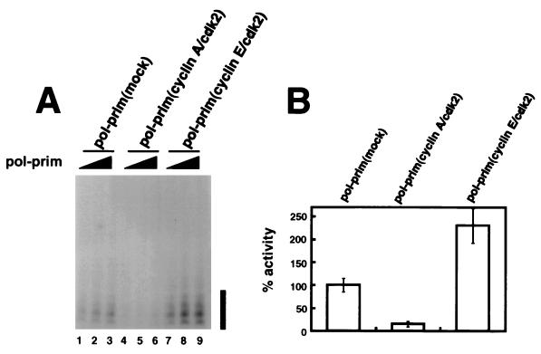
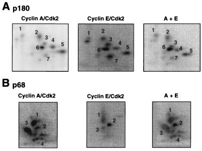
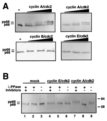
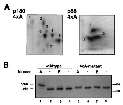
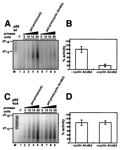
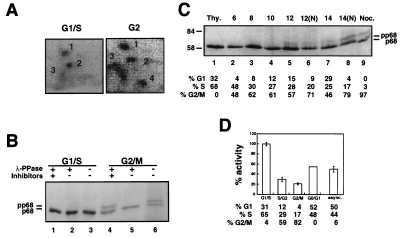
DNA polymerase alpha-primase is known to be phosphorylated in human and yeast cells in a cell cycle-dependent manner on the p180 and p68 subunits. Here we show that phosphorylation of purified human DNA polymerase alpha-primase by purified cyclin A/cdk2 in vitro reduced its ability to initiate simian virus 40 (SV40) DNA replication in vitro, while phosphorylation by cyclin E/cdk2 stimulated its initiation activity. Tryptic phosphopeptide mapping revealed a family of p68 peptides that was modified well by cyclin A/cdk2 and poorly by cyclin E/cdk2. The p180 phosphopeptides were identical with both kinases. By mass spectrometry, the p68 peptide family was identified as residues 141 to 160. Cyclin A/cdk2- and cyclin A/cdc2-modified p68 also displayed a phosphorylation-dependent shift to slower electrophoretic mobility. Mutation of the four putative phosphorylation sites within p68 peptide residues 141 to 160 prevented its phosphorylation by cyclin A/cdk2 and the inhibition of replication activity. Phosphopeptide maps of the p68 subunit of DNA polymerase alpha-primase from human cells, synchronized and labeled in G1/S and in G2, revealed a cyclin E/cdk2-like pattern in G1/S and a cyclin A/cdk2-like pattern in G2. The slower-electrophoretic-mobility form of p68 was absent in human cells in G1/S and appeared as the cells entered G2/M. Consistent with this, the ability of DNA polymerase alpha-primase isolated from synchronized human cells to initiate SV40 replication was maximal in G1/S, decreased as the cells completed S phase, and reached a minimum in G2/M. These results suggest that the replication activity of DNA polymerase alpha-primase in human cells is regulated by phosphorylation in a cell cycle-dependent manner.


Figures

FIG. 1
FIG. 1
Initiation of SV40 DNA replication in vitro by DNA polymerase α-primase (pol-prim) prephosphorylated by cyclin A/cdk2 or cyclin E/cdk2. (A) Increasing amounts of the indicated preparations of DNA polymerase α-primase were tested for initiation of SV40 DNA replication. The initiation products of each reaction were separated by denaturing PAGE and quantitated by PhosphorImager analysis, and the background (data not shown) was subtracted to give the number of SV40 initiation units per microliter of DNA polymerase α-primase. The bar to the right indicates primers of 8 to 10 nucleotides. (B) The SV40 initiation activity in each reaction in A was divided by the number of primase units per microliter (data not shown) determined for the corresponding preparation of phosphorylated DNA polymerase α-primase. The bar graph shows the average relative initiation activity of each preparation; that of the mock-phosphorylated preparation was set to 100%. The error bars represent the variation obtained from three different reactions with each preparation of DNA polymerase α-primase.
FIG. 2
FIG. 2
Tryptic phosphopeptide maps of in vitro-phosphorylated DNA polymerase α-primase. Two micrograms of DNA polymerase α-primase bound to SJK 132-20–Sepharose was phosphorylated by either cyclin A/cdk2 or cyclin E/cdk2 at 200 pmol/h for 30 min. After removal of the cyclin/cdk complexes, the subunits were separated by SDS-PAGE and transferred to a PVDF membrane. The labeled subunits were detected by autoradiography and excised from the membrane. The p180 (A) and p68 (B) subunits were digested twice with 10 μg of tolylsulfonyl phenylalanyl chloromethyl ketone-treated trypsin. The digested peptides were loaded onto thin-layer chromatography plates and separated by electrophoresis at pH 1.9 in the first dimension, followed by ascending chromatography in the second dimension. The labeled peptides were detected by PhosphorImager analysis. To verify identical peptides, the digests of cyclin E/cdk2- and cyclin A/cdk2-treated p180 were loaded onto the same spot and subjected to two-dimensional separation (A, far right), or the maps from the differently phosphorylated p68 subunits were superimposed (B, far right).
FIG. 3
FIG. 3
Cyclin A-dependent kinases induce a p68 mobility shift which is reversible with λ-phosphatase. (A) Purified recombinant DNA polymerase α-primase (2 μg) was incubated without (−) or with increasing amounts (200, 400, and 600 pmol/h) of cyclin/cdk kinases for 20 min. The subunits of DNA polymerase α-primase were separated by SDS–7.5% PAGE, and the p68 subunit was detected by Western blotting using monoclonal antibody 9D5. (B) Two micrograms of DNA polymerase α-primase, immobilized on SJK 132-20–Sepharose, was prephosphorylated with the indicated cyclin/cdk complexes or mock phosphorylated (mock), washed with phosphate-buffered saline, and incubated with (+) λ-phosphatase (λ-PPase) in the presence (+) or absence (−) of phosphatase inhibitors, as indicated, or without phosphatase (−). The p68 bands were separated by SDS-PAGE and detected by immunoblotting with monoclonal antibody 9D5. The values on the right are molecular sizes in kilodaltons.
FIG. 4
FIG. 4
The 4× alanine mutant form of p68 lacks peptide 4 in the cyclin A/cdk2-generated pattern and shows no mobility shift after phosphorylation. (A) Purified recombinant 4× alanine (4×A) DNA polymerase α-primase was phosphorylated with cyclin A/cdk2 at 200 pmol/h, and tryptic peptide maps of p180 and p68-4×A were prepared as described in the legend to Fig. 2. (B) Wild-type or mutant polymerase α-primase (4 μg) was incubated with cyclin A/cdk2 (A) or cyclin E/cdk2 (E) (700 pmol/h) or without kinase (−). The subunits were separated by SDS–7.5% PAGE, and the p68 bands were detected by immunoblotting using monoclonal antibody 9D5. The values on the right are molecular sizes in kilodaltons.
FIG. 5
FIG. 5
The SV40 initiation activity of DNA polymerase α-primase (pol-prim) containing the 4× alanine mutant form of p68 is resistant to inhibition by cyclin A/cdk2. Prephosphorylated and mock-phosphorylated wt (A) and mutant (C) forms of DNA polymerase α-primase were assayed for the ability to initiate SV40 DNA replication. The reaction products were separated on 20% urea gels as described in Materials and Methods and analyzed by PhosphorImager. A control reaction was carried out in the absence of SV40 T antigen (−T). The SV40 initiation activities of the mock- and cyclin A/cdk2-phosphorylated forms of DNA polymerase α-primase (units per microliter) were divided by the number of primase units per microliter of the corresponding preparation. The bar graphs show the SV40 initiation activities of the cyclin A/cdk2-phosphorylated wt (B) and mutant (D) forms of DNA polymerase α-primase relative to the activity of the corresponding mock-phosphorylated preparation, which was set to 100%. Error bars indicate the variation observed in three different reactions with each preparation of DNA polymerase α-primase.
FIG. 6
FIG. 6
Phosphorylation pattern and replication activity of DNA polymerase α-primase purified from human cells at different stages of the cell cycle. (A) 293S cells were blocked in G1/S or blocked and released into fresh medium for 9 h prior to labeling. Cells in G1/S and late G2 were then labeled with [32P]orthophosphate for 3 h prior to preparation of cell extracts. DNA polymerase α-primase was precipitated from the extracts on SJK 132-20–Sepharose beads, separated by denaturing PAGE, and transferred to a PVDF membrane. Membrane slices containing phosphorylated p68 were digested with trypsin. Peptides were resolved by electrophoresis at pH 1.9 (left to right), followed by ascending chromatography, and detected by PhosphorImager (4 days). (B) 293S cells synchronized in G1/S with thymidine and in G2/M with nocodazole were lysed under hypotonic conditions, and DNA polymerase α-primase was immunoprecipitated with antibody SJK 132-20–Sepharose. After washing, the beads were incubated with λ-phosphatase, as indicated (+), in either the absence (−) or the presence (+) of phosphatase inhibitors. The p68 bands were separated by SDS-PAGE and detected by immunoblotting with monoclonal antibody 9D5. (C) 293S cells were blocked in G1/S and released into S phase for the times indicated. At 10 h after release, 500-ng/ml nocodazole was added to the indicated cultures (N) to prevent passage through mitosis into G1. DNA polymerase α-primase was isolated on SJK 132-20–Sepharose beads, and the p68 bands were detected after separation on denaturing 7.5% gels and immunoblotting with monoclonal antibody 9D5. Flow cytometry was performed on parallel cultures at each time point, and the cell cycle distributions are given under each lane. Thy., thymidine. The values to the left are molecular sizes in kilodaltons. (D) DNA polymerase α-primase purified from cells at different stages of the cell cycle was tested for SV40 initiation activity. Primase assays and SV40 initiation assays were performed and quantitated as described in the legends to Fig. 1 and 5 (data not shown). The bar graph shows the average SV40 initiation activity per primase unit determined for each preparation, relative to the activity of DNA polymerase α-primase in G1/S, which was set to 100%. The error bars represent the variation observed in three separate SV40 initiation assays with each preparation, except the G1/G0 preparation, which was tested only once. The cell cycle distribution of the cells from which DNA polymerase α-primase was purified was determined by flow cytometry and is indicated under each bar. async., asynchronous.

References

    1. Aparicio O M, Weinstein D M, Bell S P. Components and dynamics of DNA replication complexes in S. cerevisiae: redistribution of MCM proteins and Cdc45p during S phase. Cell. 1997;91:59–69. - PubMed
    1. Boyle W J, van der Geer P, Hunter T. Phosphopeptide mapping and phosphoamino acid analysis by two-dimensional separation on thin-layer cellulose plates. Methods Enzymol. 1991;201:110–149. - PubMed
    1. Bradford M. A rapid and sensitive method for the quantitation of microgram quantities of protein utilizing the principle of protein-dye binding. Anal Biochem. 1976;72:248–254. - PubMed
    1. Braun K A, Lao Y, He Z, Ingles C J, Wold M S. Role of protein-protein interactions in the function of replication protein A (RP-A): RP-A modulates the activity of DNA polymerase α by multiple mechanisms. Biochemistry. 1997;36:8443–8454. - PubMed
    1. Bullock P A. The initiation of simian virus 40 DNA replication in vitro. Crit Rev Biochem Mol Biol. 1997;32:503–568. - PubMed

Publication types

LinkOut - more resources