. 1999 Jan;19(1):751-63.
doi: 10.1128/MCB.19.1.751.

Withdrawal of survival factors results in activation of the JNK pathway in neuronal cells leading to Fas ligand induction and cell death

Affiliations

Withdrawal of survival factors results in activation of the JNK pathway in neuronal cells leading to Fas ligand induction and cell death

H Le-Niculescu et al. Mol Cell Biol. 1999 Jan.

Abstract

The JNK pathway modulates AP-1 activity. While in some cells it may have proliferative and protective roles, in neuronal cells it is involved in apoptosis in response to stress or withdrawal of survival signals. To understand how JNK activation leads to apoptosis, we used PC12 cells and primary neuronal cultures. In PC12 cells, deliberate JNK activation is followed by induction of Fas ligand (FasL) expression and apoptosis. JNK activation detected by c-Jun phosphorylation and FasL induction are also observed after removal of either nerve growth factor from differentiated PC12 cells or KCl from primary cerebellar granule neurons (CGCs). Sequestation of FasL by incubation with a Fas-Fc decoy inhibits apoptosis in all three cases. CGCs derived from gld mice (defective in FasL) are less sensitive to apoptosis caused by KCl removal than wild-type neurons. In PC12 cells, protection is also conferred by a c-Jun mutant lacking JNK phosphoacceptor sites and a small molecule inhibitor of p38 mitogen-activated protein kinase and JNK, which inhibits FasL induction. Hence, the JNK-to-c-Jun-to-FasL pathway is an important mediator of stress-induced neuronal apoptosis.


Figures

FIG. 1
FIG. 1
JNK and p38 activation and ERK inhibition following MEKK1 expression in PC12 cells. (A) Individual clones of PC12 cells stably cotransfected with pJ5Ω-MEKK1Δ and RSV-Neo were examined for induction of JNK activity by Dex. After 24 h of incubation in the absence or presence of 10−8 M Dex, cell lysates were prepared and JNK activity was measured by immunocomplex kinase assay with GST–c-Jun(1-79) as a substrate. (B) Clone 21 cells were incubated with the indicated concentrations of Dex for 24 h, at which time they were lysed and the contents of MEKK1Δ, activated JNK, JNK1, activated p38, c-Jun phosphorylated at S63, and total c-Jun were determined by immunoblotting with specific antibodies. (C) Clone 21 cells were incubated in the absence or presence (10−8 and 10−7 M) of Dex for 24 h and then treated with or without NGF (50 ng/ml) for 15 and 30 min as indicated. Cell lysates were prepared, and the levels of MEKK1Δ, activated ERK, and total ERK2 were determined by immunoblotting. ERK enzymatic activity was determined by immunocomplex kinase assay with MBP as a substrate. (D) MMTV plasmid mock-transfected cells were incubated in the absence or presence of 10−7 M Dex for 24 h and then treated with or without NGF (50 ng/ml) for 15 and 30 min as indicated. Cell lysates were prepared, and the levels of MEKK1Δ, activated ERK and total ERK2 were determined by immunoblotting. ERK enzymatic activity was determined by immunocomplex kinase assay with MBP as a substrate.
FIG. 2
FIG. 2
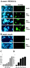
Induction of MEKK1Δ expression causes apoptotic cell death. (A) Clone 21 cells (2 × 10⁴ per well) were cultured in the absence or presence of Dex (10−7 M) for the indicated times. DNA fragmentation was examined at the time indicated, using an in situ cell death detection assay (TUNEL). Nuclei were stained with Hoechst dye H33258. The cells were examined in a Zeiss Axioskop microscope with the appropriate filters. EtoH, ethanol. (B) PC12 cells stably transfected with empty pJ5Ω expression vector were incubated with ethanol or Dex (10−7 M) for 5 days and examined as described above. (C) Clone 21 cells were incubated with 10−7 M Dex for the indicated duration, at which point they were stained with a mixture of acridine orange and ethidium bromide and the percentage of apoptotic cells was determined. The results shown are averages of three experiments. (D) Clone 21 cells were cultured in the absence or presence of Dex (10−7 M). At the indicated times (in days) the induction medium was removed, and the cells were washed twice with PBS and then cultured in normal growth medium until day 7, at which time they were stained with acridine orange-ethidium bromide. A minimum of 200 cells were placed in a hemocytometer, and the relative numbers of apoptotic cells were determined. The results shown are averages of two experiments.
FIG. 3
FIG. 3
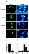
MEKK1Δ-induced cell death is blocked by phosphorylation-defective c-Jun mutant and SB202190. (A) Clone 21 cells (5 × 10⁴) were cultured on chamber slides and transiently transfected with HA-tagged wt c-Jun or c-Jun(A63/73) expression vector. After 16 h, the cells were washed twice with PBS and incubated in the presence of 10−7 M Dex. After 3 more days, the cells were fixed with 4% paraformaldehyde and permeabilized with 0.1% Triton X-100. Expression of FITC-labeled HA-c-Jun proteins was detected by indirect immunofluorescence using a monoclonal HA antibody. Nuclear morphology was determined by staining with Hoechst dye H33258. The arrows indicate cells with apoptotic morphology, which in the case of wt c-Jun are also positive for HA-c-Jun expression. However, in the case of c-Jun(A63/73), only 5% of the HA-positive cells have apoptotic morphology. (B) The results of the experiments shown above were quantitated by counting six independently transfected cultures, 80 to 90 cells each. The total numbers of transfected cells that were counted are indicated above each column. (C) Clone 21 cells were induced with or without 10−7 M Dex in the absence or presence of SB202190 (SB; 3 or 30 μM) for 6 h, followed by two washes to remove the drug. Dex-containing medium was added for an additional 18 h; then the cells were stained with acridine orange-ethidium bromide and the percentage of apoptotic cells was determined. The results are averages of three experiments.
FIG. 4
FIG. 4
Expression of MEKK1Δ causes induction of FasL and thereby leads to apoptosis. (A) Clone 21 cells were incubated with 10−7 M Dex, and total cellular RNA was extracted at the indicated times. Expression of Fas, FasL, and GAPDH mRNAs was determined by quantitative RT-PCR using specific primers. To confirm the PCR product as a fragment of FasL cDNA, the reaction products were blotted and detected with a rat FasL cDNA probe (bottom panel). In addition, the nucleotide sequence of this fragment was determined and found to correspond to that of rat FasL cDNA (data not shown). d, days. (B) Clone 21 cells were incubated with or without 10−7 M Dex in the absence or presence of SB202190 (SB; 3 or 30 μM) for 6 h, followed by two washes to remove the drug. Dex-containing medium was added for an additional 18 h, at which time the cells were collected and lysed. After separation by SDS-PAGE, c-Jun phosphorylation was examined by immunoblotting with antibody against c-Jun phosphorylated at S63. Expression of FasL and GAPDH mRNAs was determined by RT-PCR (bottom two panels). (C) To determine the role of FasL in MEKK1Δ-induced apoptosis, clone 21 cells were cultured for the indicated times with Dex (10−7 M) in the presence of chimeric Fas-Fc protein (20 μg/ml) or purified IgG (20 μg/ml). At the indicated times, the cells were stained with acridine orange-ethidium bromide and the percentage of apoptotic cells was determined. The results shown are averages of two experiments done in duplicate.
FIG. 5
FIG. 5
JNK and p38 activation and FasL induction in response to NGF withdrawal. (A) PC12 cells cultured on poly-l-lysine/laminin-coated plates were incubated with or without (lane C) NGF (50 ng/ml) for 11 days, at which time the cells were carefully washed with NGF-free medium and incubated for 6 h (6hr), 1 day (1d), 2 days (2d), and 3 days (3d) with neutralizing NGF (2.5S) antibodies (1:4,000). As an additional control, PC12 cells were incubated with NGF for 11 days before harvesting (lane NGF, 11d). The cells were collected and lysed, and the level of JNK activity was determined by immunocomplex kinase assays with GST–c-Jun(1-79) as a substrate. The levels of activated p38, c-Jun phosphorylated at S63, JNK1, and ERK2 and of FasL expression were determined by immunoblotting with specific antibodies (top six panels). The levels of FasL and GAPDH mRNAs were determined by RT-PCR using extracts from cultures treated in parallel to those used for protein analysis (bottom two panels). (B) PC12 cells were subjected to the same treatment as for panel A. The lane labeled Control represents PC12 cells cultured without NGF. One hour prior to NGF withdrawal, the cells were incubated with or without SB202190 (SB; 3 or 30 μM) for 6 h. The cells were then washed to remove the drug, and NGF-free medium was added for an additional 18 h. After 1 day of NGF withdrawal, the cells were stained with acridine orange-ethidium bromide and the percentage of apoptotic cells was determined. The level of c-Jun phosphorylated at S63 was determined by immunoblotting, and FasL and GAPDH mRNA levels were determined by RT-PCR. The results in the top panel are averages of three experiments. (C) PC12 cells were treated as described for panel A. Purified IgG or chimeric Fas-Fc protein (20 μg/ml) was added 12 h prior to and during the time of NGF withdrawal or to cells that were continuously incubated with NGF. The lane labeled Con represents PC12 cells cultured without NGF. After 1 day, the cells were stained with acridine orange-ethidium bromide and the percentage of apoptotic cells was determined. The levels of c-Jun phosphorylated at S63, FasL, and GAPDH mRNAs were examined as described above. The results in the top panel are averages of three experiments.
FIG. 6
FIG. 6
Neurons from gld mice are less sensitive to induction of apoptosis following KCl withdrawal. (A) Detection of apoptotic nuclei by hematoxylin staining 12 h after KCl withdrawal. CGCs of wt type and gld C3H/HeJ mice were cultured in 25 mM KCl for 7 days, at which time the KCl concentration was lowered to 5 mM. (B) Time course of appearance of apoptotic nuclei in wt and gld CGC cultures after KCl deprivation, with and without addition of Fas-Fc (40 μM). Apoptotic nuclei were detected by hematoxylin staining, and their frequency as a percentage of the total neuronal population was determined. Five fields were counted for each experiment; all experiments were repeated five times in triplicate. (C) RT-PCR analysis of FasL mRNA expression in wt and gld CGC cultures under control conditions (25 mM KCl) and 12 h after KCl deprivation (5 mM KCl). The number of PCR cycles was determined as described in Materials and Methods, and β-actin mRNA was used as a control. (D) Parallel samples of CGCs treated as described above were collected and lysed, and the levels of c-Jun phosphorylation at S63, activated JNK, and total c-Jun were determined by immunoblotting.
FIG. 7
FIG. 7
FasL and c-Jun mRNA are upregulated upon KCl withdrawal. In situ hybridization using probes for FasL (A to C) and c-jun (D to F) mRNAs was performed on CGCs cultured in the presence of 25 mM KCl (A and D) or on CGCs incubated for 12 h in medium containing only 5 mM KCl (B, C, E, and F). As a specificity control, the cells in panels C and F were hybridized to the probes in the presence of a 10-fold excess of unlabeled competitor.

Similar articles

Cited by

References

    1. Angel P, Hattori K, Smeal T, Karin M. The jun proto-oncogene is positively autoregulated by its product, Jun/AP-1. Cell. 1988;55:875–885. - PubMed
    1. Angel P, Karin M. The role of Jun, Fos and the AP-1 complex in cell proliferation and transformation. Biochim Biophys Acta. 1991;1072:129–157. - PubMed
    1. Ben-Ari Y. Limbic seizure and brain damage produced by kainic acid: mechanisms and relevance to human temporal lobe epilepsy. Neuroscience. 1985;14:375–403. - PubMed
    1. Binétruy B, Smeal T, Karin M. Ha-Ras augments c-Jun activity and stimulates phosphorylation of its activation domain. Nature. 1991;351:122–127. - PubMed
    1. Bonfoco E, Leist M, Zhivotovsky B, Orrenius S, Lipton S A, Nicotera P. Cytoskeletal breakdown and apoptosis elicited by NO donors in cerebellar granule cells require NMDA receptor activation. J Neurochem. 1996;67:2484–2493. - PubMed

Publication types

MeSH terms

Substances

LinkOut - more resources