. 1999 Jan;19(1):764-76.
doi: 10.1128/MCB.19.1.764.

CREB binding protein interacts with nucleoporin-specific FG repeats that activate transcription and mediate NUP98-HOXA9 oncogenicity

Affiliations

CREB binding protein interacts with nucleoporin-specific FG repeats that activate transcription and mediate NUP98-HOXA9 oncogenicity

L H Kasper et al. Mol Cell Biol. 1999 Jan.

Abstract

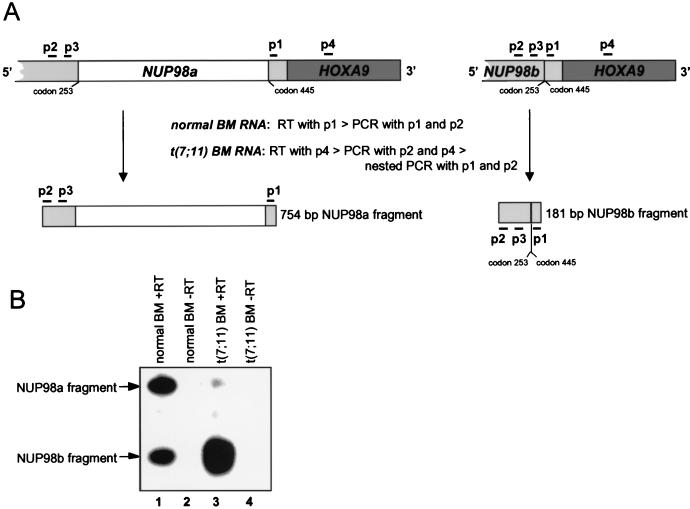
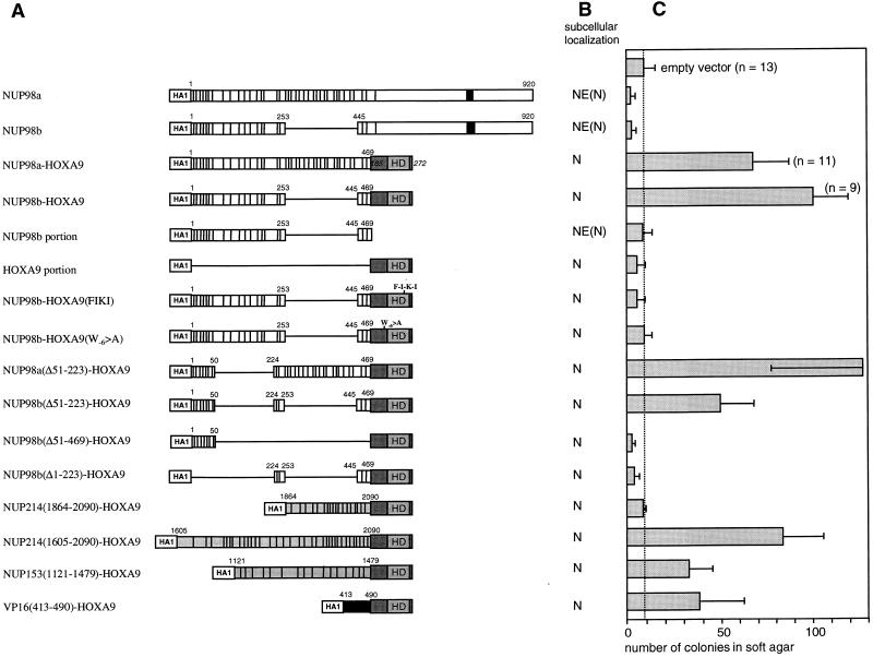
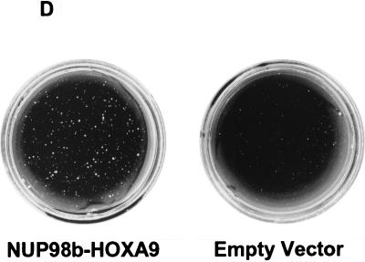
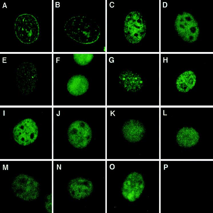
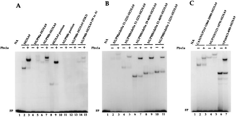
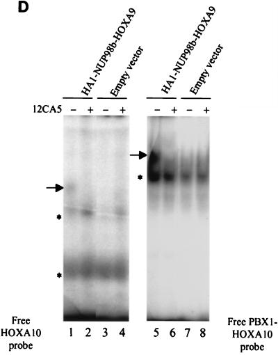
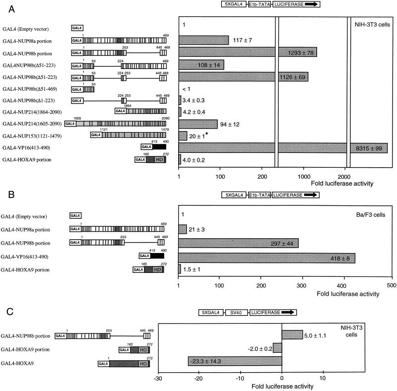
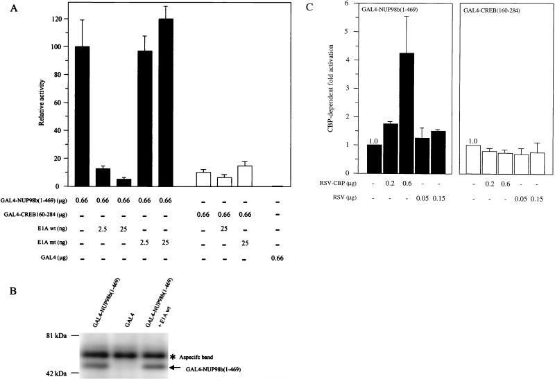
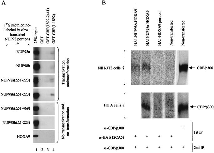
Genes encoding the Phe-Gly (FG) repeat-containing nucleoporins NUP98 and CAN/NUP214 are at the breakpoints of several chromosomal translocations associated with human acute myeloid leukemia (AML), but their role in oncogenesis is unclear. Here we demonstrate that the NUP98-HOXA9 fusion gene encodes two nuclear oncoproteins with either 19 or 37 NUP98 FG repeats fused to the DNA binding and PBX heterodimerization domains of the transcription factor HOXA9. Both NUP98-HOXA9 chimeras transformed NIH 3T3 fibroblasts, and this transformation required the HOXA9 domains for DNA binding and PBX interaction. Surprisingly, the FG repeats acted as very potent transactivators of gene transcription. This NUP98-derived activity is essential for transformation and can be replaced by the bona fide transactivation domain of VP16. Interestingly, FG repeat-containing segments derived from the nucleoporins NUP153 and CAN/NUP214 functioned similarly to those from NUP98. We further demonstrate that transactivation by FG repeat-rich segments of NUP98 correlates with their ability to interact functionally and physically with the transcriptional coactivators CREB binding protein (CBP) and p300. This finding shows, for the first time, that a translocation-generated fusion protein appears to recruit CBP/p300 as an important step of its oncogenic mechanism. Together, our results suggest that NUP98-HOXA9 chimeras are aberrant transcription factors that deregulate HOX-responsive genes through the transcriptional activation properties of nucleoporin-specific FG repeats that recruit CBP/p300. Indeed, FG repeat-mediated transactivation may be a shared pathogenic function of nucleoporins implicated human AML.


Figures

FIG. 1
FIG. 1
NUP98 and NUP98-HOXA9 genes generate an alternatively spliced transcript in human BM cells. (A) Overview of the RT-PCR procedure with representations of relevant NUP98-HOXA9 cDNA portions and the expected PCR products. The positions of the primers used in the various RT reactions and PCRs are indicated by horizontal bars. To detect NUP98a and NUP98b transcripts in normal BM cells, we used primer 1 (p1) to generate cDNA and primer set p1 and p2 for PCR amplification (40 cycles). Seminested PCR was used to detect NUP98-HOXA9-specific transcripts in BM cells from a patient with t(7;11)-positive leukemia; p4 was used for the RT step, p2 and p4 were used in the first round of amplification (40 cycles), and p1 and p2 were used in the seminested round of PCR (30 additional cycles). The oligonucleotide probe for detection of NUP98a- and NUP98b-derived PCR fragments is depicted as p3. (B) Autoradiogram of RT-PCR products detected by probe p3. Lanes: 1, 754- and 181-bp fragments from NUP98a and NUP98b transcripts in normal human BM; 2, negative control for lane 1 (same sample as in lane 1 but without reverse transcriptase); 3, 754- and 181-bp fragments from NUP98a-HOXA9 and NUP98b-HOXA9 transcripts in BM from a patient with t(7;11)-positive leukemia; 4, negative control for lane 3. Since our RT-PCR technique was not quantitative, the intensities of the 181- and 754-bp PCR bands may not be correlated with the actual transcript levels. Moreover, Northern blot analysis revealed that the BM RNA from the t(7;11) patient was partially degraded.
FIG. 2
FIG. 2
Structural and functional properties of NUP98-HOXA9 isoforms and mutants. (A) Schematic representations of the NUP98 and NUP98-HOXA9 isoforms and mutants used in this study. Indicated NUP98 motifs include the nucleoporin-specific FG repeats (each repeat is denoted by a vertical bar), the NUP98 RNP-binding motif (solid box), and NUP153 and CAN/NUP214 sequences (indicated in grey). Numbers correspond to the amino acids of the full-length NUP98 (7). Indicated in the HOXA9 portion of the fusion protein is the homeodomain (HD). Numbers in italics correspond to the amino acids of the full-length HOXA9 (44). (HA1) represents two consecutive HA1 epitopes recognized by monoclonal antibody 12CA5. (B) Summary of the subcellular localization studies (N, nuclear localization; NE, nuclear envelope localization) of the NUP98 and NUP98-HOXA9 isoforms and mutants. (C) Anchorage-independent growth of NIH 3T3 cells expressing NUP98-HOXA9 isoforms and mutants. The number of colonies (per 2 × 10⁴ CD8+ cells with a diameter of at least 90 to 100 μm) was determined after 3 weeks of growth in soft agar. The values are the means and standard deviations of 4 to 13 experiments (if more than 4 experiments were performed, the actual number of experiments is indicated in the figure). (D) Soft agar dishes demonstrating efficient colony formation by NIH 3T3 cells infected with NUP98b-HOXA9. In contrast, NIH 3T3 cells infected with pSRαMSVtkCD8 (empty vector) displayed only minimal anchorage-independent growth.
FIG. 2
FIG. 2
Structural and functional properties of NUP98-HOXA9 isoforms and mutants. (A) Schematic representations of the NUP98 and NUP98-HOXA9 isoforms and mutants used in this study. Indicated NUP98 motifs include the nucleoporin-specific FG repeats (each repeat is denoted by a vertical bar), the NUP98 RNP-binding motif (solid box), and NUP153 and CAN/NUP214 sequences (indicated in grey). Numbers correspond to the amino acids of the full-length NUP98 (7). Indicated in the HOXA9 portion of the fusion protein is the homeodomain (HD). Numbers in italics correspond to the amino acids of the full-length HOXA9 (44). (HA1) represents two consecutive HA1 epitopes recognized by monoclonal antibody 12CA5. (B) Summary of the subcellular localization studies (N, nuclear localization; NE, nuclear envelope localization) of the NUP98 and NUP98-HOXA9 isoforms and mutants. (C) Anchorage-independent growth of NIH 3T3 cells expressing NUP98-HOXA9 isoforms and mutants. The number of colonies (per 2 × 10⁴ CD8+ cells with a diameter of at least 90 to 100 μm) was determined after 3 weeks of growth in soft agar. The values are the means and standard deviations of 4 to 13 experiments (if more than 4 experiments were performed, the actual number of experiments is indicated in the figure). (D) Soft agar dishes demonstrating efficient colony formation by NIH 3T3 cells infected with NUP98b-HOXA9. In contrast, NIH 3T3 cells infected with pSRαMSVtkCD8 (empty vector) displayed only minimal anchorage-independent growth.
FIG. 3
FIG. 3
NUP98-HOXA9 isoforms and engineered mutants are localized in the nucleus. NIH 3T3 cells were transduced with retroviral expression vectors and immunostained with 12CA5 monoclonal antibodies that recognize the HA1 epitope encoded by the 5′ end of the various cDNA constructs. (A) NUP98a; (B) NUP98b; (C) NUP98a-HOXA9; (D) NUP98b-HOXA9; (E) NUP98b portion; (F) HOXA9 portion; (G) NUP98b-HOXA9(FIKI); (H) NUP98b-HOXA9(W−6>A); (I) NUP98a(Δ51–223)-HOXA9; (J) NUP98b(Δ51–223)-HOXA9; (K) NUP98b(Δ51–4693)-HOXA9; (L) NUP98a(Δ1–223)-HOXA9; (M) CAN/NUP214(1864–2090)-HOXA9; (N) NUP153(1121–1479)-HOXA9; (O) VP16(413–490)-HOXA9; (P) empty pSRαMSVtkCD8 vector (negative control).
FIG. 4
FIG. 4
NUP98-HOXA9 fusion proteins bind cooperatively with PBX1a to a PBX1-HOXA10 bipartite DNA sequence. (A to C) EMSAs of in vitro-translated proteins whose identities are indicated above the gel lanes. EMSAs were performed with a radiolabeled probe containing a PBX1-HOXA10 bipartite binding site in the absence (−) or presence (+) of in vitro-translated PBX1a. Cooperative DNA binding was observed for fusion proteins with an intact HOXA9 homeodomain. Typically, the binding affinity for the bipartite probe increased when the NUP98 portion fused to HOXA9 became smaller. The differences in intensities of the protein-DNA complexes within each panel (A or B) represent true variations in DNA binding activity. FP, free probe. (D) NUP98b-HOXA9 and PBX form heterodimers in NIH 3T3 cells. Radiolabeled probe containing the PBX1-HOXA10 or HOXA10 binding site was added to lysates of cells expressing the HA1-tagged NUP98-HOXA9 fusion proteins, and the formation of protein-DNA complexes in the absence (−) or presence (+) of 12CA5 monoclonal antibody was studied. For each lysate, the shifted complexes formed with the PBX1-HOXA10 probe (arrow to the left of lane 5) are larger than those formed with the HOXA10 probe (arrow to the left of lane 1), indicating that NUP98-HOXA9 and PBX form heterodimers in vivo. The shifted complex ablates when incubated with 12CA5 antibody, confirming the presence of HA1-NUP98b-HOXA9 in such complexes.
FIG. 4
FIG. 4
NUP98-HOXA9 fusion proteins bind cooperatively with PBX1a to a PBX1-HOXA10 bipartite DNA sequence. (A to C) EMSAs of in vitro-translated proteins whose identities are indicated above the gel lanes. EMSAs were performed with a radiolabeled probe containing a PBX1-HOXA10 bipartite binding site in the absence (−) or presence (+) of in vitro-translated PBX1a. Cooperative DNA binding was observed for fusion proteins with an intact HOXA9 homeodomain. Typically, the binding affinity for the bipartite probe increased when the NUP98 portion fused to HOXA9 became smaller. The differences in intensities of the protein-DNA complexes within each panel (A or B) represent true variations in DNA binding activity. FP, free probe. (D) NUP98b-HOXA9 and PBX form heterodimers in NIH 3T3 cells. Radiolabeled probe containing the PBX1-HOXA10 or HOXA10 binding site was added to lysates of cells expressing the HA1-tagged NUP98-HOXA9 fusion proteins, and the formation of protein-DNA complexes in the absence (−) or presence (+) of 12CA5 monoclonal antibody was studied. For each lysate, the shifted complexes formed with the PBX1-HOXA10 probe (arrow to the left of lane 5) are larger than those formed with the HOXA10 probe (arrow to the left of lane 1), indicating that NUP98-HOXA9 and PBX form heterodimers in vivo. The shifted complex ablates when incubated with 12CA5 antibody, confirming the presence of HA1-NUP98b-HOXA9 in such complexes.
FIG. 5
FIG. 5
Transcriptional regulatory properties of HOXA9 and FG repeat-containing segments of nucleoporins. (A) Schematic representation of the fusion proteins containing the GAL4 DNA binding domain and HOXA9, NUP98, NUP153, CAN/NUP214, and VP16 protein regions corresponding to the amino acids indicated (the various protein domains are indicated as described in the legend to Fig. 2). Transfection assays were performed in NIH 3T3 cells with reporter plasmid p5GB/GL2. Synthesis of the GAL4 fusion proteins was verified by Western blot analysis (data not shown). The asterisk indicates that there were no major differences in the levels of the various effector proteins, with the exception of NUP153(1121–1479), which was consistently expressed at approximately 10-fold-lower levels (data not shown). (B) Schematic representation of GAL4 fusion proteins tested for transcriptional properties in Ba/F3 cells (IL-3-dependent myeloid progenitor cells). The reporter used was p5GB/GL2. (C) HOXA9 is a strong repressor of gene transcription in NIH 3T3 cells. The reporter was the pGL2-promoter vector with five GAL4 DNA-binding sites ligated upstream of the SV40 promoter (43). Data in all panels are expressed as the fold difference in luciferase activity obtained with the GAL4 fusion proteins compared to that obtained with the GAL4 DNA binding domain alone. Values are the means and standard deviations of three independent experiments.
FIG. 6
FIG. 6
NUP98 transactivation function requires CBP/p300. (A) GAL4-NUP98b(1–469) transcriptional activity is repressed by E1A, an inhibitor of CBP/p300. NIH 3T3 cells were transfected with p5GB/GL2 firefly luciferase reporter plasmid, and expression vectors for GAL4(1–147), GAL4-NUP98b(1–469), or GAL4-CREB(160–284) (a CBP/p300-independent-glutamine-rich activator from CREB; amino acids 160 to 284), and wild-type or mutant E1A [mutant E1A(Δ2–36)]. Luciferase activity derived from the p5GB/GL2 luciferase reporter was normalized to Renilla luciferase derived from the internal control reporter pRL-CMV. The mean activity and standard deviation is presented relative to the activity of GAL4-NUP98b(1–469) in the absence of E1A. (B) GAL4-NUP98 levels do not change in the presence of E1A. Western blot analysis of proteins precipitated with 12CA5 antibody from lysates of UCLA cells transiently transfected with GAL4-NUP98(1–469) (predicted molecular mass, ∼50 kDa), GAL4, or NUP98(1–469) plus E1A. Precipitated proteins were visualized with 12CA5. The asterisk indicates a protein precipitated nonspecifically with 12CA5. (C) Full-length CBP potentiates GAL4-NUP98b(1–469) transcriptional activity in NIH 3T3 cells. The p5GB/GL2 luciferase reporter was cotransfected with expression vectors for GAL4-NUP98b(1–469) or GAL4-CREB(160–284), and equimolar amounts of CBP (RSV-CBP) or empty vector (RSV). Luciferase activity derived from the p5GB/GL2 luciferase reporter was normalized to Renilla luciferase derived from the internal control reporter pRL-TK. Mean activation and standard deviation is expressed as the ratio of luciferase activity in the presence of RSV-CBP plasmid DNA divided by the level of luciferase activity in its absence. The results represent at least two independent experiments.
FIG. 7
FIG. 7
Transcriptionally active FG repeat-rich portions of NUP98 bind CBP/p300. (A) GAL4-NUP98 transcriptional activity correlates with binding to CBP in vitro. GST pull-down assays were performed with the indicated [35S]methionine-labeled NUP98 or HOXA9 portions produced in vitro and GST-CBP fusion proteins purified from E. coli. The 25% input (lane) shows 25% of the NUP98 or HOXA9 segments used in each pull-down assay. Typically, in vitro-translated NUP98 portions appear as doublets, representing fragments with and without a HA1 tag (the NUP98 portions were cloned in pSP73 as HA1 fusion genes that retained the endogenous NUP98 translation initiation codon). GST acts as a negative control for binding. Comparable amounts of GST and GST-CBP fusion proteins were used in each pull-down assay. The experiment shown is representative of three independent experiments. (B) In vivo interaction between NUP98-HOXA9 oncoproteins and CBP/p300. Total-cell lysates were prepared from [35S]methionine-labeled HtTA or NIH 3T3 cells expressing the indicated HA1-tagged proteins. Two sequential immunoprecipitations were performed as detailed in Materials and Methods. The first and second immunoprecipitations (IP) were performed with the indicated antisera. A CBP/p300 antiserum cocktail was used for most efficient detection of these transcriptional coactivators. The positions of CBP/p300 and a molecular mass marker are indicated.

References

    1. Adler H T, Nallaseth F S, Walter G, Tkachuk D C. HRX leukemic fusion proteins form a heterocomplex with the leukemia-associated protein SET and protein phosphatase 2A. J Biol Chem. 1997;272:28407–28414. - PubMed
    1. Arai Y, Hosoda F, Kobayashi H, Arai K, Hayashi Y, Kamada N, Kaneko Y, Ohki M. The inv(11)(p15q22) chromosome translocation of de novo and therapy-related myeloid malignancies results in fusion of the nucleoporin gene, NUP98, with the putative RNA helicase gene, DDX10. Blood. 1997;89:3936–3944. - PubMed
    1. Bastos R, Lin A, Enarson M, Burke B. Targeting and function in mRNA export of nuclear pore complex protein Nup153. J Cell Biol. 1996;134:1141–1156. - PMC - PubMed
    1. Bedigian H G, Johnson D A, Jenkins N A, Copeland N G, Evans R. Spontaneous and induced leukemias of myeloid origin in recombinant inbred BXH mice. J Virol. 1984;51:586–594. - PMC - PubMed
    1. Bedigian H G, Taylor B A, Meier H. Expression of murine leukemia viruses in the highly lymphomatous BXH-2 recombinant inbred mouse strain. J Virol. 1981;39:632–640. - PMC - PubMed

Publication types

MeSH terms

LinkOut - more resources