. 1999 Jan;19(1):941-7.
doi: 10.1128/MCB.19.1.941.

Interactions between the class II transactivator and CREB binding protein increase transcription of major histocompatibility complex class II genes

Affiliations

Interactions between the class II transactivator and CREB binding protein increase transcription of major histocompatibility complex class II genes

J D Fontes et al. Mol Cell Biol. 1999 Jan.

Abstract

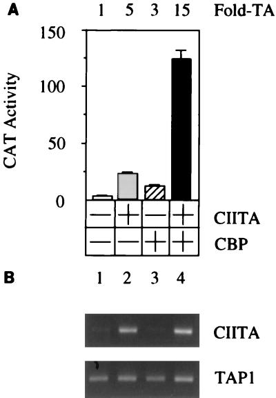
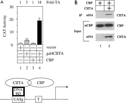
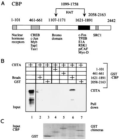
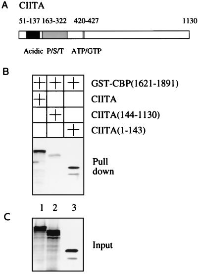
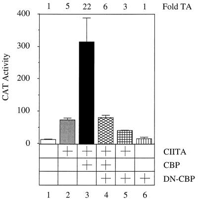
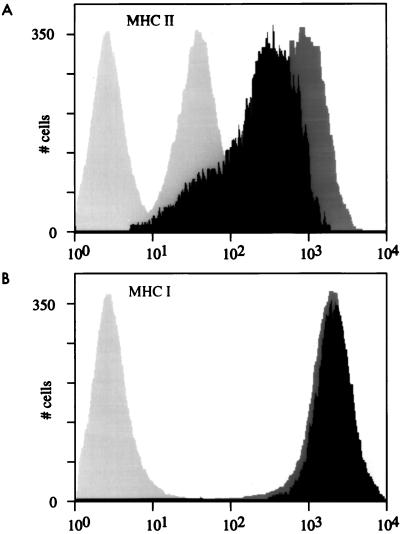
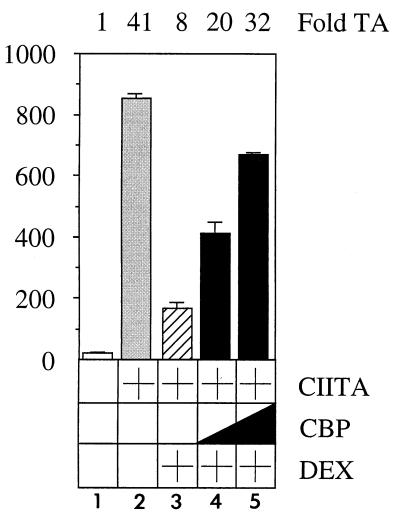
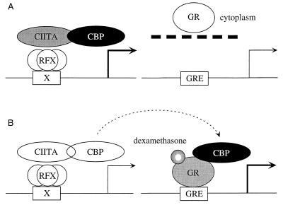
Class II major histocompatibility (class II) genes are regulated in a B-cell-specific and gamma interferon-inducible fashion. The master switch for the expression of these genes is the class II transactivator (CIITA). In this report, we demonstrate that one of the functions of CIITA is to recruit the CREB binding protein (CBP) to class II promoters. Not only functional but also specific binding interactions between CIITA and CBP were demonstrated. Moreover, a dominant negative form of CBP decreased the activity of class II promoters and levels of class II determinants on the surface of cells. Finally, the inhibition of class II gene expression by the glucocorticoid hormone could be attributed to the squelching of CBP by the glucocorticoid receptor. We conclude that CBP, a histone acetyltransferase, plays an important role in the transcription of class II genes.


Figures

FIG. 1
FIG. 1
CIITA and CBP act synergistically to increase expression from the DRA promoter. The plasmid target (pDRASCAT) was coexpressed with CIITA and/or CBP in 293T cells (+ denotes protein expression). Amounts of cotransfected plasmid DNA were held constant (0.25 μg of each plasmid effector and target, balanced to a total of to 1.0 μg with the empty plasmid vector DNA). pDRASCAT was coexpressed with empty plasmid vector (white bar), CIITA (gray bar), CBP (striped bar), or CIITA and CBP (black bar). Fold transactivation (Fold-TA) was calculated from the bar graph and represents values of top bars over the value obtained with pDRASCAT alone. CAT activities represent mean values of three independent experiments performed in triplicate with indicated standard errors of the mean. RT-PCR was performed on total RNA isolated from 293T cells transfected as noted above. Primers for CIITA mRNA (B, top; from nucleotide positions 3309 to 3507) or human TAP1 (bottom; from nucleotide positions 1904 to 2247) were used in the PCR.
FIG. 2
FIG. 2
CBP interacts with CIITA in cells. (A) CBP interacts with CIITA in a modified one-hybrid assay in vivo. The plasmid target (pG1bCAT) was coexpressed with Gal4-CIITA and/or CBP in 293T cells (+ denotes protein expression). Amounts of cotransfected plasmid DNA were held constant (0.25 μg of each plasmid effector and target, balanced to a total of to 1.0 μg with the empty plasmid vector DNA). pG1bCAT was coexpressed with empty plasmid vector (white bar), Gal4-CIITA (grey bar), CBP (striped bar), or Gal4-CIITA and CBP (black bar). Fold transactivation (Fold-TA) was calculated from the bar graph and represents values of top bars over the value obtained with pG1bCAT alone. CAT activities represent mean values of three independent experiments performed in triplicate with indicated standard errors of the mean. BD, binding domain; T, TATA box. (B) CIITA can be immunoprecipitated with CBP in cells. COS cells were transfected with pCMVCBP (1.0 μg) alone (lane 1) or with pCMVCBP (1.0 μg) and pSVCIITA (1.0 μg) (lane 2). The empty plasmid vector was included to maintain the total amount of transfected plasmid at 2.0 μg. One-quarter of the cellular lysate used for the immunoprecipitation (IP) was examined for the presence of expressed proteins (Input). Anti-CBP (αCBP) immunoprecipitates were also probed for the presence of CIITA by Western blotting, αHA, anti-hemagglutinin epitope antibody.
FIG. 3
FIG. 3
CIITA binds to CBP via residues from positions 1621 to 1891. (A) Schematic representation of CBP. Domains which interact with other proteins that regulate transcription of various genes are depicted as gray and black rectangles (22). (B) In vitro binding assays were carried out with the transcribed and translated CIITA and GST-CBP fusion proteins containing amino acids from positions 1 to 101 (lane 4), 461 to 661 (lane 5), 1621 to 1891 (lane 6), and 2058 to 2163 (lane 7). Glutathione-Sepharose beads (lane 2) and GST alone (lane 3) did not bind to CIITA. Lane 1 contains 10% of the input CIITA protein. (C) Coomassie blue-stained SDS-polyacrylamide gel demonstrating that equivalent amounts of GST-CBP fusion proteins were included in the binding reactions.
FIG. 4
FIG. 4
CBP binds to the N-terminal transcriptional activation domain of CIITA. (A) Schematic representation of CIITA (33). Structural domains: Acidic, region rich in acidic amino acids; P/S/T, region rich in proline, serine, and threonine residues; ATP/GTP, putative purine ribonucleotide binding site. (B and C) In vitro binding assays were carried out with the transcribed and translated full-length CIITA (lane 1), CIITA(144-1103) (lane 2), and CIITA(1-143) (lane 3). (B) GST-CBP(1621-1891) pull-down assay; (C) input CIITA and truncated CIITA proteins used in the binding reaction.
FIG. 5
FIG. 5
DN-CBP inhibits the transcriptional synergy between CIITA and CBP. pDRASCAT was coexpressed with CIITA individually and in combination with CBP in 293T cells. In addition, DN-CBP, which contains amino acids from positions 1903 to 2441 in CBP, was coexpressed in these cells. Amounts of cotransfected plasmids were kept constant (0.5 μg of pDRASCAT and pCRDN-DBP; 0.25 μg of pCMVCBP and pSVCIITA; empty plasmid vector was included to maintain the total amount of transfected plasmid at 2.0 μg). pDRASCAT was coexpressed with empty plasmid vector (white bar), CIITA (gray bar), CIITA and CBP (black bar), CIITA, CBP, and DN-CIITA (cross-hatched bar), CIITA and DN-CBP (small-squares bar), or DN-CBP (striped bar). Fold transactivation (TA) was calculated from the bar graph and represents values of top bars over the value obtained with pDRASCAT alone. CAT activities represent mean values of three different experiments performed in triplicate with indicated standard errors of the mean.
FIG. 6
FIG. 6
DN-CBP inhibits the expression of class II determinants on the surface of B-lymphoblastoid RM3 cells. RM3 cells were transfected with the pSVCIITA (5 and 15 μg of empty plasmid vector) (dark gray peaks), with pSVCIITA (5 μg) and pCRDN-CBP (15 μg) (black peaks), or with the empty plasmid vector (20 μg) (light gray peaks). Cells were incubated with the anti-HLA-DP (MHC II) or anti-HLA-A, -B, and -C (MHC I) antibodies and subsequently visualized with a phycoerythrin-conjugated rat anti-mouse antibody. As pEGFP (1 μg) was cotransfected into these cells, only 10% of the brightest cells were analyzed by FACS. In panel A, two light gray peaks on the left represent unstained and anti-HLA-DP-stained RM3 cells that were transfected with the plasmid vector alone; the light gray peak in panel B represents unstained RM3 cells.
FIG. 7
FIG. 7
Increasing amounts of CBP relieve the inhibition of class II transcription by dexamethasone. pDRASCAT was coexpressed with CIITA, and transfected cells were treated with 1 μM dexamethasone (DEX; striped bar) or dimethyl sulfoxide vehicle (gray bar). Increasing amounts of CBP were coexpressed in these cells (0.25 and 0.5 μg of pCMVCBP; black bars). Amounts of cotransfected plasmids were kept constant (0.5 μg of pDRASCAT and 0.75 of pSVCIITA; 0.25 μg and 0.5 μg of pCMVCBP; empty plasmid vector was included to maintain the total amount of transfected plasmid at 2.0 μg). pDRASCAT was coexpressed with empty plasmid vector (white bar), CIITA (gray bar), CIITA and dexamethasone (striped bar), or CIITA and different amounts of CBP (0.25 and 0.5 μg) in the presence of dexamethasone (black bar). Fold transactivation (TA) was calculated from the bar graph and represents values of top bars over the value obtained with pDRASCAT alone. CAT activities represent mean values of three independent experiments performed in triplicate with indicated standard errors of the mean.
FIG. 8
FIG. 8
Diagrammatic representation of how dexamethasone inhibits the transcription of class II genes. (A) In the absence of dexamethasone, CBP binds to CIITA. In turn, CIITA is attracted to class II promoters via RFX, which binds to the X box. (B) The administration of dexamethasone results in the translocation of the GR to the nucleus, where it binds to CBP. In this manner, GR sequesters a limited amount of CBP in the nucleus, leaving little to no free CBP to interact with CIITA on class II promoters. GRE, glucocortoid response element.

References

    1. Aarnisalo P, Palvimo J J, Janne O A. CREB-binding protein in androgen receptor-mediated signaling. Proc Natl Acad Sci USA. 1998;95:2122–2127. - PMC - PubMed
    1. Abdulkadir S A, Ono S J. How are class II MHC genes turned on and off? FASEB J. 1995;9:1429–1435. - PubMed
    1. Abraham S E, Lobo S, Yaciuk P, Wang H G, Moran E. p300, and p300-associated proteins, are components of TATA-binding protein (TBP) complexes. Oncogene. 1993;8:1639–1647. - PubMed
    1. Arany Z, Newsome D, Oldread E, Livingston D M, Eckner R. A family of transcriptional adaptor proteins targeted by the E1A oncoprotein. Nature. 1995;374:81–84. - PubMed
    1. Arias J, Alberts A S, Brindle P, Claret F X, Smeal T, Karin M, Feramisco J, Montminy M. Activation of cAMP and mitogen responsive genes relies on a common nuclear factor. Nature. 1994;370:226–229. - PubMed

Publication types

LinkOut - more resources