. 1998 Dec 22;95(26):15189-93.
doi: 10.1073/pnas.95.26.15189.

Structure-based assignment of the biochemical function of a hypothetical protein: a test case of structural genomics

Affiliations

Structure-based assignment of the biochemical function of a hypothetical protein: a test case of structural genomics

T I Zarembinski et al. Proc Natl Acad Sci U S A. .

Abstract

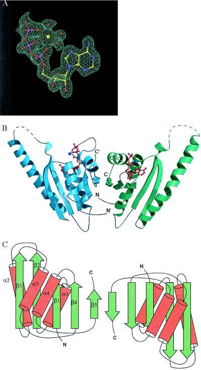
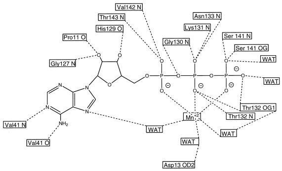
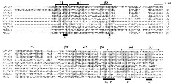
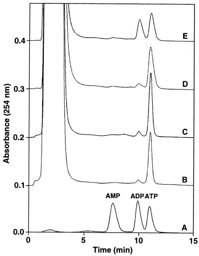
Many small bacterial, archaebacterial, and eukaryotic genomes have been sequenced, and the larger eukaryotic genomes are predicted to be completely sequenced within the next decade. In all genomes sequenced to date, a large portion of these organisms' predicted protein coding regions encode polypeptides of unknown biochemical, biophysical, and/or cellular functions. Three-dimensional structures of these proteins may suggest biochemical or biophysical functions. Here we report the crystal structure of one such protein, MJ0577, from a hyperthermophile, Methanococcus jannaschii, at 1.7-A resolution. The structure contains a bound ATP, suggesting MJ0577 is an ATPase or an ATP-mediated molecular switch, which we confirm by biochemical experiments. Furthermore, the structure reveals different ATP binding motifs that are shared among many homologous hypothetical proteins in this family. This result indicates that structure-based assignment of molecular function is a viable approach for the large-scale biochemical assignment of proteins and for discovering new motifs, a basic premise of structural genomics.


Figures

Figure 1
Figure 1
(A) A sample of the electron density map for ATP in the ATP-binding pocket of MJ0577. The multiwavelength anomalous diffraction-phased electron density map at 1.8-Å resolution is contoured at 1 sigma, with the current model displayed for comparison. A Mn ion and three waters bound to ATP are also shown. (B) Ribbon diagram of the crystal structure of the MJ0577 dimer drawn by using molscript (29) and raster3d (30). The secondary structure assignment is based on dssp (31). Dashed lines represent loops not modeled in the structure. The N and C termini of each monomer are labeled. (C) Schematic structure, where β-strands and α-helices are represented by arrows and cylinders, respectively.
Figure 2
Figure 2
Schematic drawing showing all of the hydrogen bonds (dashed lines) involving ATP and coordination bonds (dotted lines) involving the Mn+2 ion. The protein residues and atoms involved in each hydrogen bond are shown in the boxes.
Figure 3
Figure 3
Multiple alignment of conserved regions of the MJ0577 superfamily showing four sequence motifs. It was constructed by using psi-blast (38) followed by clustalw (39). The first column shows the protein identifier with the letters designating the organism code and the subsequent numbers designating the gene. Organism codes: MJ, M. jannaschii; MTH, Methanobacterium thermoautotrophicum; AF, Archaeoglobus fulgidus; BS, Bacillus subtilis; AQ, Aquifex aeolicus. Conserved residues are shaded. The black boxes below the sequence indicate conserved residues contacting ATP (A: adenine; R: ribose; P: phosphate; D: dimer interface). α-helices and β-strands are represented by brackets above the sequences. * at the C terminus designate those sequences with tandem homologs of MJ0577. Percent identities between the sequences are at far right.
Figure 4
Figure 4
Measurement of ATP hydrolysis. Anion exchange chromatograms from four different MJ0577 reactions processed as described in Materials and Methods are shown. The samples are: A, a reference sample containing 10 nmol each of AMP, ADP, and ATP; B, MJ0577 alone (4° C); C, MJ0577 alone (80° C); D, MJ0577 plus M. jannaschii extract (4° C); E, MJ0577 plus M. jannaschii extract (80° C). Migration positions of AMP, ADP, and ATP are indicated above sample A. The initial large peak is the result of residual phenol/chloroform/isoamyl alcohol during the nucleotide extraction step (see Materials and Methods).

References

    1. Hodgkin J, Plasterk R H A, Waterston R H. Science. 1995;270:410–414. - PubMed
    1. Fleischmann R D, Adams M D, White O, Clayton R A, Kirkness E F, Kerlavage A R, Bult C J, Tomb J F, Dougherty B A, Merrick J M, et al. Science. 1995;269:496–512. - PubMed
    1. Fraser C M, Gocayne J D, White O, Adams M D, Clayton R A, Fleischmann R D, Bult C J, Kerlavage A R, Sutton G, Kelley J M, et al. Science. 1995;270:397–403. - PubMed
    1. Bult C J, White O, Olsen G J, Zhou L, Fleischmann R D, Sutton G G, Blake J A, FitzGerald L M, Clayton R A, Gocayne J D, et al. Science. 1996;273:1058–1073. - PubMed
    1. Kaneko T, Sato S, Kotani H, Tanaka A, Asamizu E, Nakamura Y, Miyajima N, Hirosawa M, Sugiura M, Sasamoto S, et al. DNA Res. 1996;3:109–136. - PubMed

Publication types

MeSH terms

Associated data

LinkOut - more resources