. 1998 Dec 22;95(26):15303-8.
doi: 10.1073/pnas.95.26.15303.

Gap junctional coupling in lenses lacking alpha3 connexin

Affiliations

Gap junctional coupling in lenses lacking alpha3 connexin

X Gong et al. Proc Natl Acad Sci U S A. .

Abstract

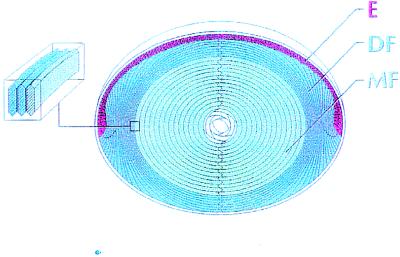
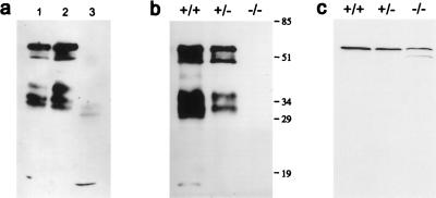
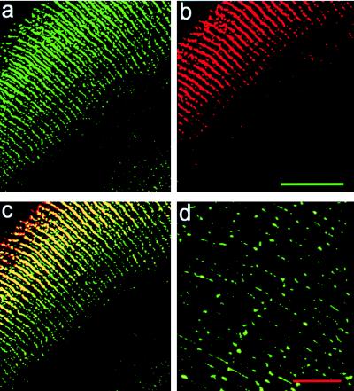
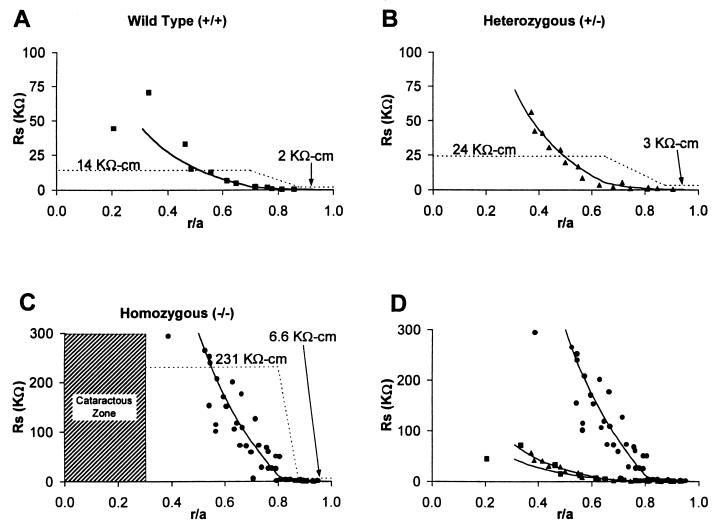
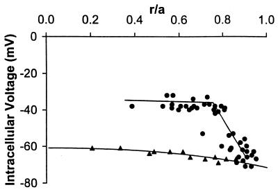
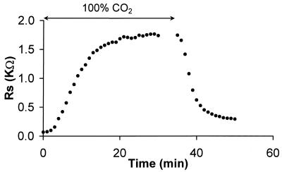
Fiber cells of the lens are interconnected by an extensive network of gap junctions containing alpha3 (Cx46) and alpha8 (Cx50) connexins. A specific role for these connexins in lens homeostasis is not known. To determine the contribution of these connexins to lens function, we used impedance techniques to study cell-to-cell coupling in lenses from homozygous alpha3 knockout (-/-), heterozygous (+/-), and wild-type (+/+) mice. Western blots and immunofluorescence data indicated that alpha8 remained at similar levels in the three classes of lenses, whereas alpha3 was approximately 50% of the normal level in the +/- lenses, and it was absent from the -/- lenses. Moreover, the data from +/+ lenses suggest that a cleavage of connexins occurs abruptly between the peripheral shell of differentiating fibers (DF) and the inner core of mature fibers (MF). The appearance of the cleaved connexins was correlated to a change in the coupling conductance. In -/- lenses the coupling conductance of MF was zero, and these fibers were depolarized by about 30 mV from normal (approximately -65 mV). The DF remained coupled, but the conductance was reduced to 30-35% of normal. However, the gap junctions in the DF of alpha3 -/- lenses remained sensitive to pH. We conclude that alpha3 connexin is necessary for the coupling of central fibers to peripheral cells, and that this coupling is essential for fiber cell homeostasis because uncoupled MF depolarize and subsequently become opaque.


Figures

Figure 1
Figure 1
The cellular structure of the lens. The anterior surface of the lens is capped by a single layer of typical epithelial cells (E). At the equator, the epithelial cells elongate and differentiate into fiber cells. This process occurs in the outer layer of DF. MF have no organelles, very little active membrane transport, and a low level of metabolic activity. All of these cells are connected through an extensive network of gap junctions. When the epithelial cells begin to differentiate, new cytoplasmic and membrane proteins are synthesized, and many of the epithelial proteins are deleted. As the DF change to MF, there is a rather abrupt cleavage of most membrane proteins, including the gap junction proteins of the fibers. In this study, we have defined the DF and MF zones based on where the gap junction proteins are cleaved. (Inset) The fiber cell cross section has the shape of a flattened hexagon with a long side dimension of about 10 μm and a short side of about 3 μm. Each MF extends from anterior to posterior pole.
Figure 2
Figure 2
Western blots of α3 and α8 connexins. (a) Membrane proteins were isolated from the lenses of 6-week-old wild-type mice and analyzed by using anti-α3 connexin antibodies that bind to an epitope on the inner cytoplasmic loop; the same antibodies were used for immunocytochemical staining. Lane 1, membrane proteins from total lens; lane 2, membrane proteins from fibers of the DF zone and some outer fraction of the MF zone; lane 3, membrane proteins from nuclear fibers. (b) A comparison of α3 in lenses from the litter mates of +/+, +/−, and −/− mice at 2 months of age. (c) A comparison of α8 in the cortex of lenses from the litter mates of +/+, +/−, and −/− mice at the age of 2 months. The same α8 antibodies were used for immunocytochemical staining shown in Fig. 3.
Figure 3
Figure 3
The distribution of α3 and α8 connexins in mouse lenses. The scale bar in b is 50 μm and ac are at the same magnification. (a and b) Fluorescent antibody double staining of the α3 (green) and α8 (red) connexins in a frozen section of the lens from a 6-week-old +/+ mouse. (c) The merged picture of a and b. Orange/yellow staining represents the colocalization of α3 and α8 connexins to the same gap junctional plaques. The plaques are preferentially located in the long sides of the hexagonal fibers in the DF zone. Small plaques also were detected in the edges of the narrow sides of fibers in the DF zone. (d) Fluorescent signals of the α3 connexin in MF of the same mouse lens in ac by using the same anti-α3 connexin antibodies. This pattern of staining was consistent throughout the MF right up to the DF. The scale bar in d is 10 μm.
Figure 4
Figure 4
The cell-to-cell coupling resistance in +/+, +/−, and −/− lenses. The Rs data represent the cumulative resistance between the point of recording (r/a, where r is the distance from the center of the lens and a is the radius of the lens) and the surface of the lens (r/a = 1). The smooth curves are generated by using Eq. 1 with the values of Rp in Eq. 1 shown as the dashed lines. We assumed Rp increased linearly between the DF and MF zones; however, equally good fits to the data could have been achieved with other assumptions (e.g., a sigmoidal increase). Rp represents the parallel combination of the cell to cell intracellular resistivity and extracellular resistivity (Eq. 2). In normal circumstances Rp is approximately equal to Ri; however, in the MF zone of −/− lenses, Ri is essentially infinite so in this case, Rp is equal to Re. (A) Wild-type (+/+) lenses. Data are from two lenses. (B) Heterozygous (+/−) lenses. Data are from two lenses. (C) Homozygous (−/−) lenses. Data are from 10 lenses. (D) An overplot of Rs from +/+, +/−, and −/− lenses.
Figure 5
Figure 5
The radial distribution of resting voltages in +/+ and −/− lenses. Comparison with the coupling data (Rp) in Fig. 4C suggests that as the coupling of MF to the lens surface is lost, the fibers depolarize.
Figure 6
Figure 6
The effect of pH on junctional coupling in DF from a −/− lens. The graph shows the time dependence of the cumulative coupling resistance of junctions between the point of recording (r = 0.09 cm) and the lens surface (a = 0.10 cm). When the bathing solution is bubbled with 100% CO2, the junctions uncouple, then recover when the CO2 is removed. Thus, DF junctions lacking the α3 connexin are capable of gating similarly to normal DF junctions.

References

    1. Bassnett S, Beebe D C. Dev Dyn. 1992;194:85–93. - PubMed
    1. Rae J L, Bartling C, Mathias R T. J Membr Biol. 1996;150:89–103. - PubMed
    1. Goodenough D A. Semin Cell Biol. 1992;3:49–58. - PubMed
    1. Goodenough D A, Dick J S, Lyons J E. J Cell Biol. 1980;86:576–589. - PMC - PubMed
    1. Mathias R T, Rae J L, Baldo G J. Phys Rev. 1997;77:21–50. - PubMed

Publication types

LinkOut - more resources