. 1998 Dec 22;95(26):15345-50.
doi: 10.1073/pnas.95.26.15345.

Otoconin-90, the mammalian otoconial matrix protein, contains two domains of homology to secretory phospholipase A2

Affiliations

Otoconin-90, the mammalian otoconial matrix protein, contains two domains of homology to secretory phospholipase A2

Y Wang et al. Proc Natl Acad Sci U S A. .

Abstract

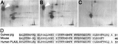
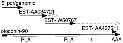
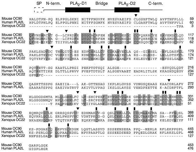
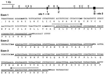
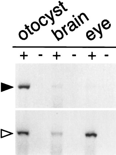
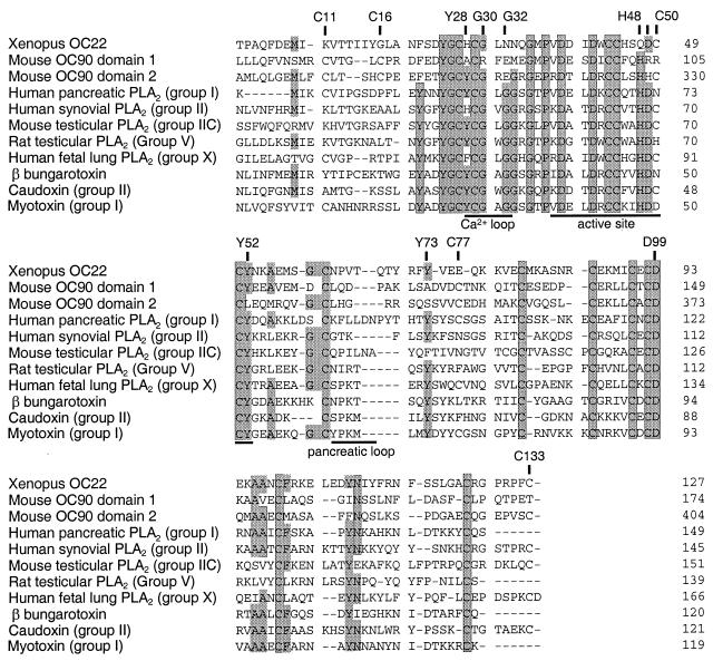
The ability to sense orientation relative to gravity requires dense particles, called otoconia, which are localized in the vestibular macular organs. In mammals, otoconia are composed of proteins (otoconins) and calcium carbonate crystals in a calcite lattice. Little is known about the mechanisms that regulate otoconial biosynthesis. To begin to elucidate these mechanisms, we have partially sequenced and cloned the major protein component of murine otoconia, otoconin-90 (OC90). The amino acid sequence identified an orphan chimeric human cDNA. Because of its similarity to secretory phospholipase A2 (sPLA2), this gene was referred to as PLA2-like (PLA2L) and enabled the identification of human Oc90. Partial murine cDNA and genomic clones were isolated and shown to be specifically expressed in the developing mouse otocyst. The mature mouse OC90 is composed of 453 residues and contains two domains homologous to sPLA2. The cloning of Oc90 will allow an examination of the role of this protein in otoconial biosynthesis and in diseases that affect the vestibular system.


Figures

Figure 1
Figure 1
Identification of mouse and guinea pig OC90. Two-dimensional SDS/PAGE separation of proteins (10 μg dry weight) from isolated otoconia of the mouse (A) and guinea pig (B). Isoelectric focusing was used in the first dimension (left to right) and SDS/PAGE was used in the second dimension (top to bottom). Protein standards (carbonic anhydrase, ovalbumin, and β-galactosidase) were run alone (C) and spiked into the otoconial preparations shown in A and B. Arrows identify OC90. (D) N-terminal sequence of partially purified mouse and guinea pig otoconins were aligned and used to identify an orphan human cDNA, PLA2L.
Figure 2
Figure 2
cDNA assembly and structure of mouse Oc90. Diagram showing the strategy used to assemble the Oc90 cDNA. Solid lines show sequences obtained by using PCR amplification or from ESTs. Additional sequencing to extend EST sequences is indicated by dashed lines. Solid box, the region corresponding to N-terminal OC90 sequence; open boxes, the two domains of homology to sPLA2; ∗, translation stop codon; AAA, poly(A) tail.
Figure 3
Figure 3
Primary structure and sequence alignment of mouse Oc90 and PLA2L (human OC90) with Xenopus laevis OC22. (Upper) line diagram of the OC90 ORF. SP, putative signal peptide; N-term., leader domain; PLA2-D1 and PLA2-D2, regions of homology to PLA2 domains; bridge, region linking the two PLA2 domains; C-term, carboxyl-terminal domain. (Lower) sequence alignment between mouse and human OC90 and Xenopus laevis OC22 showing alignment of OC22 with both PLA2 domains of OC90. Residues conserved between all three sequences are shaded. Conserved cysteine residues are marked with a solid bar. Cysteine residues present in mammalian OC90 but not amphibian OC22 are marked with an arrowhead.
Figure 4
Figure 4
Partial genomic structure of Oc90. (Upper) Restriction map of the cloned 8.1-kb genomic locus showing the location of three coding exons (solid boxes). Arrows indicate the primers (oto 1 and oto 2) used for RT-PCR amplification of the 5′ Oc90 cDNA. (Lower) sequence of exons 1–3 and flanking intron sequence. Potential splice donor (GT) and splice acceptor (AG) nucleotides are shown in bold. ORFs are indicated with single-letter amino acid notation and those encoding Oc90 are indicated by a solid line. B, BamHI; C, ClaI; E, EcoRI; H, HindIII; K, KpnI, S, SfiI; X, XbaI.
Figure 5
Figure 5
Expression of Oc90 in E17.5 mouse embryo otocysts, brain (minus otocysts), and eyes. Total RNA (5 μg) was reverse-transcribed in vitro. cDNA was amplified by using primers Oto1 and Oto2 (marked by arrows in Fig. 4, Upper) or by using primers specific for the β-actin cDNA as the positive control (Lower). +, with RTase; −, without RTase.
Figure 6
Figure 6
Alignment of the PLA2-like domains of mouse OC90 and Xenopus lavius OC22 with sPLA2s. Alignments of the indicated amino acid sequences were made by using gene works software and refined manually. Shaded residues are present in at least 7 of the 11 sequences shown. These sequences represent the mammalian sPLA2 Groups IA (human pancreatic PLA2; ref. 37), IIA (human synovial PLA2; ref. 38), IIC (mouse testicular PLA2; ref. 39), V (rat testicular PLA2; ref. 40) and X (human fetal lung PLA2; ref. 30), and the snake venom PLA2s, caudoxin (group II; ref. 41), myotoxin (group I; ref. 42), and β1 bungarotoxin A1 (43).The calcium-binding loop, catalytic site, and pancreatic loop (29, 30, 44, 45) are underlined. Labeled residues are numbered as in ref. .

References

    1. Lim D J. Birth Defects. 1980;16:111–146. - PubMed
    1. Mann S, Parker S B, Ross M D, Skarnulis A J, Williams R J. Proc R Soc London B. 1983;218:415–424. - PubMed
    1. Ross M D, Pote K G, Perini F. In: Auditory Biochemistry. Drescher D G, editor. Springfield, IL: Thomas; 1985. pp. 500–514.
    1. Ross M D, Pote K G. Philos Trans R Soc London B. 1984;304:445–452. - PubMed
    1. Carlstrom D. Biol Bull (Woods Hole, Mass) 1963;125:441–463.

Publication types

MeSH terms

Associated data