. 1998 Dec 22;95(26):15502-7.
doi: 10.1073/pnas.95.26.15502.

RNA as a target of double-stranded RNA-mediated genetic interference in Caenorhabditis elegans

Affiliations

RNA as a target of double-stranded RNA-mediated genetic interference in Caenorhabditis elegans

M K Montgomery et al. Proc Natl Acad Sci U S A. .

Abstract

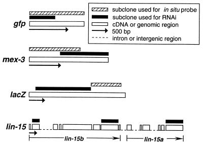
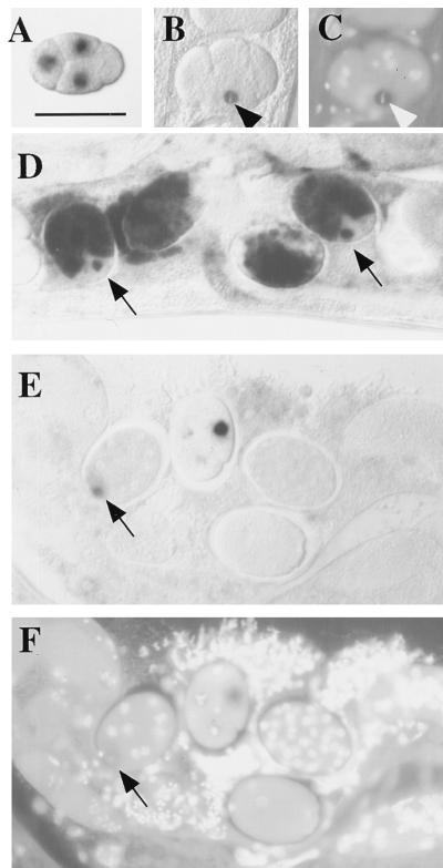
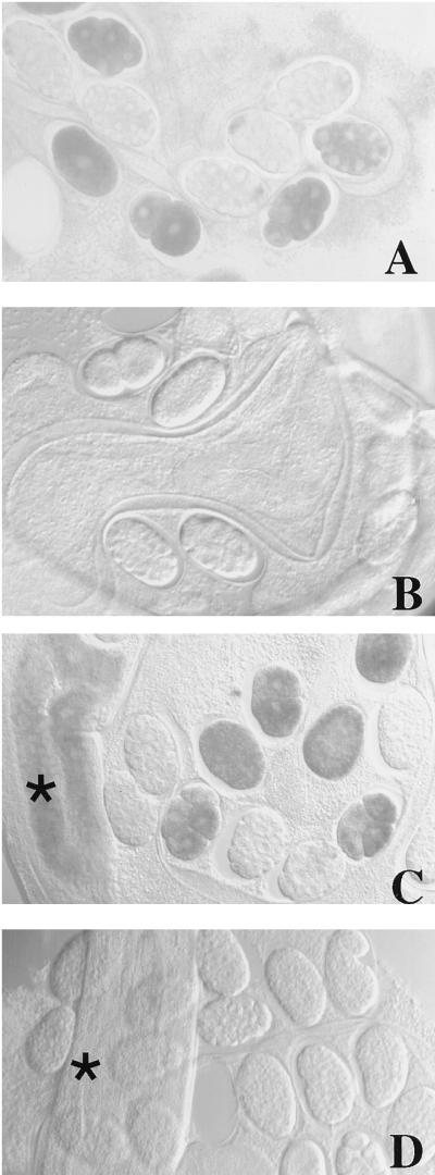
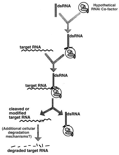
Introduction of exogenous double-stranded RNA (dsRNA) into Caenorhabditis elegans has been shown to specifically and potently disrupt the activity of genes containing homologous sequences. In this study we present evidence that the primary interference effects of dsRNA are post-transcriptional. First, we examined the primary DNA sequence after dsRNA-mediated interference and found no evidence for alterations. Second, we found that dsRNA-mediated interference with the upstream gene in a polar operon had no effect on the activity of the downstream gene; this finding argues against an effect on initiation or elongation of transcription. Third, we observed by in situ hybridization that dsRNA-mediated interference produced a substantial, although not complete, reduction in accumulation of nascent transcripts in the nucleus, while cytoplasmic accumulation of transcripts was virtually eliminated. These results indicate that the endogenous mRNA is the target for interference and suggest a mechanism that degrades the targeted RNA before translation can occur. This mechanism is not dependent on the SMG system, an mRNA surveillance system in C. elegans responsible for targeting and destroying aberrant messages. We suggest a model of how dsRNA might function in a catalytic mechanism to target homologous mRNAs for degradation.


Figures

Figure 1
Figure 1
Structure of clones used to generate dsRNA for RNAi and probes for in situ hybridization.
Figure 2
Figure 2
Analysis of target RNA distribution in RNAi-treated embryos. Embryos shown are from an integrated (homozygous) transgenic line carrying a pes-10gfp fusion. The pattern of RNA products was analyzed by whole-mount in situ hybridization, with and without prior injection of dsRNA covering the 5′ half of the gfp coding region. (A) Control (uninjected) 4-cell embryo showing transcripts of a pes-10gfp translational fusion as they first accumulate in the nuclei of the 3 somatic blastomeres of the 4-cell embryo (anterior at left; dorsal at top). This corresponds to the pattern of pes-10 expression described by Seydoux et al. (15). [Bar = 50 μm (for all panels).] (B) An equivalent embryo after injection of dsRNA targeting gfp. Transcripts are still present in the nucleus of cells that have recently commenced transgene expression. As shown in this figure, a relatively strong signal was often detected in the EMS blastomere (arrowhead) of the 4- to 6-cell stage embryo, even while undergoing early stages of mitosis. These transcripts generally do not accumulate to the same levels observed in untreated embryos. (C) Image of embryo in B stained with the DNA-binding dye 4′,6-diamidino-2-phenylindole (DAPI), showing EMS in metaphase (arrowhead). (D) Untreated embryos. Transcripts are eventually transported out of the nucleus and accumulate in the cytoplasm. Nascent transcripts continue to be detected in later emerging somatic blastomeres (arrows). (E) RNAi-treated embryos of stages similar to those depicted in D. Whereas nascent transcripts from a recently emerging somatic blastomere are detected (arrow), transcripts do not accumulate to high levels and are never detected in the cytoplasm. (F) Image of DAPI-stained embryos shown in E. Images of in situ results were obtained with Nomarski differential interference contrast (DIC) microscopy; images of DAPI-stained embryos were obtained with epifluorescence microscopy.
Figure 3
Figure 3
Loss of maternal mex-3 RNA in RNAi-treated smg-3 and wild-type hermaphrodites and their progeny as revealed by whole-mount in situ hybridization. mex-3 message is abundant and correctly localized in the embryos of a smg-3 mutant (A) but disappears from the progeny of smg-3 mutant hermaphrodites injected with mex-3 dsRNA (B). (C) In wild-type hermaphrodites mex-3 message is detected throughout the gonad (indicated by the asterisk at far left) as well as in embryos. (D) Injection of mex-3 dsRNA results in complete loss of detection of endogenous mex-3 in the maternal germ line (here splayed out) as well as in all F1 progeny. Images were obtained with Nomarski differential interference contrast microscopy. As a control for the in situ hybridization experiments, we determined that loss of hybridization signal was specific to the targeted mRNA: levels of another germ-line mRNA, cey-2, were unaffected when adult hermaphrodites were injected with mex-3 dsRNA (data not shown). (×240)
Figure 4
Figure 4
Possible model for dsRNA-mediated genetic interference in C. elegans. Upon introduction into the cell, dsRNA is proposed to complex with a (hypothetical) protein or ribonucleoprotein complex that allows unwinding of an arbitrary segment of the duplex. The complex would then search by homology for corresponding segments of cellular RNA. Recognition of cellular RNAs would be followed by a process marking the target RNA for degradation. Possible “marking” mechanisms include direct cleavage of the target RNA, covalent modification (e.g., by adenosine deaminase), or the recruitment or removal of specific RNA-binding proteins. Whatever the mechanism of this initial interaction, our data suggest a rapid subsequent degradation of the entire targeted transcript. The secondary degradation machinery could involve a combination of components specific to RNA interference and/or more general catabolic mechanisms.

References

    1. Proud C. Trends Biochem Sci. 1995;20:241–246. - PubMed
    1. Jacobs B L, Langland J O. Virology. 1996;219:339–349. - PubMed
    1. Fire A, Xu S, Montgomery M K, Kostas S A, Driver S, Mello C C. Nature (London) 1998;391:806–811. - PubMed
    1. Montgomery M K, Fire A. Trends Genet. 1998;14:255–258. - PubMed
    1. Matzke M A, Matzke A J. Cell Mol Life Sci. 1998;54:94–103. - PMC - PubMed

Publication types

LinkOut - more resources