. 1998 Dec 22;95(26):15547-52.
doi: 10.1073/pnas.95.26.15547.

Altered thymic positive selection and intracellular signals in Cbl-deficient mice

Affiliations

Altered thymic positive selection and intracellular signals in Cbl-deficient mice

M Naramura et al. Proc Natl Acad Sci U S A. .

Abstract

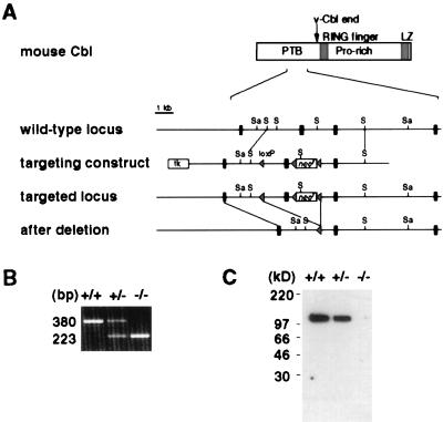
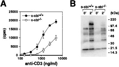
Cbl is the product of the protooncogene c-cbl and is involved in T cell antigen receptor (TCR)-mediated signaling. To understand the role of Cbl for immune system development and function, we generated a Cbl-deficient mouse strain. In Cbl-deficient mice, positive selection of the thymocytes expressing major histocompatibility complex class II-restricted transgenic TCR was significantly enhanced. Two factors may have contributed to the altered thymic selection. First, Cbl deficiency markedly up-regulated the activity of ZAP-70 and mitogen-activated protein kinases. The mitogen-activated protein kinase pathway was shown previously to be involved in thymic positive selection. Second, Cbl-deficient thymocytes expressed CD3 and CD4 molecules at higher levels, which consequently may increase the avidity of TCR/major histocompatibility complex/coreceptor interaction. Thus, Cbl plays a novel role in modulating TCR-mediated multiple signaling pathways and fine-tunes the signaling threshold for thymic selection.


Figures

Figure 1
Figure 1
Generation of Cbl-deficient mice by gene targeting. (A) Partial restriction map of the c-cbl locus, the targeting construct, and the mutated c-cbl locus. The second exon in the figure, which subsequently was deleted by Cre/loxP-mediated DNA recombination, corresponds to nucleotides 681–837 of the published mouse c-cbl cDNA sequence (GenBank accession no. X57111). Black rectangles represent exons, and triangles represent loxP sequence. Sa, SacI; S, SphI. (B) Reverse transcription–PCR analysis of c-cbl transcripts from wild-type (+/+), heterozygous (+/−), and homozygous (−/−) c-cbl mutant thymocytes. The sequences of the primers used in the PCRs correspond to nucleotides 640–663 (sense orientation) and to nucleotides 996-1019 (antisense orientation) of mouse c-cbl cDNA. (C) Immunoblot analysis of the Cbl protein. Thymocyte lysate was immunoblotted with a polyclonal antibody against the C terminus of the Cbl protein.
Figure 2
Figure 2
Flow-cytometric analysis of thymocytes. (A) CD4 and CD8 staining of thymocytes from c-cbl+/+ and c-cbl−/− mice. (B) Expression of surface markers on thymocytes. Shaded patterns and the thick lines represent c-cbl wt and homozygous mutant cells, respectively.
Figure 3
Figure 3
Positive selection in Cbl-deficient mice. (A) Positive selection of 5C.C7 TCR transgenic thymocytes. Freshly isolated thymocytes of indicated genotypes were stained with antibodies and analyzed by flow cytometry. The percentage of each cell population is indicated in the 5% contour profiles. More than 95% of thymocytes expressed transgenic TCR α and β chains. Total thymocyte numbers are: Eb/b wt, 2.3 × 10⁸; Eb/b c-cbl−/−, 1.7 × 10⁸; Eb/k wt, 4.7 × 10⁸; Eb/k c-cbl−/−, 2.5 × 10⁸; Ek/k wt, 1.8 × 10⁸; Ek/k c-cbl−/−, 0.9 × 10⁸. There was no statistically significant difference in total cell numbers between groups. (B) Positive selection of H-Y TCR transgenic thymocytes in female mice. Total thymocyte numbers of the wt and c-cbl−/− mice are 1.0 × 10⁸ and 1.3 × 10⁸, respectively. More than five pairs of mice were analyzed, and a representative experiment is shown here. There was no statistically significant difference in total thymocyte numbers.
Figure 4
Figure 4
Negative selection in Cbl-deficient mice. (A) Negative selection of self-reactive T cells in H-Y male mice. Thymocytes from age-matched wt and c-cbl−/− male mice with transgenic H-Y TCR were compared. Total thymocyte numbers of the wt and c-cbl−/− male mice are 1.7 × 10⁷ and 2.4 × 10⁷, respectively. A total of four pairs of mice were analyzed, and there was no significant difference in total thymocyte numbers between wt and c-cbl−/− mice. (B) Clonal deletion by superantigen. Peripheral blood lymphocytes were stained with anti-CD4, anti-CD8, and anti-Vβ3 antibodies and analyzed by flow cytometry. Integration of MMTV-3 and/or -13 genomes was detected by PCR (PCR does not distinguish between MMTV-3 and -13). The percentage of Vβ3+ cells out of total T cells is plotted.
Figure 5
Figure 5
Altered TCR-mediated signal transduction in Cbl-deficient thymocytes. (A) Tyrosine phosphorylation of cellular proteins upon CD3ɛ stimulation. Thymocytes were stimulated with anti-CD3ɛ antibody for indicated time periods. Equal amount of protein from cell lysate was separated by SDS/PAGE and immunoblotted with 4G10 antiphosphotyrosine antibody. The positions of Cbl and molecular size standards are indicated. (B) Analysis of individual signaling molecules. Thymocytes were stimulated for 2 min with antibodies against CD3ɛ and CD4 as indicated. Equal amount of protein from cell lysate was immunoprecipitated (IP) with indicated antibodies and immunoblotted with antibodies specific for phosphotyrosine (pY), Lck, ZAP-70, or PLCγ1. For MAP kinase (MAPK), total cell lysate was immunoblotted with antibodies specific for active MAP kinase or total MAP kinase. (C) PI-3K activity of thymocytes. Thymocytes were stimulated with anti-CD3ɛ and anti-CD28 antibodies for 2 min. PI-3K activity was measured as described previously (34). The data are presented as fold increase over control (unstimulated) and represent the average ± range from two separate experiments.
Figure 6
Figure 6
Function of Cbl-deficient peripheral T cells. (A) Proliferative response of peripheral T cells. Purified CD4+ T cells from 5C.C7 transgenic mice were stimulated with plate-bound anti-CD3 ɛ antibody. Culture was pulsed with [3H]thymidine for the last 12 hr of incubation. (B) Total tyrosine phosphorylation of splenocytes. Splenocytes were enriched for T cells by passing through nylon wool columns. T cells were stimulated with anti-CD3ɛ and anti-CD4 antibodies for 2 min. Equal amount of protein from total cell lysate was separated by SDS/PAGE and immunoblotted with 4G10 antibody.

References

    1. Robey E, Fowlkes B J. Annu Rev Immunol. 1994;12:675–705. - PubMed
    1. Jameson S C, Hogquist K A, Bevan M J. Annu Rev Immunol. 1995;13:93–126. - PubMed
    1. Kisielow P, von Boehmer H. Adv Immunol. 1995;58:87–209. - PubMed
    1. Benoist C, Mathis D. Curr Opin Immunol. 1997;9:245–249. - PubMed
    1. Ashton-Rickardt P G, Van Kaer L, Schumacher T N, Ploegh H L, Tonegawa S. Cell. 1993;73:1041–1049. - PubMed

MeSH terms

Substances