. 1998 Dec 22;95(26):15592-7.
doi: 10.1073/pnas.95.26.15592.

Targeted disruption of mouse long-chain acyl-CoA dehydrogenase gene reveals crucial roles for fatty acid oxidation

Affiliations

Targeted disruption of mouse long-chain acyl-CoA dehydrogenase gene reveals crucial roles for fatty acid oxidation

D M Kurtz et al. Proc Natl Acad Sci U S A. .

Abstract

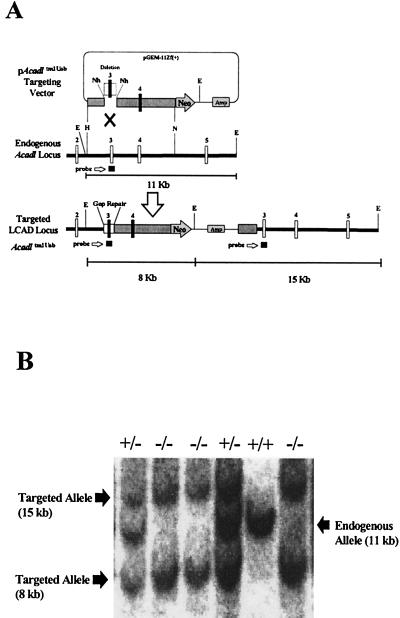
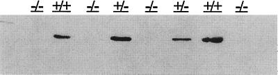
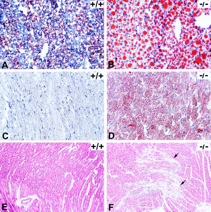
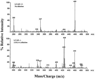
Abnormalities of fatty acid metabolism are recognized to play a significant role in human disease, but the mechanisms remain poorly understood. Long-chain acyl-CoA dehydrogenase (LCAD) catalyzes the initial step in mitochondrial fatty acid oxidation (FAO). We produced a mouse model of LCAD deficiency with severely impaired FAO. Matings between LCAD +/- mice yielded an abnormally low number of LCAD +/- and -/- offspring, indicating frequent gestational loss. LCAD -/- mice that reached birth appeared normal, but had severely reduced fasting tolerance with hepatic and cardiac lipidosis, hypoglycemia, elevated serum free fatty acids, and nonketotic dicarboxylic aciduria. Approximately 10% of adult LCAD -/- males developed cardiomyopathy, and sudden death was observed in 4 of 75 LCAD -/- mice. These results demonstrate the crucial roles of mitochondrial FAO and LCAD in vivo.


Figures

Figure 1
Figure 1
Targeting of the mouse Acadl gene. (A) Schematic diagram of the targeting vector, pAcadltm1Uab, the endogenous Acadl locus, and the targeted Acadltm1Uab locus. Gap repair of the deleted region (an 821-bp NheI deletion of exon 3 and flanking intron sequence) in the targeting vector occurs on homologous recombination and was the basis for genotype screening by Southern blot analysis of EcoRI-digested DNA. E, EcoRI; H, HindIII; N, NotI; Nh, NheI. (B) Representative Southern blot from Acadl-targeted mice showing three genotypes. EcoRI-digested genomic DNA was probed with a 304-bp Acadl genomic fragment containing exon 3 and flanking intron sequence. Banding patterns are as follows: Acadl normal controls (+/+), −11 kb; heterozygous (+/−), −15 kb, 11 kb, and 8 kb; and homozygous mutant (−/−), −15 kb and 8 kb.
Figure 2
Figure 2
LCAD immunoblot. Immunoblot of liver mitochondrial proteins (50 μg) isolated from normal control (+/+), heterozygous mutant (+/−), and homozygous mutant (−/−) mice and probed with a rat anti-LCAD antibody. No detectable LCAD protein was observed in samples from the homozygous mutant mice.
Figure 3
Figure 3
Histopathology on LCAD +/+ (normal control) and LCAD −/− (deficient mice). (A) +/+ and (B) −/−: Oil-Red-O staining of frozen liver sections from 4- to 5-wk-old mice after an 18- to 20-hr fast. Abundant large lipid droplets present in the LCAD-deficient mice compared with minimal lipid accumulation in normal control. (C) +/+ and (D) −/−: Oil-Red-O staining of frozen hearts from 14- to 16-wk-old mice after an 18- to 20-hr fast. Note lipid accumulation in all cardiomyocytes of the LCAD-deficient mouse as compared with the LCAD-normal control. (E) +/+ and (F) −/−: Hematoxylin/eosin-stained heart sections from 16-wk-old, nonfasted male mice. Note prominent myocardial degeneration and fibrosis (arrows) present in the LCAD −/− mice (F). (A and B, ×25, C and D, ×40, and E and F, ×10.)
Figure 4
Figure 4
Butyl-ester acylcarnitine profiles of bile specimens obtained by using precursor-ion scanning (parent of m/z 85) electrospray/tandem mass spectrometry. Mice were sacrificed after overnight fast (18–20 hr), and bile specimens were collected as spots on filter paper. (Top) LCAD +/+ (normal control), no dilution. (Bottom) LCAD −/− (LCAD-deficient), 1:10 (vol/vol) dilution. Internal standards: m/z 263, d3-acetylcarnitine; m/z 347, d3-octanoylcarnitine; and m/z 459, d3-palmitoylcarnitine. Peak identification: m/z 260, acetylcarnitine (C2); m/z 372, decanoylcarnitine (C10); m/z 400, dodecanoylcarnitine (C12); m/z 424, tetradecadienoylcarnitine (C14:2); m/z 426, tetradecenoylcarnitine (C14:1); m/z 456, palmitoylcarnitine (C16); m/z 480, linoleylcarnitine (C18:2); m/z 482, oleylcarnitine (C18:1).

References

    1. Mannaerts G P, Debeer L J. Ann NY Acad Sci. 1982;386:30–39. - PubMed
    1. Nicholls D G, Locke R M. Physiol Rev. 1984;64:1–64. - PubMed
    1. Roe C R, Coates P M. In: The Metabolic and Molecular Bases of Inherited Disease. Scriver C R, Beaudet A L, Sly W S, Valle D, editors. New York: McGraw–Hill; 1995. pp. 1501–1533.
    1. Boles R G, Buck E A, Blitzer M G, Platt M S, Cowan T M, Martin S K, Yoon H, Madsen J A, Reyes-Mugica M, Rinaldo P. J Pediatr (Berlin) 1998;132:924–933. - PubMed
    1. Naito E, Ozasa H, Ikeda Y, Tanaka K. J Clin Invest. 1989;83:1605–1613. - PMC - PubMed

Publication types

MeSH terms

Substances