. 1998 Dec 22;95(26):15647-52.
doi: 10.1073/pnas.95.26.15647.

Lamellar lipoproteins uniquely contribute to hyperlipidemia in mice doubly deficient in apolipoprotein E and hepatic lipase

Affiliations

Lamellar lipoproteins uniquely contribute to hyperlipidemia in mice doubly deficient in apolipoprotein E and hepatic lipase

N Bergeron et al. Proc Natl Acad Sci U S A. .

Abstract

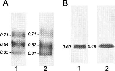
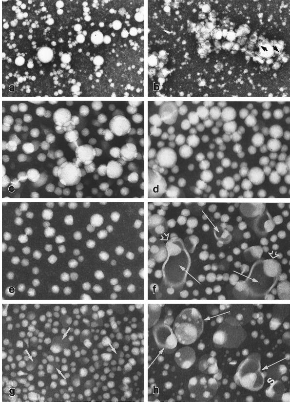
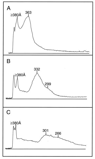
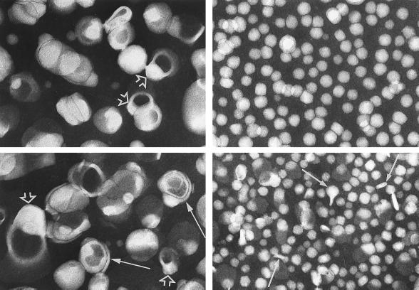
Remnants of triglyceride-rich lipoproteins containing apolipoprotein (apo) B-48 accumulate in apo E-deficient mice, causing pronounced hypercholesterolemia. Mice doubly deficient in apo E and hepatic lipase have more pronounced hypercholesterolemia, even though remnants do not accumulate appreciably in mice deficient in hepatic lipase alone. Here we show that the doubly deficient mice manifest a unique lamellar hyperlipoproteinemia, characterized by vesicular particles 600 A-1,300 A in diameter. As seen by negative-staining electron microscopy, these lipoproteins also contain an electron-lucent region adjacent to the vesicle wall, similar to the core of typical lipoproteins. Correlative chemical analysis indicates that the vesicle wall is composed of a 1:1 molar mixture of cholesterol and phospholipids, whereas the electron-lucent region appears to be composed of cholesteryl esters (about 12% of the particle mass). Like the spherical lipoproteins of doubly deficient mice, the vesicular particles contain apo B-48, but they are particularly rich in apo A-IV. We propose that cholesteryl esters are removed from spherical lipoproteins of these mice by scavenger receptor B1, leaving behind polar lipid-rich particles that fuse to form vesicular lipoproteins. Hepatic lipase may prevent such vesicular lipoproteins from accumulating in apo E-deficient mice by hydrolyzing phosphatidyl choline as scavenger receptor B1 removes the cholesteryl esters and by gradual endocytosis of lipoproteins bound to hepatic lipase on the surface of hepatocytes.


Figures

Figure 1
Figure 1
Agarose gel electropherograms of IDL (1) and LDL (2) from an apo E- and HL-doubly deficient mouse (A) and an apo E-deficient mouse (B). A single component with β-mobility is evident in both fractions from the apo E-deficient mouse, whereas three components with slow β, β, and pre-β mobilities are evident in fractions from the doubly deficient mouse. (The latter pattern was observed with IDL and LDL from two other mice.) Rf values relative to albumin marker (not shown) are indicated for each lipoprotein component.
Figure 2
Figure 2
Plasma lipoproteins, visualized by negative-staining electron microscopy from apo E-deficient (Left: a, c, e, g) and doubly deficient mice (Right: b, d, f, h). Whole plasma from apo E-deficient mice (a) contains mainly large spherical particles of 200 Å–1,600 Å in diameter; that from doubly deficient mice (b) contains, in addition, clusters of polymorphic structures, some of which are flattened (arrows). VLDL fractions from apo E-deficient mice (c) and doubly deficient mice (d) contain spherical particles of 250 Å–1,600 Å in diameter with electron-lucent cores as seen in whole plasma. IDL from apo E-deficient mice (e) contain round to rectangular particles of 225 Å–450 Å diameter with electron-lucent cores whereas IDL from doubly deficient mice (f) contain, in addition, larger polymorphic particles with electron-opaque cores (arrows); in many of these an electron-lucent region (open arrowhead) is seen abutting the wall. The LDL fraction from apo E-deficient mice (g) contains somewhat smaller particles (170 Å–300 Å diameter) than those in e; some larger particles are partially electron-opaque (arrows). LDL from doubly deficient mice (h) contain a similar mixture of particles to those seen in (f) but with a greater proportion of the larger species (arrows). (×87,300, except for a and b (×29,100.)
Figure 3
Figure 3
Densitometric tracings of lipid-stained 2%–l4% polyacrylamide gels after electrophoresis of VLDL (A), IDL (B), and LDL (C) from an apo E- and HL-doubly deficient mouse. Components near the top of the gel are evident in all cases, together with included components of progressively decreasing size. Comparable patterns were observed with lipoproteins from two other doubly deficient mice. The early components for VLDL represent large spherical particles that were excluded from, or barely entered, the gel, whereas those for IDL and LDL represent lamellar particles. The more rapidly migrating components represent spherical microemulsion particles in all cases (see Fig. 4) and may include lipoprotein species containing or lacking apo B.
Figure 4
Figure 4
Species of lipoproteins in IDL (Upper) and LDL (Lower) fractions from apo E- and HL-doubly deficient mice, separated by gel filtration. The larger species of 600 Å–1,300 Å diameter (Left) are characterized by an electron-opaque core, with an electron-lucent region abutting the wall (open arrowheads). Some particles are multilamellar (arrows). The smaller species (Right) resemble the spherical/rectangular particles of total IDL and LDL (see Fig. 2), with a few oblong or irregular particles in the latter (arrows). (×87,300.)

Similar articles

Cited by

References

    1. Havel R J. Curr Opin Lipidol. 1995;6:312–316. - PubMed
    1. Zhang S H, Reddick R L, Piedrahita J A, Maeda N. Science. 1992;258:468–471. - PubMed
    1. Plump A, Smith J, Hayek T, Aalto-Setala K, Walsh A, Verstuyft J, Rubin E, Breslow J. Cell. 1992;71:343–353. - PubMed
    1. Ghiselli G, Schaefer E J, Gascon P, Brewer H B., Jr Science. 1981;214:1239–1241. - PubMed
    1. Qiu S, Bergeron N, Kotite L, Krauss R M, Bensadoun A, Havel R J. J Lipid Res. 1998;39:1661–1668. - PubMed

Publication types