. 1998 Dec 22;95(26):15769-74.
doi: 10.1073/pnas.95.26.15769.

Tetracyclines inhibit microglial activation and are neuroprotective in global brain ischemia

Affiliations

Tetracyclines inhibit microglial activation and are neuroprotective in global brain ischemia

J Yrjänheikki et al. Proc Natl Acad Sci U S A. .

Abstract

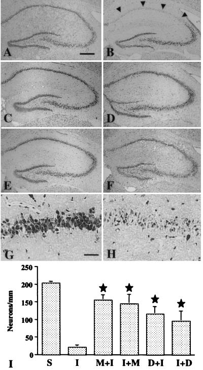
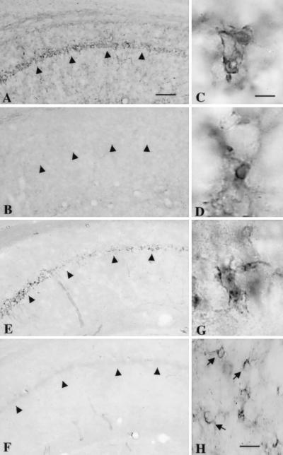
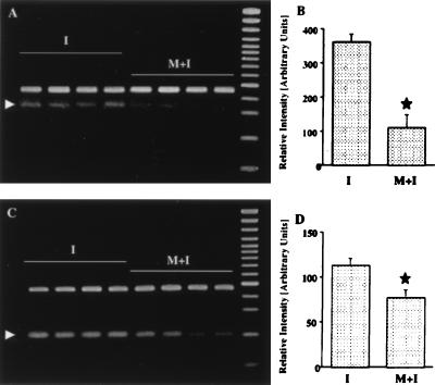
Ischemic stroke is the most common life-threatening neurological disease and has limited therapeutic options. One component of ischemic neuronal death is inflammation. Here we show that doxycycline and minocycline, which are broad-spectrum antibiotics and have antiinflammatory effects independent of their antimicrobial activity, protect hippocampal neurons against global ischemia in gerbils. Minocycline increased the survival of CA1 pyramidal neurons from 10.5% to 77% when the treatment was started 12 h before ischemia and to 71% when the treatment was started 30 min after ischemia. The survival with corresponding pre- and posttreatment with doxycycline was 57% and 47%, respectively. Minocycline prevented completely the ischemia-induced activation of microglia and the appearance of NADPH-diaphorase reactive cells, but did not affect induction of glial acidic fibrillary protein, a marker of astrogliosis. Minocycline treatment for 4 days resulted in a 70% reduction in mRNA induction of interleukin-1beta-converting enzyme, a caspase that is induced in microglia after ischemia. Likewise, expression of inducible nitric oxide synthase mRNA was attenuated by 30% in minocycline-treated animals. Our results suggest that lipid-soluble tetracyclines, doxycycline and minocycline, inhibit inflammation and are neuroprotective against ischemic stroke, even when administered after the insult. Tetracycline derivatives may have a potential use also as antiischemic compounds in humans.


Figures

Figure 1
Figure 1
Effect of minocycline and doxycycline treatment on the CA1 pyramidal neurons after global brain ischemia. The hippocampus is shown in a cresyl violet-stained section from a sham-operated gerbil (A); and from ischemia-operated gerbils treated with saline (B, H); minocycline, starting 12 h before operation (C, G); minocycline, starting 30 min after operation (D); doxycycline, starting 12 h before operation (E); and doxycycline, starting 30 min after operation (F). A substantial number of the stained pyramidal neurons remain intact in the CA1 pyramidal cell layer in minocycline- and doxycycline-treated gerbils compared to the CA1 pyramidal cells in saline-treated gerbils (arrowheads). Bars = 400 μm (A–F) and 40 μm (G, H). The graph (I) summarizes the quantitative results of CA1 neuronal counts: S, sham-operated; I, saline-treated; M+I/D+I, minocycline/doxycycline treatment started before ischemia; I+M/I+D, minocycline/doxycycline treatment started after ischemia. star, P < 0.05, when compared with saline-treated ischemic (I) gerbils.
Figure 2
Figure 2
Minocycline treatment does not alter GFAP expression in the hippocampus of gerbils exposed to global ischemia. Northern blotting (A) demonstrates a similar induction of GFAP mRNA in saline-treated (I) and minocycline-pretreated (M+I) gerbils when compared to sham-operated controls (C). Samples from two different animals in each group are shown. A 32P-dATP-labeled cyclophilin oligonucleotide probe (CP) was used to demonstrate equal amounts of loaded RNAs. GFAP immunostaining (B, C) in the CA1 hippocampal field shows approximately the same distribution and number of positively labeled astrocytes (arrows) in saline- (B) and minocycline-pretreated (C) ischemic gerbils. PCL = pyramidal cell layer. Bar = 30 μm.
Figure 3
Figure 3
Activation of microglial cells (A–D) and induction of NADPH-diaphorase (NOS) reactivity (E–H) in the CA1 hippocampus 6 days after global ischemia are prevented by minocycline pretreatment. Phosphotyrosine immunoreactivity (A–C) and isolectin B4-binding (D) were used as markers of microglial activation. In a saline-treated gerbil, phosphotyrosine immunoreactivity is evident, especially in the CA1 pyramidal cell layer (A), while in a minocycline-treated gerbil (B), no clear staining is seen. High magnification of phosphotyrosine staining (C) and isolectin B4 binding (D) reveals a cell morphology typical of microglia. NADPH-diaphorase reactivity is induced in the CA1 pyramidal cell layer of a saline-treated (E) but not in a minocycline-treated (F) gerbil. The NADPH-diaphorase-stained cells in the pyramidal cell layer send out short processes with an irregular shape, resembling microglia (G). In the stratum radiatum, the NADPH-diaphorase-reactive cells show staining in the cell body and send out thick processes, which is typical of astrocyte morphology (H, arrows). Arrowheads point to the CA1 pyramidal cell layer. Bars = 250 μm (A, B, E, F); 8 μm (C, D, G) and 25 μm (H).
Figure 4
Figure 4
RT-PCR analysis of ICE mRNA (A, B) and iNOS mRNA (C, D) in the hippocampus of saline-treated (I) and minocycline-treated (M+I) gerbils 4 days after ischemia. Arrowheads in A and C point to the amplified ICE and iNOS mRNA products. mRNA isolated from the same tissues was subjected to RT-PCR by using primers for the GAPDH gene. No significant alteration in mRNA expression is observed for this gene (upper bands in A and C). B and D represent quantitation of amplified mRNA products, corrected by corresponding GADPH hybridization signals. (mean ± standard error). Star, statistically significant difference (P < 0.05).

References

    1. Klein N C, Cunha B A. Med Clin N Am. 1995;79:789–801. - PubMed
    1. Cunha B A, Sibley C, Ristuccia P A. Ther Drug Monit. 1982;4:115–130. - PubMed
    1. Sande M A, Mandell G L. In: The Pharmacological Basis of Therapeutics. Goodman L S, Gilman A, editors. New York: Macmillan; 1985. pp. 1170–1192.
    1. Kramer P A, Chapron D J, Benson J, Mercik S A. Clin Pharmacol Ther. 1978;23:467–472. - PubMed
    1. Barza M, Brown R B, Shanks C, Gamble C, Weinstein L. Antimicrob Agents Chemother. 1975;8:713–720. - PMC - PubMed

Publication types

MeSH terms