. 1998 Dec 22;95(26):15781-6.
doi: 10.1073/pnas.95.26.15781.

Conserved structural features of the synaptic fusion complex: SNARE proteins reclassified as Q- and R-SNAREs

Affiliations

Conserved structural features of the synaptic fusion complex: SNARE proteins reclassified as Q- and R-SNAREs

D Fasshauer et al. Proc Natl Acad Sci U S A. .

Abstract

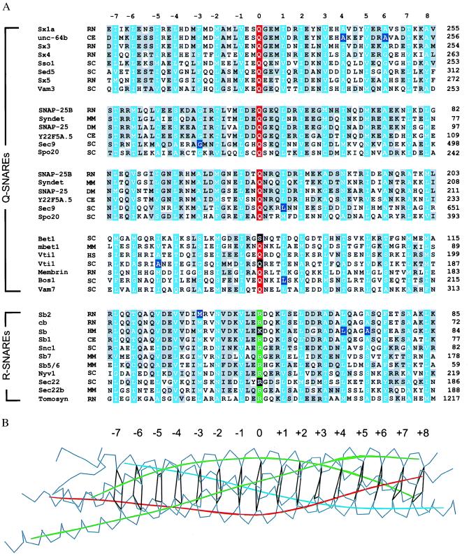
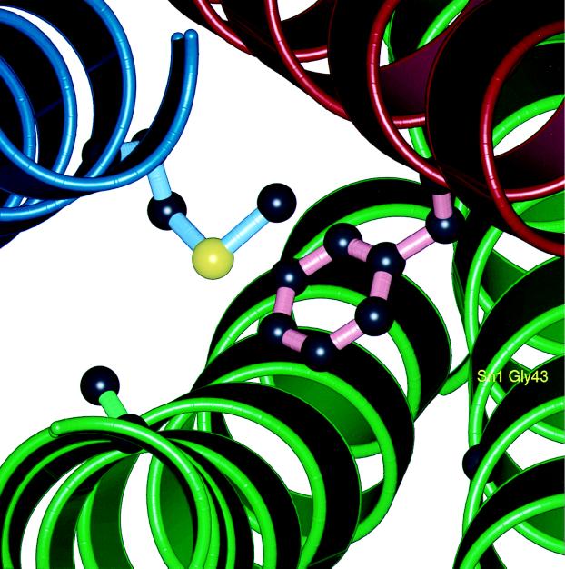
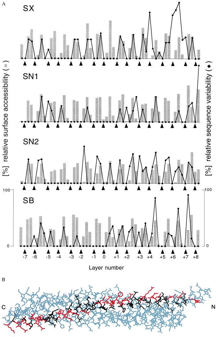
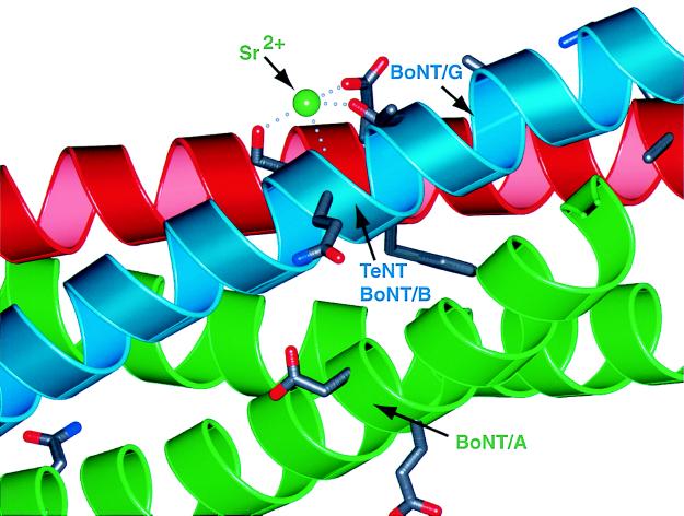
SNARE [soluble NSF (N-ethylmaleimide-sensitive fusion protein) attachment protein receptor] proteins are essential for membrane fusion and are conserved from yeast to humans. Sequence alignments of the most conserved regions were mapped onto the recently solved crystal structure of the heterotrimeric synaptic fusion complex. The association of the four alpha-helices in the synaptic fusion complex structure produces highly conserved layers of interacting amino acid side chains in the center of the four-helix bundle. Mutations in these layers reduce complex stability and cause defects in membrane traffic even in distantly related SNAREs. When syntaxin-4 is modeled into the synaptic fusion complex as a replacement of syntaxin-1A, no major steric clashes arise and the most variable amino acids localize to the outer surface of the complex. We conclude that the main structural features of the neuronal complex are highly conserved during evolution. On the basis of these features we have reclassified SNARE proteins into Q-SNAREs and R-SNAREs, and we propose that fusion-competent SNARE complexes generally consist of four-helix bundles composed of three Q-SNAREs and one R-SNARE.


Figures

Figure 1
Figure 1
(A) Sequence alignment (16) of the four-helix bundle region of the synaptic fusion complex (5) for a representative subset of the entire SNARE family. The sequence analysis was restricted to 16 layers (blue) of the four-helix bundle in the synaptic fusion complex (5), including 7 layers upstream (layers −1 to −7) and 8 layers downstream (layers +1 to +8) of the ionic layer (layer 0). Conserved residues are shaded in gray. The conserved glutamine and arginine residues forming the ionic 0 layer are indicated in red and green, respectively. The mutations discussed in the text are indicated by black shaded boxes. There are two alignment tables for the SNAP-25 family that correspond to the two SNAP-25 α-helices in the synaptic fusion complex (5). GenBank accession numbers for the syntaxin family are as follows: sx1a, RN, P32851; unc-64b, CE, AF047885; sx3, RN, Q08849; sx4, RN, Q08850; Sso1, SC, P32867; sed5, SC, Q01590; sx5, RN, Q08851; vam3, SC, Q12241. Accession numbers for the SNAP-25 family are SNAP-25B, HS, P13795; syndet, MM, U73143; SNAP-25, DM, U81153; Y22F5A.5, CE, AL021479; sec9, SC, L34336; and spo20, SC, Z49211. Accession numbers for the bos-group are bet1, SC, P22804; mbet1, MM, AF007552; vti1, HS, AF035824; vti1, SC, AF006074; membrin, RN, U91539; bos1, SC, P25385; and vam7, SC, P32912. Accession numbers for the synaptobrevin/vamp family (R-SNAREs) are sb2, RN, M24105; cb/cellubrevin, RN, S63830; sb1 CE AF003281; sb, HM, U85805; Snc1, SC, M91157; sb5/6, HS, AA222692; sb7, MM, X96737; sec22 (Sly2), SC, L8479; sec22b, MM, U91538; nyv1, SC, Z73265; and tomosyn, RN, U92072. The two-letter species abbreviations after the protein name are as follows: HS, Homo sapiens; MM, Mus musculus; RN, Rattus norvegicus; SC, Saccharomyces cerevisiae; DM, Drosophila melanogaster; TM, Torpedo marmorata; CE, Caenorhabditis elegans; and HM, Hirudo medicinalis. (B) Layers of the synaptic fusion complex crystal structure (5). Indicated are Cα traces (gray), local helical axes (blue, red, and green for synaptobrevin-II, syntaxin-1A, and SNAP-25b, respectively), and layers (black) by virtual bonds between corresponding Cα positions.
Figure 2
Figure 2
Picture of the −3 layer in the synaptic fusion complex crystal structure (5). This layer consists of synaptobrevin-II Met-46, syntaxin-1A Phe-216, and SNAP-25B Gly-43 and Ala-164. The interactions within this layer explain the destabilizing effect of a mutation involving the corresponding glycine residue in the sec9–4 allele. Side-chain atoms in the layer are shown as “licorice bonds.” The polypeptide backbone is shown as a ribbon (blue, synaptobrevin-II; red, syntaxin-1A; green, SNAP-25B). This figure was prepared by Bobscript (39) and rendered using PovRay (http://www.povray.org).
Figure 3
Figure 3
Conservation of SNARE complex structure. (A) Sequence variation (black line) for selected syntaxin (SX), synaptobrevin (SB), and SNAP-25 (residues 27–82, SN1; residues 148–203, SN2) families and surface accessibilities (gray bars) in the synaptic fusion complex crystal structure. The analysis was restricted to the 16 layers (indicated by arrowheads) of the synaptic fusion complex crystal structure (5) consisting of syntaxin-1A residues 201–255 (SX), SNAP-25B residues 28–82 (SN1) and residues 149–203 (SN2), and synaptobrevin residues 31–85 (SB). The layer numbers refer to Fig. 1B. The GenBank accession numbers used for the syntaxin-1 through -4 family are as follows: sx-1b, RN, P32853; sx1b, BS, P41414; sx-1a, RN, P32851; sx-1a, HS, L37792; sx-1a, MM, D45208; sx-A, HS, U12918; sx1a, BS, P32850; sx1, HM, U85807; sx, AC, U03123; sx1a, DM, L37732; unc-64b, CE, AF047885; sx, SP, AF014122; sx2, RN, P50279; sx3, RN, Q08849; sx3a, HS, AJ002076; sx4, RN, Q08850; sx4, HS, X85784; and sx4, MM, U76832. The accession numbers used for the SNAP-25 family are SNAP-25B, HS, P13795; SNAP-25, TM, P36976; syndet, MM, U73143; SNAP-25, SP, AF036902; SNAP-25, DM, U81153; SNAP-25, HM, U85806; SNAP-25, GG, L09253; SNAP-25A, CA, L22973; SNAP-25D, CA, L22976; and Y22F5A.5, CE, AL021479. The accession numbers used for the synaptobrevin family members in the second branch of the dendrogram (not shown) are Sb1, RN, M24104; Sb1B, RN, U74621; Sb1, MM, AF007167; Sb1, TM, P13701; Syb-B, DM, L14270; Syb-A, DM, L14270; N-Syb, DM, S66686; Sb, AC, P35589; cellubrevin (Sb3), MM U61751; cellubrevin (Sb3), RN S63830; Sb, SP, AF014119; Sb2, RN, M24105; Sb2, HS, AJ5044; Sb2, MM, AF007168; Sb, HM, U85805; Sb, FR, AF016494; Sb, LP, X74748; XSybI, XL, AF035016; Syb2, XL, U16801; Sb, BT, X76199; Snb1, CE, AF003281; and Sb, SM, U30182. The following two-letter species abbreviations are used: CA, Carrassius auratus; HS, Homo sapiens; MM, Mus musculus; RN, Rattus norvegicus; SC, Saccharomyces cerevisiae; DM, Drosophila melanogaster; TM, Torpedo marmorata; CE, Caenorhabditis elegans; SP, Strongylocentrotus purpuratus; HM, Hirudo medicinalis; GG, Gallus gallus; BT, Bos taurus; AC, Aplysia californica; SM, Schistosoma mansoni; FR, Fugu rubripes; XL, Xenopus laevis; and LP, Loligo pealei. (B) Model of a synaptic fusion complex consisting of syntaxin-4, SNAP-25B, and synaptobrevin-II, based on the crystal structure of the synaptic fusion complex (5), which contains syntaxin-1A. Syntaxin-4 residues are shown in black and red for matching and different residue types between syntaxin-1A and syntaxin-4, respectively. Most substituted residues occur on the surface of the complex.
Figure 4
Figure 4
A binding site for Sr2+ is close to the TeNT and BoNT/A toxin cleavage sites of the synaptic fusion complex (5). This binding site for Sr2+ is found in two of the three complexes in the asymmetric unit of the crystal structure of the synaptic fusion complex (5). The divalent cation is coordinated by the conserved (among the synaptobrevin family selected in Fig. 3A) synaptobrevin-II Ser-75, Glu-78, and partially conserved Thr-79 residues. The protein backbone is represented as a ribbon drawing. Blue, synaptobrevin-II; red, syntaxin-1A; green, SNAP-25B. The indicated toxin cleavage sites are located at the peptide bonds between Gln-76 and Phe-77 (TeNT and BoNT/B), and between Ala-81 and Ala-82 (BoNT/G) for synaptobrevin-II, between Lys-253 and Ala-254 (BoNT/C) for syntaxin-1A, and between Gln-197 and Arg-198 (BoNT/A) for SNAP-25B. The residues adjacent to the scissile bond are shown as licorice bonds. This figure prepared by Bobscript (36) and rendered by using PovRay.

References

    1. Ferro-Novick S, Jahn R. Nature (London) 1994;370:191–193. - PubMed
    1. Hanson P I, Heuser J E, Jahn R. Curr Opin Neurobiol. 1997;7:310–315. - PubMed
    1. Rothman J E. Nature (London) 1994;372:55–63. - PubMed
    1. Südhof T C. Nature (London) 1995;375:645–653. - PubMed
    1. Sutton R B, Fasshauer D, Jahn R, Brunger A T. Nature (London) 1998;395:347–353. - PubMed

Publication types

LinkOut - more resources