. 1998 Dec 22;95(26):15849-54.
doi: 10.1073/pnas.95.26.15849.

The Arabidopsis thaliana RPM1 disease resistance gene product is a peripheral plasma membrane protein that is degraded coincident with the hypersensitive response

Affiliations

The Arabidopsis thaliana RPM1 disease resistance gene product is a peripheral plasma membrane protein that is degraded coincident with the hypersensitive response

D C Boyes et al. Proc Natl Acad Sci U S A. .

Abstract

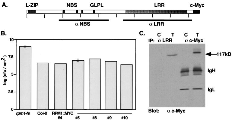
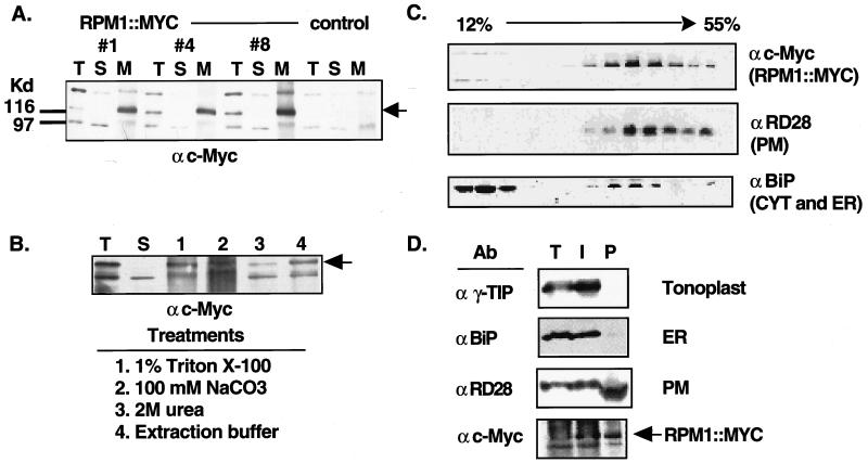
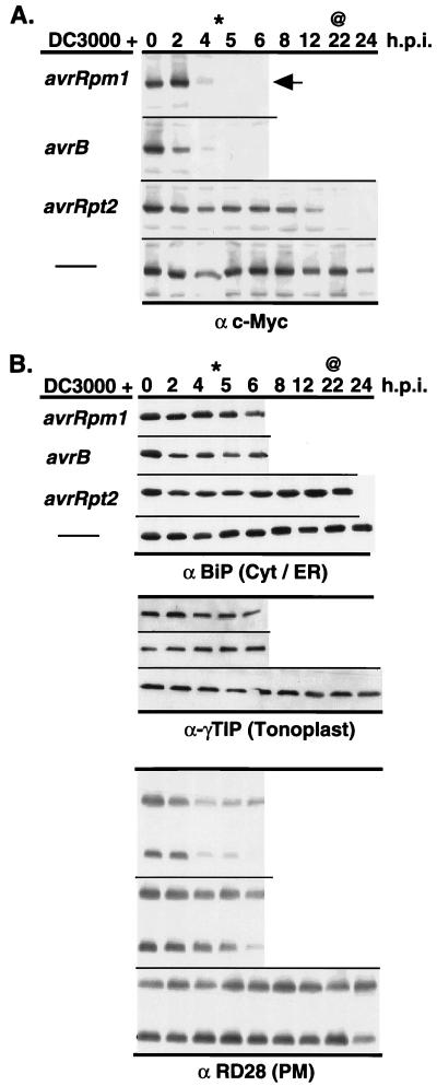
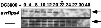
Disease resistance in plants is often controlled by a gene-for-gene mechanism in which avirulence (avr) gene products encoded by pathogens are specifically recognized, either directly or indirectly, by plant disease resistance (R) gene products. Members of the NBS-LRR class of R genes encode proteins containing a putative nucleotide binding site (NBS) and carboxyl-terminal leucine-rich repeats (LRRs). Generally, NBS-LRR proteins do not contain predicted transmembrane segments or signal peptides, suggesting they are soluble cytoplasmic proteins. RPM1 is an NBS-LRR protein from Arabidopsis thaliana that confers resistance to Pseudomonas syringae expressing either avrRpm1 or avrB. RPM1 protein was localized by using an epitope tag. In contrast to previous suggestions, RPM1 is a peripheral membrane protein that likely resides on the cytoplasmic face of the plasma membrane. Furthermore, RPM1 is degraded coincident with the onset of the hypersensitive response, suggesting a negative feedback loop controlling the extent of cell death and overall resistance response at the site of infection.


Figures

Figure 1
Figure 1
Structure, function, and immunodetection of RPM1∷MYC. (A) The deduced RPM1 protein indicating the position of the leucine zipper (L-ZIP), putative NBS, and LRR domains. GLPL denotes the core of a conserved sequence motif of unknown function found in all NBS-LRR proteins. Vertical ticks occur every 100 aa. Bars below the schematic define the regions of the protein used to generate the anti-NBS and anti-LRR polyclonal antisera. (B) Complementation of the rpm1-fs mutation with RPM1∷MYC. Bacterial growth in leaves infiltrated with DC3000(avrRpm1) at a density of ≈5.5 × 10³ cfu/cm2 was assayed 72 hpi. In planta bacterial titers are given for nontransformed rpm1-fs, wild-type Col-0, and five independent rpm1-fs (RPM1∷MYC) transgenic lines. (C) Immunodetection of RPM1∷MYC by using monoclonal anti-c-Myc antibody after immunoprecipitation with either anti-LRR antisera or monoclonal anti-c-Myc antibody. C is extract from nontransformed control plants, T from transgenic #4 of B. Anti-mouse IgG second step antibody detects the IgG heavy (IgH) and light (IgL) chains of the monoclonal.
Figure 2
Figure 2
RPM1∷MYC is a peripheral PM protein. (A) Protein blot of total (T), soluble (S), and microsomal membrane (M) fractions from nontransformed rpm1-fs control plants and three rpm1-fs (RPM1∷MYC) transgenic lines reacted with the anti-c-Myc monoclonal antibody. Equal amounts of protein were loaded in each lane. Arrowhead indicates the position of RPM1∷MYC. Anti-c-Myc crossreacting bands at ≈98 and ≈150 Kd were present in nontransformed control lines. (B) Protein blot reacted with the anti-c-Myc monoclonal antibody demonstrating peripheral association of RPM1∷MYC with the membrane. Total extract (T) was centrifuged at 100,000 × g to generate soluble (S) and microsomal membrane fractions. Membranes were treated as specified to release peripheral membrane proteins. Remaining membranes were pelleted and the newly soluble proteins were analyzed. (C) Fractionation of RPM1∷MYC on sucrose gradients. A 12–55% (wt/vol) linear sucrose gradient was used to fractionate total extract from transgenic plants. Aliquots of each fraction were blotted to nitrocellulose and were analyzed with either anti-c-Myc or the subcellular compartment marker antibodies listed at the right of each panel. (D) Protein blot analysis of membranes fractions obtained by aqueous two-phase partitioning. Total extract (T), intracellular membrane (I), and plasma membrane (P) vesicle fractions were separated by SDS/PAGE, transferred to nitrocellulose and reacted sequentially with antibodies against the c-Myc epitope, RD28, γ-TIP, and BiP.
Figure 3
Figure 3
RPM1∷MYC is degraded after inoculation with avirulent P. syringae isolates, which trigger LZ-NBS-LRR resistance genes. (A) Each panel represents infection of RPM1∷MYC transgenics with P. syringae DC3000 expressing a particular avirulence gene or not (thin line) listed at left. Tissue was harvested at the time, in hpi listed at the top, and total protein blots were probed with either anti-c-Myc monoclonal or (B) marker proteins for various subcellular compartments listed at the bottom of each series of blots in B. Extract from control plants (vector) are included for anti-c-Myc and anti-BiP experiments. The arrow marks RPM1∷MYC in A. Star (∗) represent earliest time point where visible RPM1-dependent HR was observed, and ampersand (@) represents the earliest time point where RPS2-dependent HR was observed. This set of data is all from one set of extracts and the experiment was repeated three times.
Figure 4
Figure 4
RPM1∷MYC is degraded after inoculation with an avirulent P. syringae isolate, which triggers a TIR-NBS-LRR resistance gene. Experimental design is as in Fig. 3, but the time course is extended to 40 hpi, at which time all proteins begin to degrade because of disease (DC3000) or complete HR. Star (∗) marks time point of RPS4-dependent HR and arrows denote RPM1∷MYC.

References

    1. Bent A. Plant Cell. 1996;8:1757–1771. - PMC - PubMed
    1. Hammond-Kosack K E, Jones J D G. Plant Cell. 1996;8:1773–1791. - PMC - PubMed
    1. Leister R T, Ausubel F M, Katagiri F. Proc Natl Acad Sci USA. 1996;93:15497–15502. - PMC - PubMed
    1. Lawrence G J, Finnegan E J, Ayliffe M A, Ellis J G. Plant Cell. 1995;7:1195–1206. - PMC - PubMed
    1. Gopalan S, Bauer D W, Alfano J R, Lonlello A O, He S Y, Collmer A. Plant Cell. 1996;8:1095–1105. - PMC - PubMed

Publication types