Clinical Trial
. 1999 Jan;44(1):112-7.
doi: 10.1136/gut.44.1.112.

Transabdominal bowel sonography for the detection of intestinal complications in Crohn's disease

Affiliations
Clinical Trial

Transabdominal bowel sonography for the detection of intestinal complications in Crohn's disease

C Gasche et al. Gut. 1999 Jan.

Abstract

Background: The course of Crohn's disease is characterised by the occurrence of intestinal complications such as strictures, intra-abdominal fistulas, or abscesses. Standard diagnostic procedures may fail to show these complications, in particular fistulas.

Aims: To test the value of transabdominal bowel sonography (T) for the detection of intestinal complications in Crohn's disease.

Methods: T was prospectively performed in 213 patients with Crohn's disease in a university based inflammatory bowel disease referral centre. Thirty three underwent resective bowel surgery and were included in this study. The accuracy of T to detect strictures, intra-abdominal fistulas, or abscesses was compared with surgical and pathological findings.

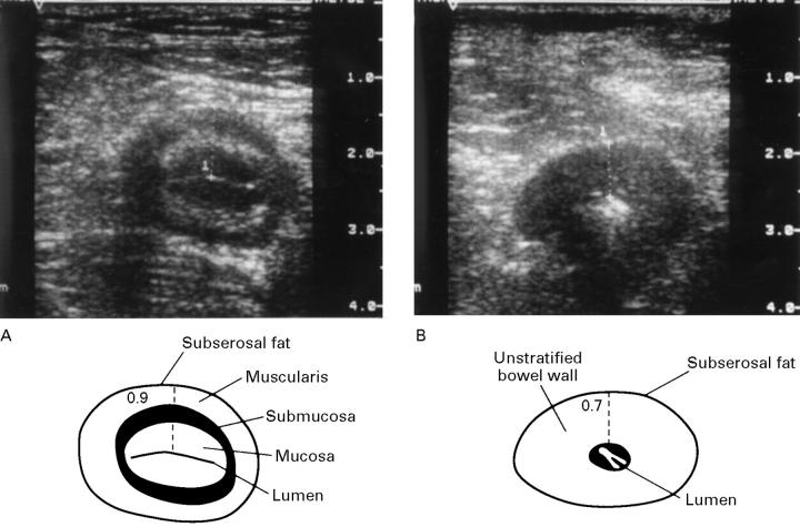
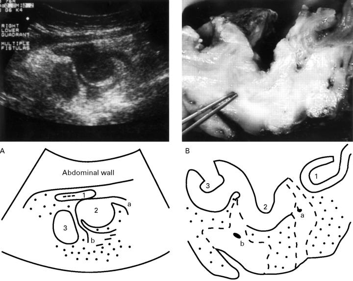
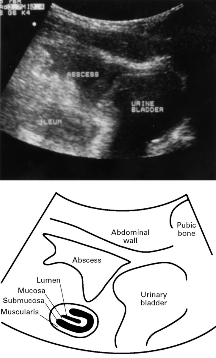
Results: T was able to identify strictures in 22/22 patients and to exclude it in 10/11 patients (100% sensitivity, 91% specificity). Fistulas were correctly identified in 20/23 patients and excluded in 9/10 patients (87% sensitivity, 90% specificity). Intra-abdominal abscesses were correctly detected in 9/9 patients and excluded in 22/24 patients (100% sensitivity, 92% specificity).

Conclusions: In experienced hands T is an accurate method for the detection of intestinal complications in Crohn's disease. T is thus recommended as a primary investigative method for evaluation of severe Crohn's disease.


Figures

Figure 1
Figure 1
Transverse TABS section (using a 10-5 MHz array) through the terminal ileum of two patients, five years (A) and eight years (B) after ileocaecal resection. Both patients show significant bowel wall thickening, 0.9 cm (A) and 0.7 cm (B). In (A), note the four layers of a typical target lesion and the hyperechoic, linear shaped collapsed lumen of the gut. No significant luminal narrowing and no other complications are present. In (B), note the loss of stratification of the bowel wall echo pattern reflecting destruction of layers after long term inflammation. The central hyperechoic, point shaped lumen corresponds to a stricture.
Figure 2
Figure 2
(A) Transverse TABS section (3.5 MHz array) through the right lower quadrant of a patient with Crohn's disease. Three adherent bowel loops of the terminal ileum (1, 2, 3, from oral to anal) are shown with hypoechoic peri-intestinal lesions (a, b) arising from loop 2 and hyperechoic wrapping mesentery (dotted area). Loop 1 is collapsed with only little thickening of the bowel wall (0.4 cm). Loops 2 and 3 show severe thickening of the bowel wall (1.2 cm and 1.0 cm), loss of bowel wall layering, and no detectable luminal hyperechoic content. These findings were regarded as high grade bowel obstruction of loops 2 and 3 with fistulas arising from loop 2. (B) Transverse section through the corresponding resection specimen after overnight fixation, longitudinal incision of the lumen, and unfolding of bowel loops (1, 2, 3, according to A) which resulted in rearrangement of loop 1. The forceps point into one of two fistulous tracts (a, b, according to A). Note perifistulous fibrotic tissue (broken lines) surrounded by fibrofatty proliferation of the mesentery (dotted area).
Figure 3
Figure 3
Sagittal TABS section through the mid lower abdomen of a patient with a two year history of uncomplicated Crohn's disease. A 4 cm hypoechoic peritoneal lesion (abscess) is present cranial to the urinary bladder. The adjacent ileum shows wall thickening (about 0.7 cm) with typical layering and a hyperechoic linear shaped central lumen without evidence of stricturing. The abscess originates from an ileal fistula (not visible on this section).

Comment in

References

    1. J Clin Gastroenterol. 1989 Apr;11(2):193-6 - PubMed
    1. J Clin Ultrasound. 1989 Jan;17(1):25-31 - PubMed
    1. Ann Surg. 1991 Jan;213(1):32-6 - PubMed
    1. Invest Radiol. 1990 Oct;25(10):1085-90 - PubMed
    1. Am J Gastroenterol. 1992 Apr;87(4):443-7 - PubMed

Publication types