

Lipopolysaccharide stimulates butyric acid-induced apoptosis in human peripheral blood mononuclear cells
- PMID: 9864191
- PMCID: PMC96272
- DOI: 10.1128/IAI.67.1.22-29.1999
Lipopolysaccharide stimulates butyric acid-induced apoptosis in human peripheral blood mononuclear cells
Abstract
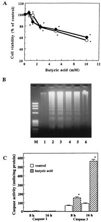
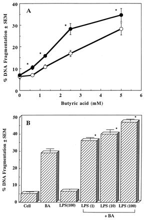
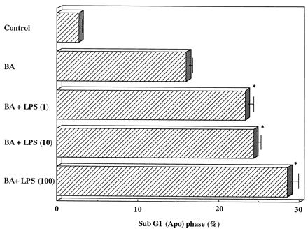
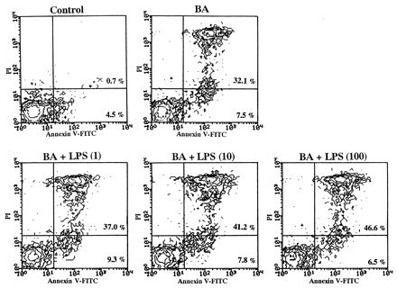
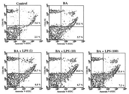
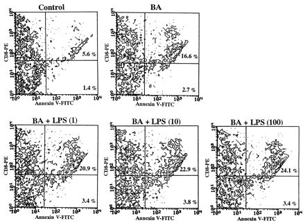
We previously reported that butyric acid, an extracellular metabolite from periodontopathic bacteria, induced apoptosis in murine thymocytes, splenic T cells, and human Jurkat T cells. In this study, we examined the ability of butyric acid to induce apoptosis in peripheral blood mononuclear cells (PBMC) and the effect of bacterial lipopolysaccharide (LPS) on this apoptosis. Butyric acid significantly inhibited the anti-CD3 monoclonal antibody- and concanavalin A-induced proliferative responses in a dose-dependent fashion. This inhibition of PBMC growth by butyric acid depended on apoptosis in vitro. It was characterized by internucleosomal DNA digestion and revealed by gel electrophoresis followed by a colorimetric DNA fragmentation assay to occur in a concentration-dependent fashion. Butyric acid-induced PBMC apoptosis was accompanied by caspase-3 protease activity but not by caspase-1 protease activity. LPS potentiated butyric acid-induced PBMC apoptosis in a dose-dependent manner. Flow-cytometric analysis revealed that LPS increased the proportion of sub-G1 cells and the number of late-stage apoptotic cells induced by butyric acid. Annexin V binding experiments with fractionated subpopulations of PBMC in flow cytometory revealed that LPS accelerated the butyric acid-induced CD3(+)-T-cell apoptosis followed by similar levels of both CD4(+)- and CD8(+)-T-cell apoptosis. The addition of LPS to PBMC cultures did not cause DNA fragmentation, suggesting that LPS was unable to induce PBMC apoptosis directly. These data suggest that LPS, in combination with butyric acid, potentiates CD3(+) PBMC T-cell apoptosis and plays a role in the apoptotic depletion of CD4(+) and CD8(+) cells.
Figures
Similar articles
-
Butyric acid-induced T-cell apoptosis is mediated by caspase-8 and -9 activation in a Fas-independent manner.Clin Diagn Lab Immunol. 2001 Mar;8(2):325-32. doi: 10.1128/CDLI.8.2.325-332.2001. Clin Diagn Lab Immunol. 2001. PMID: 11238216 Free PMC article.
-
Butyric-acid-induced apoptosis in murine thymocytes and splenic T- and B-cells occurs in the absence of p53.J Dent Res. 2000 Dec;79(12):1948-54. doi: 10.1177/00220345000790120501. J Dent Res. 2000. PMID: 11201044
-
Butyric acid-induced apoptosis of murine thymocytes, splenic T cells, and human Jurkat T cells.Infect Immun. 1997 Jan;65(1):35-41. doi: 10.1128/iai.65.1.35-41.1997. Infect Immun. 1997. PMID: 8975889 Free PMC article.
-
Rough-Form Lipopolysaccharide Increases Apoptosis in Human CD4⁺ and CD8⁺ T Lymphocytes.Scand J Immunol. 2012 Feb;75(2):193-202. doi: 10.1111/j.1365-3083.2011.02613.x. Scand J Immunol. 2012. PMID: 21854408
-
Spontaneous apoptosis of CD8+ T lymphocytes in peripheral blood of patients with advanced melanoma.Clin Cancer Res. 2000 Apr;6(4):1351-64. Clin Cancer Res. 2000. PMID: 10778963
Cited by
-
Butyric acid retention in gingival tissue induces oxidative stress in jugular blood mitochondria.Cell Stress Chaperones. 2013 Sep;18(5):661-5. doi: 10.1007/s12192-013-0409-z. Epub 2013 Feb 10. Cell Stress Chaperones. 2013. PMID: 23397230 Free PMC article.
-
Growth inhibition and apoptosis induction by (+)-Cyanidan-3-ol in hepatocellular carcinoma.PLoS One. 2013 Jul 24;8(7):e68710. doi: 10.1371/journal.pone.0068710. Print 2013. PLoS One. 2013. PMID: 23894334 Free PMC article.
-
A Novel Non-Digestible, Carrot-Derived Polysaccharide (cRG-I) Selectively Modulates the Human Gut Microbiota while Promoting Gut Barrier Integrity: An Integrated in Vitro Approach.Nutrients. 2020 Jun 29;12(7):1917. doi: 10.3390/nu12071917. Nutrients. 2020. PMID: 32610452 Free PMC article.
-
Butyric acid-induced T-cell apoptosis is mediated by caspase-8 and -9 activation in a Fas-independent manner.Clin Diagn Lab Immunol. 2001 Mar;8(2):325-32. doi: 10.1128/CDLI.8.2.325-332.2001. Clin Diagn Lab Immunol. 2001. PMID: 11238216 Free PMC article.
-
Consecutive intra-gingival injections of lipopolysaccharide and butyric acid to mice induce abnormal behavior and changes in cytokine concentrations.J Neuroinflammation. 2020 Nov 5;17(1):331. doi: 10.1186/s12974-020-02008-8. J Neuroinflammation. 2020. PMID: 33153485 Free PMC article.
References
-
- Alnemri E S, Livingston D J, Nicholson D W, Salvesen G, Thornberry N A, Wong W W, Yuan J. Human ICE/CED-3 protease nomenclature. Cell. 1996;87:171. - PubMed
-
- Eftimiadi C, Tonetti M, Cavallero A, Sacco O, Rossi G A. Short chain fatty acid produced by anaerobic bacteria inhibit phagocytosis by human lung phagocytes. J Infect Dis. 1990;161:138–142. - PubMed
-
- Egidy G, Friedman J, Viswanathan M, Wahl L M, Saavedra J M. CGP-42112 partially activates human monocytes and reduces their stimulation by lipopolysaccharides. Am J Physiol. 1997;273:C826–C833. - PubMed
-
- Eissner G, Kohlhuber F, Grell M, Ueffing M, Scheurich P, Hieke A, Multhoff G, Bornkamm G W, Holler E. Critical involvement of transmembrane tumor necrosis factor-α in endothelial programmed cell death mediated by ionizing radiation and bacterial endotoxin. Blood. 1995;86:4184–4193. - PubMed
Publication types
MeSH terms
Substances
LinkOut - more resources
Full Text Sources
Research Materials
