. 1999 Jan;67(1):94-101.
doi: 10.1128/IAI.67.1.94-101.1999.

Effect of melanin and carotenoids of Exophiala (Wangiella) dermatitidis on phagocytosis, oxidative burst, and killing by human neutrophils

Affiliations

Effect of melanin and carotenoids of Exophiala (Wangiella) dermatitidis on phagocytosis, oxidative burst, and killing by human neutrophils

N Schnitzler et al. Infect Immun. 1999 Jan.

Abstract

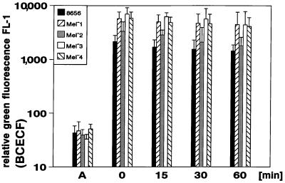
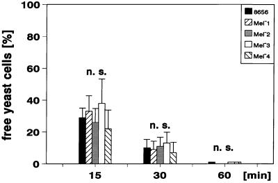
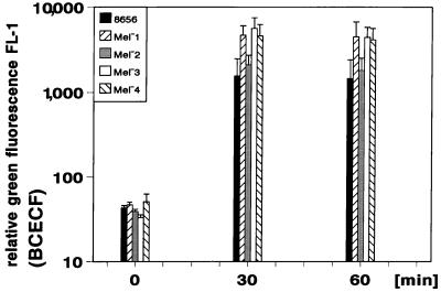
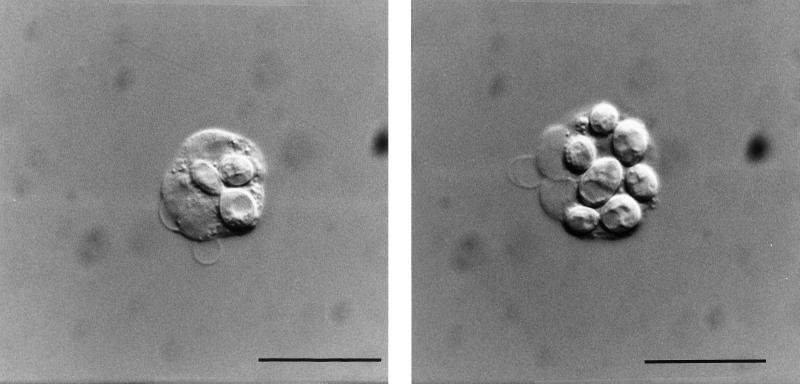
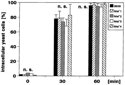
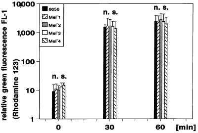
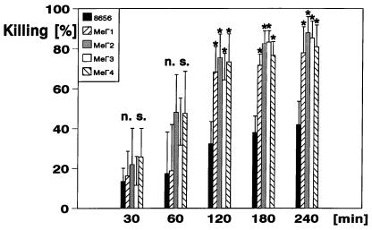
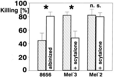
The black yeast Exophiala (Wangiella) dermatitidis is an increasingly recognized pathogen and a leading cause of severe pheohyphomycosis. Melanin is thought to contribute to the virulence of E. dermatitidis. Whereas the synthesis and the redox properties of melanin have been studied intensively, the influence of melanin and carotenoids on the phagocytosis, the oxidative burst, and the killing of E. dermatitidis by human neutrophils has not been studied. To study their effects on these phenomena, we applied a combination of flow cytometry and a colony-count-dependent method. Using E. dermatitidis wild-type strain 8565 and several melanin-deficient mutants that have been described previously, we demonstrate that melanin prevents this pathogen from being killed in the phagolysosome of the neutrophils. Melanin did not influence the phagocytosis or the oxidative burst of the neutrophils involved. The carotenoids torulene and torularhodine were not found to contribute to the prevention of killing. The ability of E. dermatitidis to block the effects of the neutrophil oxidative burst may critically impair the potential of the host to sufficiently eliminate this fungal pathogen and thus may play an important role in the pathogenesis of phaeohyphomycosis.


Figures

FIG. 1
FIG. 1
Staining of E. dermatitidis with BCECF/AM as determined by flow cytometry. Relative green fluorescence results of six independent assays are shown prior to the staining procedure (A), immediately after the staining (0 min), and at 15, 30, and 60 min after the end of coincubation with the dye (mean values ± SD are shown).
FIG. 2
FIG. 2
Decrease of free E. dermatitidis cells during incubation in heparinized blood as determined by flow cytometry. The portion of free yeast cells compared with the inoculum added to the heparinized blood is shown at the beginning (0 min) and after 15, 30, and 60 min of the incubation of the yeast cells in heparinized blood (mean values ± SD are shown: n = 6; n.s., difference not significant).
FIG. 3
FIG. 3
Kinetics of phagocytosis of E. dermatitidis as determined by flow cytometry. The subsequent increase of relative green fluorescence of neutrophils after association with the respective BCECF-labeled E. dermatitidis strains is shown. The mean values ± SD of six independent assays are displayed.
FIG. 4
FIG. 4
Intracellular location of the BCECF/AM-stained E. dermatitidis 8656 as determined by interference contrast microscopy. After 60 min of incubation in heparinized blood, the yeast cells are located within the neutrophils (Bar = 10 μm). (Left panel) Neutrophil with two yeast cells ingested, one of them budding. (Right panel) Neutrophil with maximum number of yeast cells ingested. These microorganisms could be also identified as yeast cells due to their green fluorescence in simultaneously performed epifluorescence microscopy studies.
FIG. 5
FIG. 5
Kinetics of the ingestion of E. dermatitidis as determined by epifluorescence interference contrast microscopy. The portion of intracellularly located yeast cells of 300 yeast cells judged for their association with the neutrophils is shown. The data represent mean values ± SD of three independent counts (n.s., difference not significant).
FIG. 6
FIG. 6
Kinetics of oxidative burst evoked by phagocytized E. dermatitidis as determined by flow cytometry. The results of studies of relative green fluorescence of neutrophils exhibiting an oxidative burst after the phagocytosis of nonlabeled yeast cells in the presence of DHR (n = 6; mean values ± SD; n.s., difference not significant) are shown.
FIG. 7
FIG. 7
Percent killing of E. dermatitidis as determined by colony counts prior to and after incubation in heparinized blood. The killing is displayed as a percentage of the decrease in the number of CFU during the incubation times indicated (n = 6; mean values ± SD) ∗, Significantly different (P < 0.005); n.s., not significantly different from strain 8656 as determined by the Mann-Whitney U test.
FIG. 8
FIG. 8
Effect of melanin on the killing of strains 8656, Mel3, and Mel2. Strain 8656 is compared with its albinized cells, and Mel−3 is compared with its melanized cells after scytalone cross-feeding. Mel2 cells that are not able to produce melanin from scytalone are also shown as controls. The mean values ± SD are shown; n = 6, ∗, Significantly different according to the Mann-Whitney U test (P < 0.005); n.s., differences not significant.

References

    1. Ajanee N, Alam M, Holmberg K, Kahn J. Brain abscess caused by Wangiella dermatitidis: case report. Clin Infect Dis. 1996;23:197. - PubMed
    1. Benett I, Knouse C, Johnson J, Meyer R L, Millard M W. Wangiella dermatitidis from a cystic fibrosis patient. Clin Microbiol Newsl. 1997;19:164–166.
    1. Bhakdi S, Greulich S, Muhly M, Eberspächer B, Becker H, Tiehle A, Hugo F. Potent leukocidal action of Escherichia coli hemolysin mediated by permeabilization of target cell membranes. J Exp Med. 1989;169:838–854. - PMC - PubMed
    1. Blaschke-Hellmessen, R., I. Lauterbach, K. D. Paul, K. Tintelnot, and G. Weissbach. 1994. Detection of Exophiala dermatitidis (Kano) De Hoog 1977 in septicemia of a child with acute lymphatic leukemia and in patients with cystic fibrosis. Mycoses 37(Suppl. 1):89–96. - PubMed
    1. Bruning J W, Kardol M J, Arentzen R. Carboxyfluorescein fluorochromasia assays. I. Non-radioactively labeled cell mediated lympholysis. J Immunol Methods. 1980;33:33–44. - PubMed

MeSH terms

LinkOut - more resources