. 1999 Jan;67(1):206-12.
doi: 10.1128/IAI.67.1.206-212.1999.

Gamma interferon augments macrophage activation by lipopolysaccharide by two distinct mechanisms, at the signal transduction level and via an autocrine mechanism involving tumor necrosis factor alpha and interleukin-1

Affiliations

Gamma interferon augments macrophage activation by lipopolysaccharide by two distinct mechanisms, at the signal transduction level and via an autocrine mechanism involving tumor necrosis factor alpha and interleukin-1

T K Held et al. Infect Immun. 1999 Jan.

Abstract

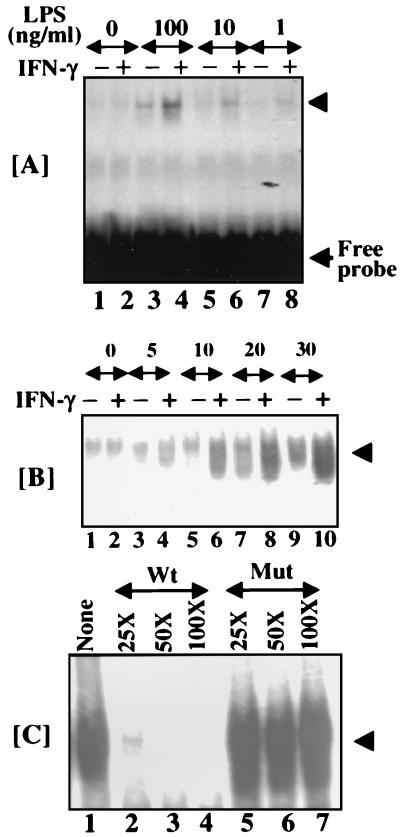
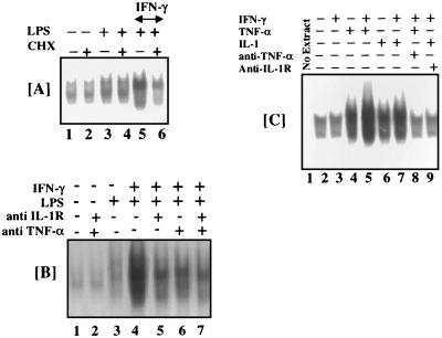
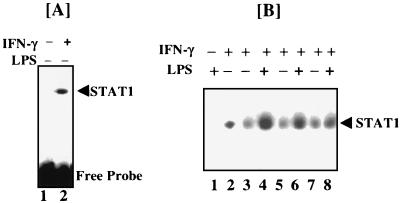
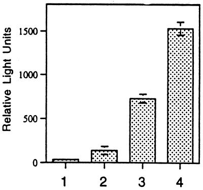
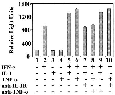
When given in the presence of gamma interferon (IFN-gamma), otherwise nontoxic doses of lipopolysaccharide (LPS or endotoxin) become highly lethal for mice. The mechanisms of this synergistic toxicity are not known. We considered the possibility that an interaction between the LPS-induced NF-kappaB and IFN-gamma-induced JAK-STAT pathways at the pretranscriptional level may enhance the LPS-induced signals. To test this hypothesis, we incubated murine macrophage RAW 264.7 cells with IFN-gamma for 2 h before addition of different doses of LPS. Consistent with the synergistic induction of inducible nitric oxide synthase mRNA and nitric oxide production by a combination of LPS and IFN-gamma, IFN-gamma strongly augmented LPS-induced NF-kappaB activation and accelerated the binding of NF-kappaB to DNA to as early as 5 min. In agreement with this, IFN-gamma pretreatment promoted rapid degradation of IkappaB-alpha but not that of IkappaB-beta. Inhibition of protein synthesis during IFN-gamma treatment suppressed LPS-initiated NF-kappaB binding. A rapidly induced protein appeared to be involved in IFN-gamma priming. Preincubation of cells with antibodies to tumor necrosis factor alpha or the interleukin-1 receptor partially reduced the priming effect of IFN-gamma. In a complementary manner, LPS enhanced the activation of signal-transducing activator of transcription 1 by IFN-gamma. These data suggest novel mechanisms for the synergy between IFN-gamma and LPS by which they cross-regulate the signal-transducing molecules. Through this mechanism, IFN-gamma may transform a given dose of LPS into a lethal stimulus capable of causing sepsis. It may also serve a beneficial purpose by enabling the host to respond quickly to relatively low doses of LPS and thereby activating antibacterial defenses.


Figures

FIG. 1
FIG. 1
(A) Pretreatment with IFN-γ enhances the DNA binding activity of NF-κB in response to LPS. RAW 264.7 cells were preincubated with IFN-γ (50 U/ml) for 2 h and then stimulated with the indicated amounts of LPS for 30 min. Nuclear extracts (20 μg) were then analyzed by EMSA using a 32P-labeled oligonucleotide specific for an NF-κB site of the murine iNOS promoter. (B) Kinetics of activation of NF-κB by LPS. Cells were incubated with IFN-γ (50 U/ml) for 2 h before addition of LPS (100 ng/ml; indicated by the horizontal arrows; the values above the arrows indicate the lengths of LPS treatment in minutes). After additional incubation with LPS for the indicated times, nuclear extracts were prepared and analyzed by EMSA using a 32P-labeled oligonucleotide based on a functional κB site of the murine iNOS promoter. Only the region corresponding to NF-κB binding is shown. The arrowheads indicate specific NF-κB complexes. (C) Specific DNA binding of NF-κB. All of the extracts are from cells stimulated with IFN-γ (50 U/ml) for 2 h and with LPS (100 ng/ml) for 20 min (similar to panel B, lane 8). Extracts in lane 1 were not pretreated with an unlabeled oligonucleotide before analysis with a 32P-labeled oligonucleotide. Nuclear extracts were preincubated with 25-, 50-, and 100-fold molar excesses of unlabeled oligonucleotides. The wild-type (Wt; lanes 2 to 4) and mutant (Mut; lanes 5 to 7) oligonucleotides used are described in Materials and Methods.
FIG. 2
FIG. 2
(A) Pretreatment with IFN-γ enhances the dose-dependent degradation of IκB-α in response to LPS. RAW cells were treated as described in the legend to Fig. 1, and cytosolic extracts were prepared in the presence of phenylmethylsulfonyl fluoride. They were analyzed for the presence of IκB-α and IκB-β by Western blotting as described in Materials and Methods. (B) Kinetics of IκB-α degradation in RAW cells induced by LPS with or without priming by IFN-γ. Cells were treated as described in the legend to Fig. 1. Cytosolic extracts were prepared at the indicated times after addition of LPS and analyzed as described above. The values above the arrows in panel B indicate the times (minutes) of LPS exposure. A minus or plus sign indicates the absence or presence, respectively, of the given agent during incubation.
FIG. 3
FIG. 3
(A) Effect of CHX on induction of the DNA binding of NF-κB by LPS with or without pretreatment with IFN-γ. RAW 264.7 cells were primed with IFN-γ (50 U/ml) for 30 min in the presence or absence of CHX (25 μg/ml) (lanes 4 and 5). LPS (100 ng/ml) was then added, and the mixture was incubated for 20 min. Nuclear extracts were prepared and analyzed by EMSA as described in the legend to Fig. 1. (B) Effect of antibodies against TNF-α or IL-1R on the induction of DNA binding of NF-κB by LPS with or without priming by IFN-γ. An anti-TNF-α antibody (2.5 μg/ml) and/or an anti-IL-1R antibody (2.5 μg/ml) were added to the culture 30 min before priming with IFN-γ (50 U/ml) for 60 min. LPS (100 ng/ml) was then added for an additional 20 min before the preparation of nuclear extracts. Extracts were then analyzed by EMSA as described in the legend to Fig. 1. (C) Synergistic activation of NF-κB by TNF-α or IL-1 in association with IFN-γ (50 U/ml). Cells were treated with the indicated agents as described above. Cells were stimulated with recombinant IL-1 (25 ng/ml) and TNF-α (10 ng/ml) for 20 min in this experiment. In all cases, only the relevant region of the autoradiogram is shown.
FIG. 4
FIG. 4
Effect of pretreatment with IFN-γ on the induction of DNA binding of STAT1 by LPS. (A) Pattern of activated STAT1 binding to the pIRE by IFN-γ. (B) Where indicated, cells were treated with IFN-γ (50 U/ml) for 2 h prior to LPS treatment for 30 min. The LPS concentrations were 100 (lanes 1 and 4), 10 (lane 6), and 1 (lane 8) ng/ml. Nuclear extracts were analyzed by EMSA using a 32P-labeled pIRE probe for STAT1 binding. Only the relevant region of the blot is shown.
FIG. 5
FIG. 5
Synergistic induction of IFN-γ-responsive gene promoter. RAW cells were transfected with a luciferase reporter gene driven by the pIRE. Cell extracts were prepared and assayed for luciferase activity as described earlier (19). The data are means ± the standard errors of the means of triplicate measurements. Bars: 1, no treatment; 2, LPS (100 ng/ml); 3, IFN-γ (50 U/ml); 4, IFN-γ plus LPS.
FIG. 6
FIG. 6
Synergistic induction of luciferase by TNF-α or IL-1 in association with IFN-γ. RAW cells were transfected with a luciferase reporter gene driven by the pIRE. Cell extracts were prepared and assayed for luciferase activity as described earlier (19). The data are means ± the standard errors of the means of triplicate measurements. The various treatments applied (for 16 h) are indicated at the bottom. A minus or plus sign indicates the absence or presence, respectively, of the given agent during incubation. An anti-TNF-α antibody (2.5 μg/ml) and/or an anti-IL-1R antibody (2.5 μg/ml) were added to the culture 30 min prior to treatment with the indicated cytokines. IFN-γ (50 U/ml), IL-1 (25 ng/ml), and TNF-α (10 ng/ml) were used in the experiment.

References

    1. Ausubel F M, Brent R, Kingston D D, Moore J G, Seidman J G, Smith J A, Struhl K. Current protocols in molecular biology. New York, N.Y: Wiley-Interscience; 1994.
    1. Baeuerle P A, Henkel T. Function and activation of NF-kappa B in the immune system. Annu Rev Immunol. 1994;12:141–179. - PubMed
    1. Bonizzi G, Dejardin E, Piret B, Piette J, Merville M P, Bours V. Interleukin-1 beta induces nuclear factor kappa B in epithelial cells independently of the production of reactive oxygen intermediates. Eur J Biochem. 1996;242:544–549. - PubMed
    1. Cannon J G, Tompkins R G, Gelfand J A, Michie H R, Stanford G G, van der Meer J W, Endres S, Lonnemann G, Corsetti J, Chernow B, et al. Circulating interleukin-1 and tumor necrosis factor in septic shock and experimental endotoxin fever. J Infect Dis. 1990;161:79–84. - PubMed
    1. Crofford L J, Tan B, McCarthy C J, Hla T. Involvement of nuclear factor kappa B in the regulation of cyclooxygenase-2 expression by interleukin-1 in rheumatoid synoviocytes. Arthritis Rheum. 1997;40:226–236. - PubMed

Publication types

MeSH terms

LinkOut - more resources