. 1999 Jan;67(1):237-43.
doi: 10.1128/IAI.67.1.237-243.1999.

Compensation for decreased expression of B7 molecules on Leishmania infantum-infected canine macrophages results in restoration of parasite-specific T-cell proliferation and gamma interferon production

Affiliations
Free PMC article

Compensation for decreased expression of B7 molecules on Leishmania infantum-infected canine macrophages results in restoration of parasite-specific T-cell proliferation and gamma interferon production

E Pinelli et al. Infect Immun. 1999 Jan.
Free PMC article

Abstract

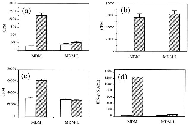
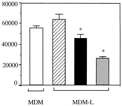
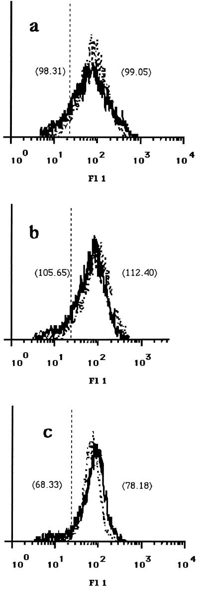
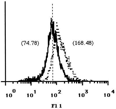
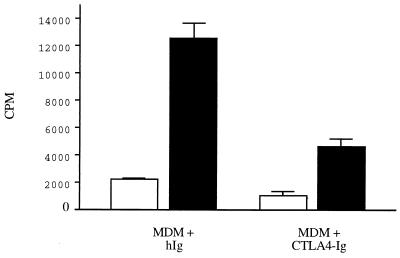
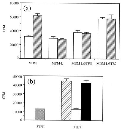
Infection of humans and dogs by Leishmania infantum may result in visceral leishmaniasis, which is characterized by impaired T-cell-mediated immune responses to parasite antigens. Dogs are natural hosts of Leishmania parasites and play an important role in the transmission of the parasites to humans. In an effort to characterize the immune response in dogs infected with this intracellular pathogen, we examined how infection with L. infantum affects canine macrophages and the consequences for T-cell activation in vitro. We showed that the proliferation of T-cell lines to cognate antigen decreases to background levels when infected autologous monocyte-derived macrophages are used as antigen-presenting cells (APC). The observed reduction of antigen-specific T-cell proliferation was shown to be dependent on the parasite load and to require cell-to-cell interaction of T cells with the infected APC. In addition, we observed a decreased expression of costimulatory B7 molecules on infected monocyte-derived macrophages. The expression of other surface molecules involved in T-cell activation, such as major histocompatibility complex class I and class II, on these cells did not change upon infection, whereas the expression of intracellular adhesion molecule 1 was marginally increased. Compensation for the decreased expression of B7 molecules by the addition of B7-transfected cells resulted in the restoration of cell proliferation and gamma interferon (IFN-gamma) production by a Leishmania-specific T-cell line. These results showed that for the activation of parasite-specific canine T cells producing IFN-gamma, which are most likely involved in protective immunity, sufficient expression of B7 molecules on infected macrophages is required. Provision of costimulatory molecules may be an approach for immunotherapy of leishmaniaisis as well as for vaccine development.


Figures

FIG. 1
FIG. 1
T-cell proliferation against cognate antigen is reduced when L. infantum-infected MDM are used as APC. Uninfected MDM or L. infantum-infected MDM (MDM-L) were used as APC. Cells were incubated in medium only (□) or in the presence of cognate antigen (▨). (a and b) A Dohyvac-specific T-cell line was stimulated with Dohyvac (a) or ConA (b). (c and d) A Leishmania-specific T-cell line was stimulated with LSA (c), and after 72 h supernatants were collected for determination of IFN-γ activity (d). Results are expressed as mean ± SD. Data are representative of three independent experiments. SU, standard units.
FIG. 2
FIG. 2
Effect of the parasite load of MDM on the proliferation of a Leishmania-specific T-cell line against LSA. Autologous uninfected MDM (□) or MDM infected at a 2:1 (▨), 10:1 (■), or 50:1 (▨) ratio were used as APC to stimulate a Leishmania-specific T-cell line. Background proliferation of T-cell lines incubated with APC and medium only ranged from 15,000 to 24,000 cpm. Results are expressed as mean ± SD counts per minute. ∗, significant difference from Leishmania-specific T cells stimulated with uninfected cells. Data are representative of three independent experiments. MDM-L, L. infantum-infected MDM.
FIG. 3
FIG. 3
Expression of MHC class I and class II and ICAM-1 on canine MDM. Monoclonal antibodies B1.1.G6 (a) directed against MHC class I, 7.5.10.1 (b) directed against MHC class II, and CL18.1D8 (c) directed against ICAM-1 were used to analyze the expression of these molecules on uninfected MDM (dotted line) and L. infantum-infected MDM (solid line) 24 h after infection. Scores of mean fluorescence intensity (MFI) are given in parentheses. MDM were infected at a 50:1 parasite-to-MDM ratio. Incubation of cells with an irrelevant mouse isotype control antibody resulted in an MFI of 13.70 (vertical broken line). The data shown are representative of four independent experiments. Fl 1, fluorescence.
FIG. 4
FIG. 4
Expression of B7 molecules on canine MDM. The expression of B7 molecules on uninfected MDM (dotted line) and L. infantum-infected MDM (solid line) was analyzed with fusion protein CTLA4-Ig. Scores of mean fluorescence intensity (MFI) are given in parentheses. As a control, human Ig was used and resulted in an MFI of 65.52 (vertical broken line). MDM were infected at a 50:1 parasite-to-MDM ratio. The data shown are representative of four independent experiments. Fl 1, fluorescence.
FIG. 5
FIG. 5
T-cell proliferation against cognate antigen is dependent on interactions with B7 molecules. A Leishmania-specific T-cell line was restimulated with MDM as APC in the presence of LSA (■) or medium only (□). Cells were incubated in the presence of 10 μg of human IgG per ml used as a control antibody (hIg) or CTLA4-Ig. The data shown are representative of two independent experiments and are expressed as mean ± SD.
FIG. 6
FIG. 6
Restoration of antigen-specific T-cell proliferation in the presence of infected APC is dependent on interactions with B7 molecules. (a) B7.1-transfected 3TB7 cells or untransfected 3TFII cells (1,000 cells/well) were added to a Leishmania-specific T-cell line incubated with L. infantum-infected MDM (MDM-L) in the presence of LSA (▨) or medium only (□). (b) A Leishmania-specific T-cell line was restimulated with LSA, infected MDM, and 3TFII cells (░⃞) or 3TB7 cells without a monoclonal antibody (▨), with a monoclonal antibody against human B7.1 molecules (□), or with a control monoclonal antibody (■). Background proliferation of T-cell lines incubated with infected MDM and medium only ranged from 13,055 to 16,062 cpm. Results are expressed as mean ± SD.

References

    1. Ashford R W, Bettini S. Ecology and epidemiology: Old World. In: Peters W, Killick-Kendrick R, editors. Leishmaniasis in biology and medicine. New York, N.Y: Academic Press, Inc.; 1987. pp. 365–424.
    1. Barral N M, Barral A, Brownell C E, Skeiky Y A, Ellingsworth L R, Twardzik D R, Reed S G. Transforming growth factor-beta in leishmanial infection: a parasite escape mechanism. Science. 1992;257:545–548. - PubMed
    1. Bretscher P. The two signal model of lymphocyte activation twenty-one years later. Immunol Today. 1992;13:74–76. - PubMed
    1. Cabral M, O’Grady J, Alexander J. Demonstration of Leishmaniaspecific cell mediated and humoral immunity in asymptomatic dogs. Parasite Immunol. 1992;14:531–539. - PubMed
    1. Carvalho E M, Teixeira R S, Johnson W D., Jr Cell-mediated immunity in American visceral leishmaniasis. Infect Immun. 1981;48:409–414. - PMC - PubMed

MeSH terms

Substances

LinkOut - more resources