. 1999 Jan;181(1):167-76.
doi: 10.1128/JB.181.1.167-176.1999.

Recruitment of ZipA to the septal ring of Escherichia coli is dependent on FtsZ and independent of FtsA

Affiliations

Recruitment of ZipA to the septal ring of Escherichia coli is dependent on FtsZ and independent of FtsA

C A Hale et al. J Bacteriol. 1999 Jan.

Abstract

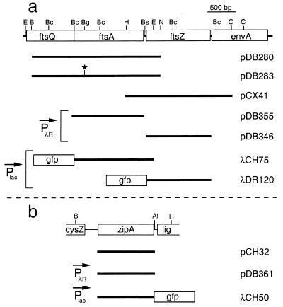
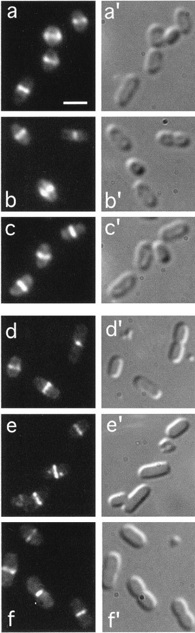
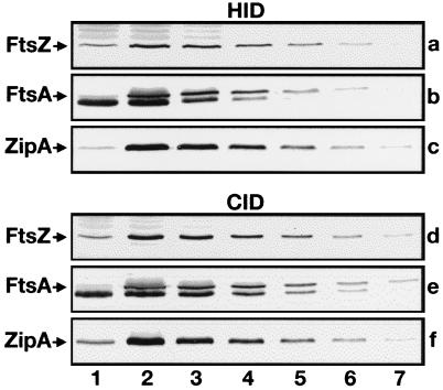
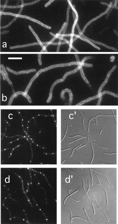
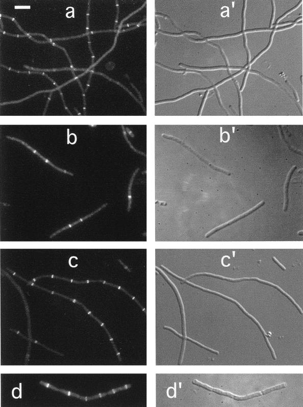
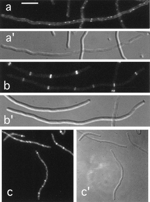
Cell division in prokaryotes is mediated by the septal ring. In Escherichia coli, this organelle consists of several essential division proteins, including FtsZ, FtsA, and ZipA. To gain more insight into how the structure is assembled, we studied the interdependence of FtsZ, FtsA, and ZipA localization using both immunofluorescence and Gfp tagging techniques. To this end, we constructed a set of strains allowing us to determine the cellular location of each of these three proteins in cells from which one of the other two had been specifically depleted. Our results show that ZipA fails to accumulate in a ring shape in the absence of FtsZ. Conversely, depletion of ZipA does not abolish formation of FtsZ rings but leads to a significant reduction in the number of rings per unit of cell mass. In addition, ZipA does not appear to require FtsA for assembly into the septal ring and vice versa. It is suggested that septal ring formation starts by assembly of the FtsZ ring, after which ZipA and FtsA join this structure in a mutually independent fashion through direct interactions with the FtsZ protein.


Figures

FIG. 1
FIG. 1
Chromosomal inserts of plasmids and phages. The physical maps of ftsA and ftsZ (a) and of zipA (b) are shown at the top of the panel. The positions of AflII (Af), BamHI (B), BclI (Bc), BglII (Bg), Bsu36I (Bs), ClaI (C), EcoRI (E), HindIII (H), and NsiI (N) restriction sites are indicated. Inserts of plasmids and phages are presented below each map. As indicated, the transcription of inserts in some of the constructs was placed under control of the lac promoter (Plac), or PλR. Note that in addition to the zipA fragment shown, pCH32 carries the ftsZ gene immediately downstream of this fragment (17).
FIG. 2
FIG. 2
Localization of ZipA, FtsA, and FtsZ in wild-type cells. Fluorescence (a through f) and corresponding differential interference contrast (a′ through f′) micrographs showing the location in normally dividing cells of both native (a to c) and Gfp tagged (d to f) ZipA, FtsA, and FtsZ proteins, respectively. Cells shown are from strains PB103 (wild type [wt]) (a to c), CH3(λCH50)/pDB355 [wt(Plac::zipA-gfp)/cI857 PλR::ftsA+] (d), PB103(λCH75) [wt(Plac::gfp-ftsA)] (e), and CH3(λDR120)/pDB361 [wt(Plac::gfp-ftsZ)/cI857 PλR::zipA+] (f). Cells in panels a to c were grown at 37°C prior to immunofluorescence staining with the appropriate antibodies. Cells in panels d to f were grown at 30°C in the presence of 25 μM (d and e) or 50 μM (f) IPTG, and were observed immediately after chemical fixation. Results with a number of other normally dividing lysogens [e.g., PB103(λCH50), CH3(λCH75), and PB103(λDR120)] were similar to those shown in panels d to f. Bar in panel a represents 2.0 μm.
FIG. 3
FIG. 3
Depletion of division proteins. Immunoblots showing cellular levels of FtsZ (a and d), FtsA (b and e), or ZipA (c and f) after growth of the corresponding HID, CID, and wild-type control strains at either 42°C (a to c) or 30°C (d to f). Lanes 1 contained 70 μg of total protein of depletion strains PB143/pCX41 (a), CH2/pDB280 (b), CH5/pCH32 (c), PB143/pDB346 (d), CH2/pDB355 (e), and CH5/pDB361 (f). Lanes 2 through 7 contained 70, 35, 18, 9, 4, and 2 μg of protein, respectively, of control strains CH3/pMAK700 (a to c), CH3/pDB346 (d), CH3/pDB355 (e), and CH3/pDB361 (f). Proteins were detected with polyclonal antisera. The anti-FtsA serum showed some reactivity to an abundant 40-kDa species (most likely EF-Tu) which migrated slightly faster than FtsA (b and e). Measurements of band intensities indicated that, in each case, the cellular concentration of the depleted division protein was less than 15% of its normal concentration.
FIG. 4
FIG. 4
Localization of ZipA in FtsZ and FtsA filaments. Fluorescence (a to d) and differential interference contrast (c′ to d′) micrographs showing the location of ZipA-Gfp (a and c) or native ZipA (b and d) in filaments depleted for FtsZ (a and b) or FtsA (c and d). Cells shown are from the FtsZCID strain PB143(λCH50)/pDB346 [ftsZ0(Plac::zipA-gfp)/cI857 PλR::ftsZ+] (a), the FtsZHID strain PB143/pCX41 [ftsZ0/repA(Ts) ftsZ+] (b), the FtsACID strain CH2(λCH50)/pDB355 [ftsA0(Plac::zipA-gfp)/cI857 PλR::ftsA+] (c), and the FtsAHID strain CH2/pDB280 [ftsA0/repA(Ts) ftsA+] (d). Cells in panels b and d were grown at 42°C prior to staining with affinity-purified anti-ZipA antiserum. Cells in panels a and c were grown at 30°C in the presence of 25 μM IPTG and were observed immediately after chemical fixation. Bar represents 5.0 (a and b) or 11.7 (c and d) μm.
FIG. 5
FIG. 5
Localization of FtsZ in ZipA and FtsA filaments. Fluorescence (a through d) and differential interference contrast (a′ to d′) micrographs showing the location of Gfp-FtsZ (a and c) or native FtsZ (b and d) in filaments depleted for ZipA (a and b) or FtsA (c and d). Cells shown are from the ZipACID strain CH5(λDR120)/pDB361 [zipA::aph(Plac::gfp-ftsZ)/cI857 PλR::zipA+] (a), the ZipAHID strain CH5/pCH32 (zipA::aph/repAts ftsZ+ zipA+) (b), the FtsACID strain CH2(λDR120)/pDB355 [ftsA0(Plac::gfp-ftsZ)/cI857 PλR::ftsA+] (c), and the FtsAHID strain CH2/pDB280 [ftsA0/repA(Ts) ftsA+] (d). Cells in panels b and d were grown at 42°C prior to staining with FtsZ-specific monoclonal antibodies. Cells in panels a and c were grown at 30°C in the presence of 50 μM IPTG and were observed immediately after chemical fixation. Bar in panel a represents 5.0 μm.
FIG. 6
FIG. 6
Localization of FtsA in FtsZ and ZipA filaments. Fluorescence (a to c) and differential interference contrast (a′ to c′) micrographs showing the location of Gfp-FtsA (a and b) or native FtsA (c) in filaments depleted of FtsZ (a) or ZipA (b and c). Cells shown are from the FtsZCID strain PB143(λCH75)/pDB346 [ftsZ0(Plac::gfp-ftsA)/cI857 PλR::ftsZ+] (a), the ZipACID strain CH5(λCH75)/pDB361 [zipA::aph(Plac::gfp-ftsA)/cI857 PλR::zipA+] (b), and the ZipAHID strain CH5/pCH32 [zipA::aph/repA(Ts) ftsZ+ zipA+] (c). Cells in panel c were grown at 42°C prior to staining with affinity-purified anti-FtsA antiserum. Cells in panels a and b were grown at 30°C in the presence of 25 μM IPTG and were observed immediately after chemical fixation. Bar represents 5.0 (a and b) or 11.0 (c) μm.

Similar articles

Cited by

References

    1. Addinall S G, Bi E, Lutkenhaus J. FtsZ ring formation in fts mutants. J Bacteriol. 1996;178:3877–3884. - PMC - PubMed
    1. Addinall S G, Cao C, Lutkenhaus J. FtsN, a late recruit to the septum in Escherichia coli. Mol Microbiol. 1997;25:303–309. - PubMed
    1. Addinall S G, Lutkenhaus J. FtsA is localized to the septum in an FtsZ-dependent manner. J Bacteriol. 1996;178:7167–7172. - PMC - PubMed
    1. Bi E, Lutkenhaus J. FtsZ ring structure associated with division in Escherichia coli. Nature. 1991;354:161–164. - PubMed
    1. Bork P, Sander C, Valencia A. An ATPase domain common to prokaryotic cell cycle proteins, sugar kinases, actin, and hsp70 heat shock proteins. Proc Natl Acad Sci USA. 1992;89:7290–7294. - PMC - PubMed

Publication types

LinkOut - more resources