. 1999 Jan;181(1):212-7.
doi: 10.1128/JB.181.1.212-217.1999.

Identification and properties of the genes encoding microcin E492 and its immunity protein

Affiliations

Identification and properties of the genes encoding microcin E492 and its immunity protein

R Lagos et al. J Bacteriol. 1999 Jan.

Abstract

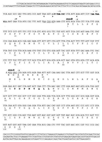
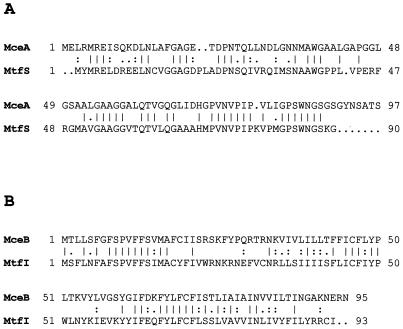
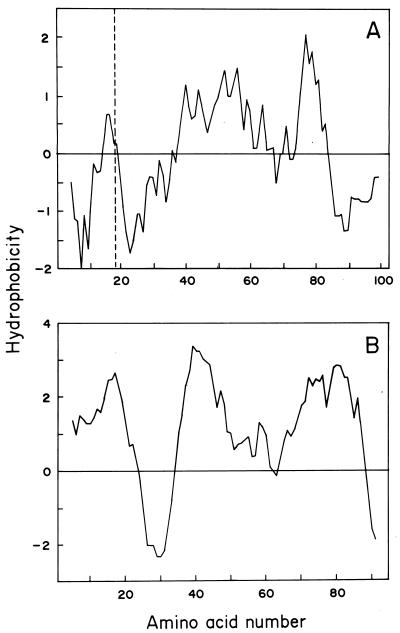
The gene coding for the immunity protein (mceB) and the structural gene of microcin E492 (mceA), a low-molecular-weight channel-forming bacteriocin produced by a strain of Klebsiella pneumoniae, have been characterized. The microcin gene codes for a precursor protein of either 99 or 103 amino acids. Protein sequencing of the N-terminal region of microcin E492 unequivocally identified this gene as the microcin structural gene and indicated that this microcin is synthesized as a precursor protein that is cleaved at either amino acid 15 or 19, at a site resembling the double-glycine motif. The gene encoding the 95-amino-acid immunity protein (mceB) was identified by cloning the DNA segment that encodes only this polypeptide into an expression vector and demonstrating the acquisition of immunity to microcin E492. As expected, the immunity protein was found to be associated with the inner membrane. Analysis of the DNA sequence indicates that these genes belong to the same family as microcin 24, and they do not share structural motifs with any other known channel-forming bacteriocin. The organization of the microcin- and immunity protein-encoding genes suggests that they are coordinately expressed.


Figures

FIG. 1
FIG. 1
Nucleotide sequence of the mceAB-containing region. The deduced amino acid sequences for the mceA (microcin E492) and mceB (immunity protein) genes from K. pneumoniae RYC492 are given below the DNA sequences. The putative transcriptional start is indicated by the curved arrow. Deduced −10 and −35 regions in DNA are in boldface, and the proposed consensus Shine-Dalgarno sequences are underlined. Two direct repeats are indicated between the arrowheads. The inferred cleavage site in MceA is represented by a vertical arrow between amino acids 19 and 20. The amino acids in boldface are those confirmed by N-terminal sequencing of the protein.
FIG. 2
FIG. 2
Comparison of the deduced amino acid sequences of nonprocessed microcin MceA with MtfS (A) and immunity protein MceB with MtfI (B). Identical matches (vertical lines) and strongly (colons) and weakly (dots) conserved amino acid residues are indicated. The comparison was performed with the GAP program. MceA and MtfS displayed an identity of 52% and a similarity of 59%, while MceB and MtfI presented an identity of 39% and a similarity of 56%.
FIG. 3
FIG. 3
Hydrophobicity profiles of MceA (A) and MceB (B). Hydropathy was calculated by the method of Kyte and Doolittle (20) with a span length of 13 amino acid residues and by using a linear weight variation model. The dashed line in panel A shows the inferred processing site for microcin E492.
FIG. 4
FIG. 4
Expression of the immunity protein from p157. (A) Expression from the T7 promoter of pT7-7 and p157 was assessed by using E. coli K-38pGP1-2 as the bacterial host. Induction of T7 RNA polymerase was performed by incubation at 42°C. Specific transcription from the T7 promoter was performed by addition of rifampin, and proteins were labeled with [35S]methionine. SDS–16% PAGE samples corresponding to E. coli K-38pGP1-2/pT7-7 are in lanes 1, 2, and 3, and those corresponding to E. coli K-38pGP1-2/p157 are in lanes 4, 5, and 6. Total (lanes 1 and 4), soluble (2 and 5), and particulate (3 and 6) fractions of the bacterial extracts were analyzed separately. The arrow indicates the band corresponding to the immunity protein. Molecular weight markers in lane 7 were myoglobin fragments with Mrs of 16,950, 14,440, 8,160, 6,210, and 3,460. (B) Autoradiography of the SDS-PAGE shown in panel A.
FIG. 5
FIG. 5
Localization of the immunity protein in the inner membrane. Induction of the T7 RNA polymerase of E. coli K-38pGP1-2 harboring pT7-7 or p157 was performed by incubation at 42°C for 5 h, and the separation of inner and outer membrane fractions was carried out as described in Materials and Methods. SDS–14% PAGE samples corresponding to E. coli K-38pGP1-2/pT7-7 are in lanes 1, 2, and 3, and those corresponding to E. coli K-38pGP1-2/p157 are in lanes 5, 6, and 7. Total (lanes 1 and 5), inner membrane (2 and 6), and outer membrane (3 and 7) fractions of the bacterial extracts were analyzed separately. The arrowhead indicates the band corresponding to the immunity protein. Molecular weight markers in lane 4 were myoglobin fragments with Mrs of 16,950, 14,440, 8,160, 6,210, and 3,460.

References

    1. Abee T. Pore-forming bacteriocins of Gram-positive bacteria and self-protection mechanisms of producer organisms. FEMS Microbiol Lett. 1995;129:1–10. - PubMed
    1. Ausubel F M, Brent R, Kingston R E, Moore D D, Seidman J G, Smith J A, Struhl K. Short protocols in molecular biology. 2nd ed. New York, N.Y: Greene Publishing Associates and John Wiley & Sons, Inc.; 1992.
    1. Baba T, Schneewind O. Instruments of microbial warfare: bacteriocin synthesis, toxicity and immunity. Trends Microbiol. 1998;6:66–71. - PubMed
    1. Baeza, M., C. Hetz, C. Villota, J. E. Villanueva, and R. Lagos. Unpublished data.
    1. Chou P Y, Fasman G D. Empirical predictions of protein conformation. Annu Rev Biochem. 1978;47:251–276. - PubMed

Publication types

MeSH terms

Associated data