Comparative Study
. 1998 Dec 28;143(7):1775-87.
doi: 10.1083/jcb.143.7.1775.

Fission yeast bub1 is a mitotic centromere protein essential for the spindle checkpoint and the preservation of correct ploidy through mitosis

Affiliations
Comparative Study

Fission yeast bub1 is a mitotic centromere protein essential for the spindle checkpoint and the preservation of correct ploidy through mitosis

P Bernard et al. J Cell Biol. .

Abstract

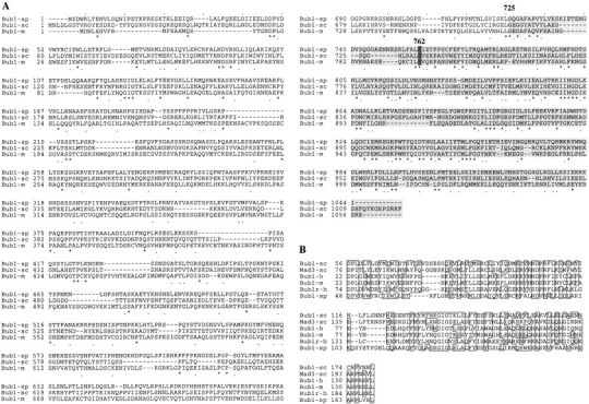
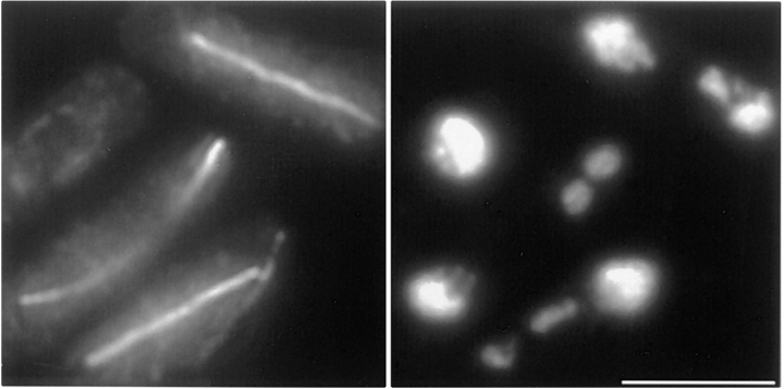
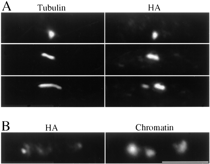
The spindle checkpoint ensures proper chromosome segregation by delaying anaphase until all chromosomes are correctly attached to the mitotic spindle. We investigated the role of the fission yeast bub1 gene in spindle checkpoint function and in unperturbed mitoses. We find that bub1(+) is essential for the fission yeast spindle checkpoint response to spindle damage and to defects in centromere function. Activation of the checkpoint results in the recruitment of Bub1 to centromeres and a delay in the completion of mitosis. We show that Bub1 also has a crucial role in normal, unperturbed mitoses. Loss of bub1 function causes chromosomes to lag on the anaphase spindle and an increased frequency of chromosome loss. Such genomic instability is even more dramatic in Deltabub1 diploids, leading to massive chromosome missegregation events and loss of the diploid state, demonstrating that bub1(+ )function is essential to maintain correct ploidy through mitosis. As in larger eukaryotes, Bub1 is recruited to kinetochores during the early stages of mitosis. However, unlike its vertebrate counterpart, a pool of Bub1 remains centromere-associated at metaphase and even until telophase. We discuss the possibility of a role for the Bub1 kinase after the metaphase-anaphase transition.


Figures

Figure 4
Figure 4
Cellular localization of Bub1 through the mitotic cell cycle. Cells from strain 411 (bub1-HA) were grown to early log phase in YES medium at 26°C, fixed, and then processed for immunofluorescence microscopy. (A) Montage of cells stained with rabbit polyclonal anti-HA (rHA in red), anti-tubulin (green), and DAPI (blue). For each cell, the corresponding panels are placed below each other. From left to right, an example is shown of cells at various stages of the cell cycle, from interphase (a) through prophase (b), prometaphase (c and d), metaphase (e), anaphase A (f), anaphase B (g), and cytokinesis (h).Yellow is produced upon colocalization of red (Bub1) and green (tubulin) signals. (B) Bub1 colocalizes with centromeres throughout mitosis. Montage of bub1-HA cells stained with mHA (red) and the digoxigenin-labeled centromere probe (green). Blue, DAPI staining. Left to right: interphase (a), prophase (b–d), prometaphase (e), metaphase (f), anaphase A (g), anaphase B (h and i), and cytokinesis (j). Bottom, merged image where yellow is produced upon colocalization of red (Bub1) and green (centromeres) signals. Bars, 5 μm.
Figure 1
Figure 1
Sequence comparison of Bub1 homologues. (A) Alignment of Bub1 protein sequences from S. pombe (sp), S. cerevisiae (sc), and mouse (m). Asterisks, identical residues; dots, similar residues; shaded area, the extent of the kinase domain. (B) Sequence alignment of the NH2-terminal domains of the Bub1 and Mad3 homologues from S. cerevisiae (sc), human (h), mouse (m), and S. pombe (sp). Boxes, identical or similar residues.
Figure 2
Figure 2
bub1 + is required for spindle checkpoint function. (A) bub1 mutants are hypersensitive to benomyl. Strains were grown in YES medium to 0.5–2 × 10⁷ cells/ml. About 200 cells were spotted onto YES plates containing the indicated concentrations of the anti-microtubule drug benomyl and incubated at 32°C for 3 d. (B–D) bub1 mutants are unable to maintain a mitotic arrest. Small G2 cells of nda3KM311 Δswi6 and nda3KM311 Δbub1 were shifted to 18°C, which disrupts spindle microtubules due to the nda3 defect, and samples were taken every hour. (B) Unlike the Δswi6 cells which arrest with high levels of histone H1 kinase activity (left), the Δbub1 cells fail to maintain such an arrest and the kinase activity drops after 3 h (right). DAPI and calcofluor staining shows that this is followed by a wave of septation (C) from 4–6 h as the Δbub1 cells (solid squares) progress through the cell cycle, and ultimately leads to a cut phenotype (D) as septation occurs without chromosome segregation. The Δswi6 cells maintain their arrest with hypercondensed chromosomes (data not shown) and do not septate (C, open circles). (E) Δbub1 cells are unable to prevent sister chromatid separation in the absence of a spindle. Cells from strain 379 (nda3KM311) and 401 (nda3KM311 Δbub1) were grown in YES medium at 32°C to 3 × 10⁶ cells/ml, shifted to 18°C for 8 h, and then fixed and processed for FISH using a probe detecting all three centromeres (cenFISH). Nuclear DNA was stained with DAPI. Bar, 5 μm.
Figure 3
Figure 3
bub1 + function is essential for high fidelity chromosome transmission through mitosis. (A) The diploid state can not be maintained in the absence of a functional bub1 + gene. Diploids were forced by intragenic complementation between the red (ade6-210) and pink (ade6-216) alleles of the ade6 locus. Ade+ cells were harvested from medium lacking adenine and plated onto YES 1/10 ade medium to allow the development of red and pink colors. The bub1 +/bub1 + (wild-type) diploid was stable, producing pure white colonies whereas the Δbub1/Δbub1 strain readily broke down to the haploid state, producing red, pink, and sectored colonies. (B) Chromosomes lag on the anaphase spindle in Δbub1 cells. Exponential growing cells in YES medium at 26°C were fixed and processed for tubulin (green) and DAPI (red, pseudocolor) staining. Right: In wild-type haploid and diploid anaphases, the chromatin masses are at the spindle poles. Left: Δbub1 haploid and diploid cells with chromosomes remaining in the midzone of the anaphase spindle. Bar, 10 μm.
Figure 5
Figure 5
Bub1 localizes at kinetochores in nda3KM311-arrested cells. Cells from strain 415 (nda3KM311 bub1-HA) were grown to early log phase at 32°C and shifted to 18°C for 8 h. Fixed cells were stained with mHA (red) and centromeric DNA was detected by FISH (green). Blue, DAPI staining. Bottom right, merged image. Two cells are shown. In some instances, Bub1 staining produces three doublets (the cell at the bottom). The merged image reveals that centromeric DNA is located precisely in between the paired dots, consistent with Bub1 being located on each sister kinetochore. Bar, 10 μm.
Figure 6
Figure 6
The synthetic lethality between Δbub1 and Δswi6 mutants is characterised by a high incidence of anaphases with lagging chromosomes. Strain 399 (nmtswi6 Δbub1) was grown to early log phase in minimal medium, then thiamine was added to repress transcription from the nmt promoter and cells were cultured for an additional 20 h to allow Swi6 to be depleted. Fixed cells were subjected to tubulin (left) and DAPI (right) staining. All the anaphase cells in the field showed chromosomes still remaining in the midzone of the spindle. Bar, 5 μm.
Figure 7
Figure 7
Bub1 localization in Δswi6 cells. Cells from strain 429 (Δswi6 bub1-HA) were grown to early log phase in YES medium at 26°C, fixed, and then stained with rHA and tubulin. (A) Top, cell in prophase; middle, cell in prometaphase; bottom, cell in metaphase. Bub1 staining in prophase and prometaphase is as in a wild-type background but the staining remains intense and punctate in about half of Δswi6 metaphase cells. (B) Bub1 appears diffuse on lagging chromosomes. An anaphase B cell is shown with a lagging chromosome lying in between the two separated chromatin masses (right). The same cell stained with mHA (left) shows a faint punctate signal leading the main chromatin masses. The lagging chromosome is diffusely stained with mHA and does not show any punctate staining.

References

    1. Allshire RC. Centromeres, checkpoints and chromatid cohesion. Curr Opin Genet Dev. 1997;7:264–273. - PubMed
    1. Allshire RC, Nimmo ER, Ekwall K, Javerzat J-P, Granston G. Mutations derepressing silent centromeric domains in fission yeast disrupt chromosome segregation. Genes Dev. 1995;9:218–233. - PubMed
    1. Allshire RC, Javerzat J-P, Redhead NJ, Granston G. Position effect variegation at fission yeast centromeres. Cell. 1994;76:157–169. - PubMed
    1. Altshul SF, Gish W, Miller W, Myers EW, Lipman DJ. Basic local alignment search tool. J Mol Biol. 1990;215:403–410. - PubMed
    1. Basi G, Schmid E, Maundrell K. TATA box mutations in the Schizosaccharomyces pombe nmt1promoter affect transcription efficiency but not the transcription start point or thiamine repressibility. Gene. 1993;123:131–136. - PubMed

Publication types