. 1998 Dec 28;143(7):1801-12.
doi: 10.1083/jcb.143.7.1801.

Functional analysis of Tpr: identification of nuclear pore complex association and nuclear localization domains and a role in mRNA export

Affiliations

Functional analysis of Tpr: identification of nuclear pore complex association and nuclear localization domains and a role in mRNA export

P Bangs et al. J Cell Biol. .

Abstract

Tpr is a 270-kD coiled-coil protein localized to intranuclear filaments of the nuclear pore complex (NPC). The mechanism by which Tpr contributes to the structure and function of the nuclear pore is currently unknown. To gain insight into Tpr function, we expressed the full-length protein and several subdomains in mammalian cell lines and examined their effects on nuclear pore function. Through this analysis, we identified an NH2-terminal domain that was sufficient for association with the nucleoplasmic aspect of the NPC. In addition, we unexpectedly found that the acidic COOH terminus was efficiently transported into the nuclear interior, an event that was apparently mediated by a putative nuclear localization sequence. Ectopic expression of the full-length Tpr caused a dramatic accumulation of poly(A)+ RNA within the nucleus. Similar results were observed with domains that localized to the NPC and the nuclear interior. In contrast, expression of these proteins did not appear to affect nuclear import. These data are consistent with a model in which Tpr is tethered to intranuclear filaments of the NPC by its coiled coil domain leaving the acidic COOH terminus free to interact with soluble transport factors and mediate export of macromolecules from the nucleus.


Figures

Figure 1
Figure 1
Table of epitope-tagged Tpr constructs and expression phenotypes. Schematic diagrams of the Tpr constructs used for transfection studies. All constructs were tagged at the NH2 termini with an HA-epitope. The localization phenotypes for all constructs are shown at right as are the RNA export phenotypes for the indicated proteins. The functional domains for Tpr and the Tpr-Met fusion domain are indicated by dotted lines.
Figure 2
Figure 2
HA-tagged full-length Tpr localizes to the NPC. (A) Double-label immunofluorescence of a COS cell expressing the full-length HA-Tpr. Anti-HA staining (green, left); staining by the antibody QE5, which recognizes a number of nucleoporins (red, center); superimposition of the two images showing overlap (yellow, right). (B) Two COS cells permeabilized with digitonin and colabeled with an anti-HA antibody and an anti-lamin B antibody. Only one of the cells is expressing HA-Tpr. Vertical series are of the same cell: DAPI staining of nuclei; HA-staining to show cytoplasmic foci of HA-Tpr but no labeling of the nuclear envelope; lamin B staining to show that the nuclear membrane is intact after digitonin extraction; and DAPI and anti-HA staining to demonstrate the lack of HA-Tpr labeling at the nuclear envelope. (C) A cell expressing HA-Tpr permeabilized with Triton X-100. The cell is depicted as in B to show: DAPI staining of the nucleus; anti-HA staining of HA-Tpr at the nuclear envelope and cytoplasmic foci; lamin B staining to show that the nuclear membrane has been permeabilized; and DAPI and anti-HA staining to show HA-Tpr labeling of the nuclear envelope. Bar, 10 μm.
Figure 3
Figure 3
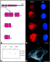
The NH2 terminus of Tpr does not localize to the nuclear envelope. (A) A schematic diagram of the HA-tagged Tpr deletion mutants used to analyze the NH2 terminus of Tpr. The bracket under HA-Tpr indicates the portion of the full-length protein that has been expanded and shown beneath. The numbers shown in the diagram indicate amino acids of the Tpr domains that are expressed. The large stippled boxes indicate areas predicted to be in a coiled-coil motif. (B) Immunofluorescence of cells expressing truncated forms of HA-Tpr. The cells are aligned directly to the right of the schematic diagram of the construct being expressed. Images of the anti-HA staining pattern (left) and DAPI stains (right) of the same cells. While HA-Tpr and HA-Tpr1-734 show labeling of the nuclear envelope, HA-Tpr1-304 and HA-Tpr1-256 show diffuse localization in the cytoplasm and nucleus. The cells were fixed before permeabilization with Triton X-100. (C) A composite of Western blot analyses of HA-Tpr proteins immunoprecipitated from Triton X-100–soluble and –insoluble cell extracts. While all three proteins are in soluble fractions (S), only HA-Tpr1-734 is in the insoluble fractions (I). Molecular mass markers on right (×103). (D) Immunofluorescence of a BHK cell expressing high levels of HA-Tpr1-304 and showing shows long filaments in the cytoplasm. Bars, 10 μm.
Figure 4
Figure 4
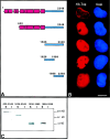
Identification of a NPC association domain. (A) Schematic diagram of the HA-Tpr clones used for the identification of an NPC association domain in Tpr. See Fig. 3 for details. (B) Immunofluorescence of COS cells expressing HA-tagged Tpr fragments. HA-Tpr400-649 and HA-Tpr435-649 localize to the NPC; HA-Tpr514-649 is diffusely scattered throughout the nucleus and cytoplasm; HA-Tpr400-605 forms large spherical foci located within the nucleus but not associated with the nuclear envelope. (C) A composite of Western blot analyses of HA-Tpr domains immunoprecipitated from Triton X-100–soluble and –insoluble cellular extracts. All proteins are present in both soluble and insoluble fractions (I) except HA-Tpr514-649, which is only detected in the soluble fraction (S). Molecular mass markers on right (×103). Bar, 10 μm.
Figure 5
Figure 5
Immunogold localization of the HA-tagged, NPC-associated domain of Tpr overexpressed in COS cells. HA-Tpr400-649 was detected with polyclonal HA antibodies and 5-nm gold goat–rabbit IgG in cells prepermeabilized with detergent (see Materials and Methods for details). Gold particles were found predominantly within 200 nm of the nucleoplasmic face of nuclear pores (A). Quantitative analysis revealed that the majority of the gold was localized to the nucleoplasmic aspect of the pore (see Results). In contrast, nup358 was localized to the cytoplasmic aspect of the pore in cells processed in parallel (B). No gold was detected if primary antibodies were omitted (C). Bar, 250 nm.
Figure 6
Figure 6
The NPC association domain of Tpr is sufficient to localize green fluorescent protein to the nuclear envelope. Two COS cells expressing GFP-Tpr435-649 (left) and the corresponding DAPI stains (right). Localization of GFP to the nuclear envelope is evident in both cells that were permeabilized with Triton X-100 before fixation to remove soluble protein that obscures the nuclear envelope signal. Bar, 10 μm.
Figure 7
Figure 7
The COOH terminus of Tpr contains a nuclear localization domain. (A) Schematic diagrams of the HA-tagged Tpr truncations used for identification of a nuclear localization domain. See Fig. 3 for details. (B) Immunofluorescence of COS cells expressing the HA-tagged Tpr domains. Full-length HA-Tpr decorates the nuclear envelope while HA-Tpr632-2348, HA-Tpr1626-2348, and HA-Tpr1626-1989 all accumulate to high levels within the nuclear interior. HA-Tpr1989-2348 is diffusely localized throughout the cytoplasm and nucleus. (C) A composite of Western blot analyses of HA-Tpr proteins immunoprecipitated from Triton X-100–soluble and –insoluble cellular fractions. HA-Tpr632-2348 is detected in soluble and insoluble fractions and all others are detected only in soluble fractions. Molecular mass markers on right (×103). Bar, 10 μm.
Figure 8
Figure 8
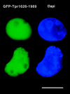
The NLS domain of Tpr localizes GFP to the nuclear interior. Two COS cells expressing GFP-Tpr1626-1989 fixed as in Fig. 3 to show that virtually all of the protein accumulates within the nuclear interior. Bar, 10 μm.
Figure 9
Figure 9
Overexpression of HA-Tpr proteins does not effect the distribution of a glucocorticoid receptor-β-galactosidase fusion protein. Immunofluorescence of BHKgrβ cells expressing (A and E) HA-Tpr, (B and F) HA-Tpr1-304, (C and G) HA-Tpr380-649, and (D and H) HA-Tpr632-2348. Cells in A–D show staining with anti-HA. Cells in E–H are the corresponding fields stained with an anti-β-galactosidase antibody. In the absence of dexamethasone, the glucocorticoid receptor-β-galactosidase fusion is largely cytoplasmic for all HA-Tpr proteins and nontransfected cells alike. Bars, 10 μm.
Figure 10
Figure 10
Overexpression of Tpr proteins does not effect nuclear import of a glucocorticoid receptor-β-galactosidase fusion protein. Immunofluorescence of BHKgrβ cells expressing (A and E) HA-Tpr, (B and F) HA-Tpr1-304, (C and G) HA-Tpr380-649, and (D and H) HA-Tpr632-2348 as described in Fig. 8. Cells were treated with 10 μg/ml dexamethasone for 30 min before fixation to induce nuclear import of the receptor. In all cases, nuclei of cells expressing the Tpr proteins accumulate the glucocorticoid receptor-β-galactosidase fusion protein to the same levels as nontransfected cells. Bar, 10 μm.
Figure 11
Figure 11
Overexpression of full-length Tpr, the NPC association domain and the nuclear localization domain interfere with poly(A)+ RNA trafficking. Poly(A)+ RNA distribution in BHK cells expressing (A and E) HA-Tpr, (B and F) HA-Tpr1-304, (C and G) HA-Tpr380-649, and (D and H) HA-Tpr632-2348. A–D Show anti-HA staining patterns for the respective domains. E–H show the corresponding fields after in situ hybridization with a biotinylated oligo-dT 45 mer. Poly(A)+ RNA accumulates in cells expressing HA-Tpr, HA-Tpr380-649, and HA-Tpr632-2348, but not HA-Tpr1-304. Bar, 10 μm.

References

    1. Akey CW, Radermacher M. Architecture of the Xenopusnuclear pore complex revealed by three-dimensional cryo-electron microscopy. J Cell Biol. 1993;122:1–19. - PMC - PubMed
    1. Aitchison JD, Rout MP, Marelli M, Blobel G, Wozniak RW. Two novel related yeast nucleoporins Nup170p and Nup157p: complementation with the vertebrate homologue Nup155p and functional interactions with the yeast nuclear pore-membrane protein Pom152p. J Cell Biol. 1995;131:1133–1148. - PMC - PubMed
    1. Bangs PL, Sparks CA, Odgren PR, Fey EG. Product of the oncogene-activating gene Tpr is a phosphorylated protein of the nuclear pore complex. J Cell Biochem. 1996;61:48–60. - PubMed
    1. Bastos R, Panté N, Burke B. Nuclear pore complex proteins. Int Rev Cytol. 1995;162:257–302. - PubMed
    1. Bastos R, Lin A, Enarson M, Burke B. Targeting and function in mRNA export of nuclear pore complex protein Nup153. J Cell Biol. 1996;134:1141–1156. - PMC - PubMed

Publication types

MeSH terms