. 1998 Dec 28;143(7):1883-98.
doi: 10.1083/jcb.143.7.1883.

Aggresomes: a cellular response to misfolded proteins

Affiliations

Aggresomes: a cellular response to misfolded proteins

J A Johnston et al. J Cell Biol. .

Abstract

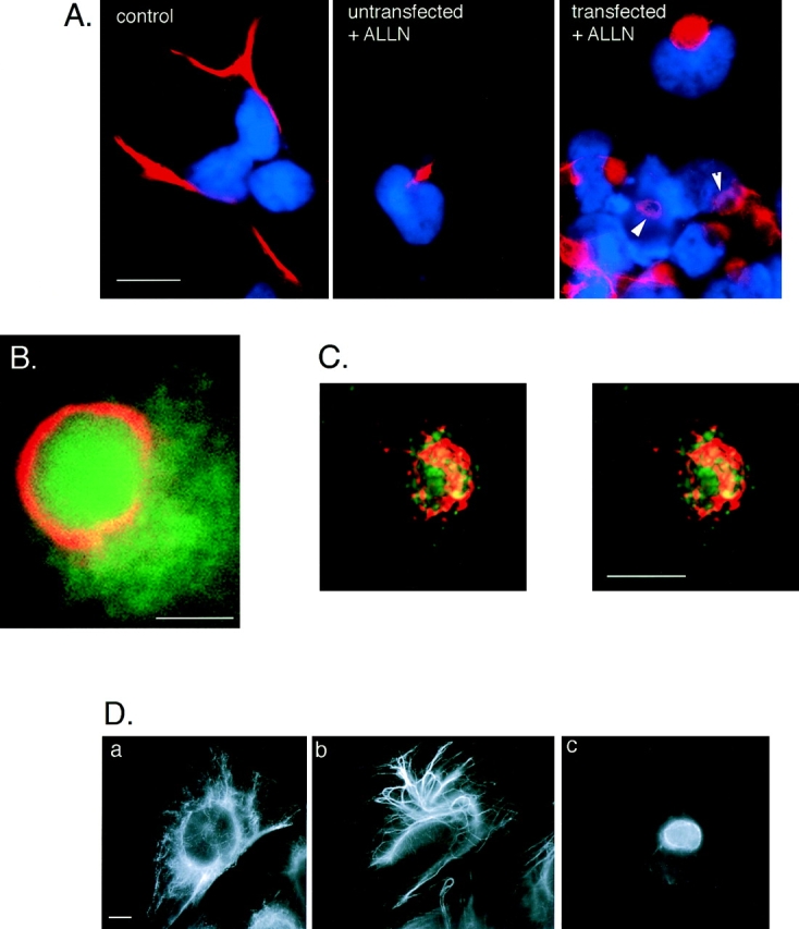
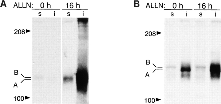
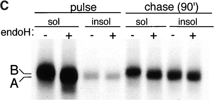
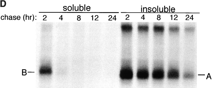
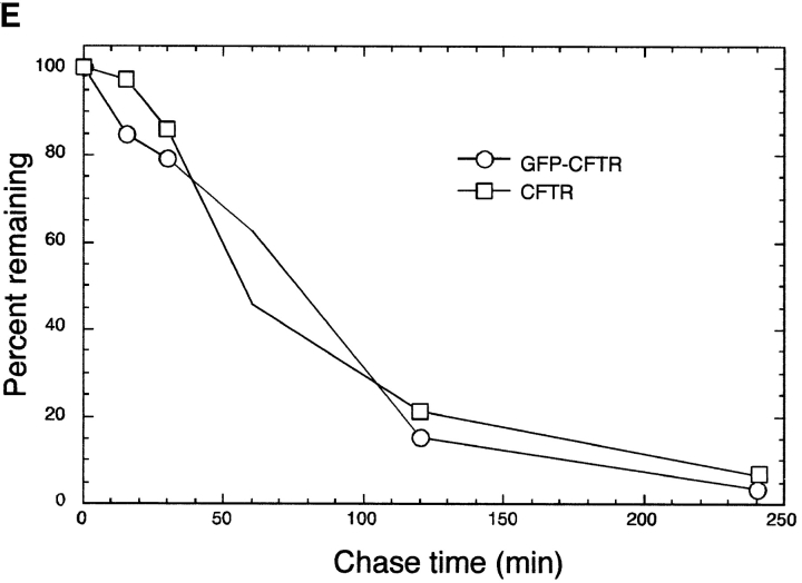
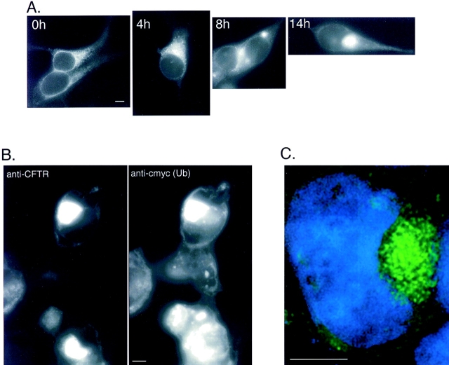
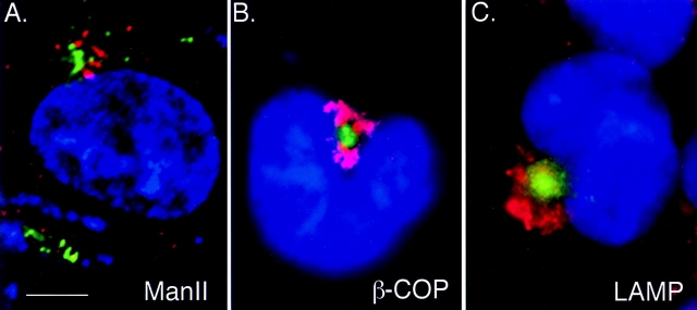
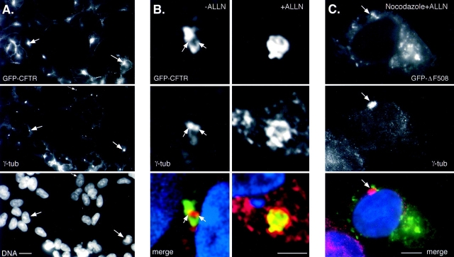
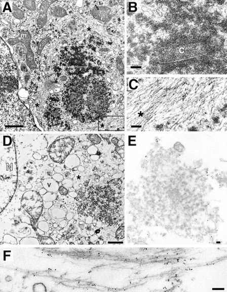
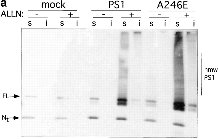
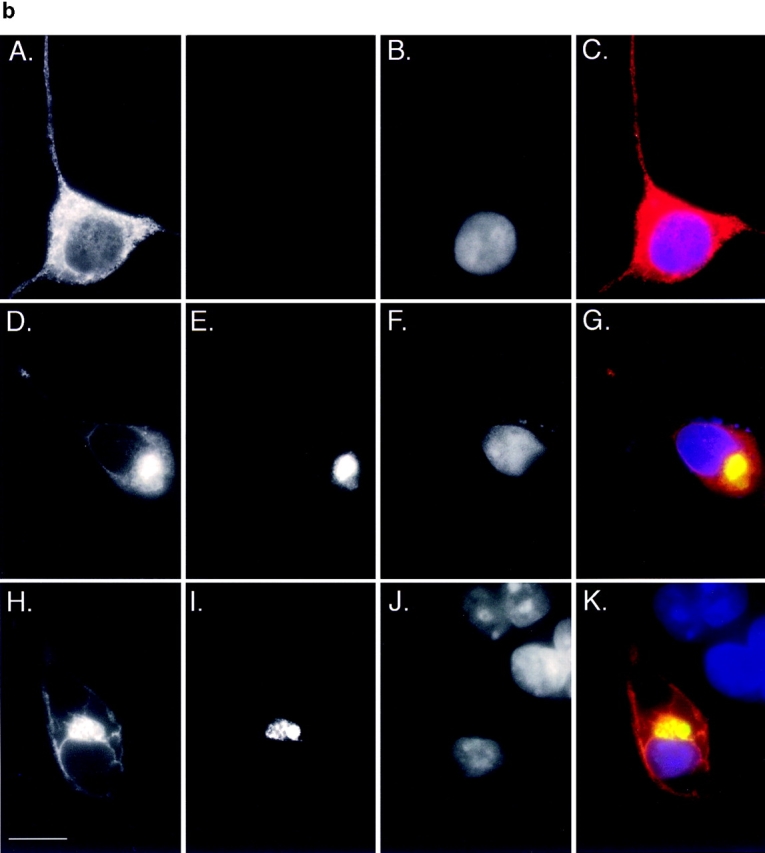
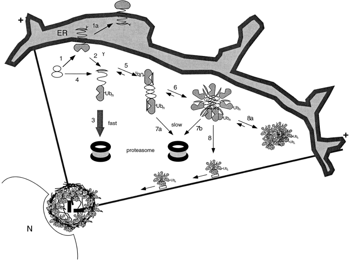
Intracellular deposition of misfolded protein aggregates into ubiquitin-rich cytoplasmic inclusions is linked to the pathogenesis of many diseases. Why these aggregates form despite the existence of cellular machinery to recognize and degrade misfolded protein and how they are delivered to cytoplasmic inclusions are not known. We have investigated the intracellular fate of cystic fibrosis transmembrane conductance regulator (CFTR), an inefficiently folded integral membrane protein which is degraded by the cytoplasmic ubiquitin-proteasome pathway. Overexpression or inhibition of proteasome activity in transfected human embryonic kidney or Chinese hamster ovary cells led to the accumulation of stable, high molecular weight, detergent-insoluble, multiubiquitinated forms of CFTR. Using immunofluorescence and transmission electron microscopy with immunogold labeling, we demonstrate that undegraded CFTR molecules accumulate at a distinct pericentriolar structure which we have termed the aggresome. Aggresome formation is accompanied by redistribution of the intermediate filament protein vimentin to form a cage surrounding a pericentriolar core of aggregated, ubiquitinated protein. Disruption of microtubules blocks the formation of aggresomes. Similarly, inhibition of proteasome function also prevented the degradation of unassembled presenilin-1 molecules leading to their aggregation and deposition in aggresomes. These data lead us to propose that aggresome formation is a general response of cells which occurs when the capacity of the proteasome is exceeded by the production of aggregation-prone misfolded proteins.


Figures

Figure 5
Figure 5
Aggresome formation is accompanied by a reorganization of the vimentin cytoskeleton. (A) Effect of ALLN on the distribution of vimentin (red) with respect to the nucleus (blue) in untransfected (left and middle) and CFTR transfected (right) HEK cells. Cells were either untreated (left) or incubated for 12 h in 10 μg/ml ALLN (middle and right). Note ring-like appearance of vimentin fluorescence in ALLN-treated CFTR-expressing cells (arrows). (B) Colocalization of GFP-CFTR (green) and vimentin (red) in ALLN-treated cells. (C) Stereo pair of optical sections from deconvolved stack of images from ALLN-treated cells showing GFP-CFTR (green autofluorescence) and vimentin (red immunofluorescence). Nuclear fluorescence signal has been omitted. (D) Distribution of vimentin in drug-treated CHO cells. CHO cells stably expressing GFP-CFTR were treated with carrier (a), 10 μg/ml nocodozole (b), or 10 μg/ml ALLN (c) for 12 h and then immunostained with antibodies to vimentin. Note the condensation and bundling of vimentin in b, as compared with the complete collapse of vimentin in c. Bars, 15 μm (A), 2.5 μm (B), and 5 μm (C and D).
Figure 1
Figure 1
Misfolded CFTR molecules form stable aggregates. (A and B) Immunoblots showing steady-state accumulation of core-glycosylated (band B), unglycosylated (band A), and high molecular weight forms of ΔF508 in HEK cells expressing low (A) or high (B) levels of the protein. Transfected cells were incubated in the presence of ALLN (10 μg/ml) or carrier for 16 h before harvesting. Cells were lysed and separated into detergent-soluble (s) and -insoluble (i) fractions as described in Materials and Methods. Blot in B was deliberately underexposed to emphasize the mobilities of the accumulated species; therefore, the intensity cannot be directly compared with the blot in A. (C) Deglycosylation of CFTR. HEK cells overexpressing ΔF508 (as in B) were pulse labeled with [35S]Met for 15 min and chased with unlabeled medium for 90 min. At the indicated time points, cell lysates were separated into detergent-soluble and -insoluble fractions before immunoprecipitation with CFTR antibody. Half of each sample was digested with endoglycosidase H (endoH) as indicated, before SDS-PAGE and autoradiography. (D) Extended pulse–chase of detergent-soluble and -insoluble ΔF508. HEK cells overexpressing ΔF508 (as in B) were pulse labeled with [35S]Met for 15 min and chased with unlabeled medium for the times indicated. At each time point, the cell lysate was separated into detergent-soluble and -insoluble fractions before immunoprecipitation with CFTR antibody. (E) Kinetics of degradation of GFP-CFTR. HEK cells expressing CFTR or GFP-CFTR were pulse labeled with [35S]Met for 15 min and chased with unlabeled medium for the times indicated. The detergent-soluble fractions were immunoprecipitated with CFTR antibody, separated by SDS-PAGE; the data were scanned and quantified from autoradiographic images.
Figure 1
Figure 1
Misfolded CFTR molecules form stable aggregates. (A and B) Immunoblots showing steady-state accumulation of core-glycosylated (band B), unglycosylated (band A), and high molecular weight forms of ΔF508 in HEK cells expressing low (A) or high (B) levels of the protein. Transfected cells were incubated in the presence of ALLN (10 μg/ml) or carrier for 16 h before harvesting. Cells were lysed and separated into detergent-soluble (s) and -insoluble (i) fractions as described in Materials and Methods. Blot in B was deliberately underexposed to emphasize the mobilities of the accumulated species; therefore, the intensity cannot be directly compared with the blot in A. (C) Deglycosylation of CFTR. HEK cells overexpressing ΔF508 (as in B) were pulse labeled with [35S]Met for 15 min and chased with unlabeled medium for 90 min. At the indicated time points, cell lysates were separated into detergent-soluble and -insoluble fractions before immunoprecipitation with CFTR antibody. Half of each sample was digested with endoglycosidase H (endoH) as indicated, before SDS-PAGE and autoradiography. (D) Extended pulse–chase of detergent-soluble and -insoluble ΔF508. HEK cells overexpressing ΔF508 (as in B) were pulse labeled with [35S]Met for 15 min and chased with unlabeled medium for the times indicated. At each time point, the cell lysate was separated into detergent-soluble and -insoluble fractions before immunoprecipitation with CFTR antibody. (E) Kinetics of degradation of GFP-CFTR. HEK cells expressing CFTR or GFP-CFTR were pulse labeled with [35S]Met for 15 min and chased with unlabeled medium for the times indicated. The detergent-soluble fractions were immunoprecipitated with CFTR antibody, separated by SDS-PAGE; the data were scanned and quantified from autoradiographic images.
Figure 2
Figure 2
Ubiquitinated CFTR molecules accumulate in aggresomes. (A) Time course of ΔF508 accumulation. HEK cells stably expressing low levels of GFP-ΔF508 were incubated in the presence of ALLN (10 μg/ml) for the times indicated. Note the absence of plasma membrane staining consistent with the inability of the ΔF508 to traffic to the cell surface. (B) Colocalization of ΔF508 with epitope-tagged ubiquitin. HEK cells were transiently cotransfected with ΔF508 and c-myc-ubiquitin cDNA and incubated in the presence of ALLN (10 μg/ ml) for 12 h and processed for immunofluorescence with antibodies to CFTR and c-myc. The left panel shows immunolocalization of ΔF508 and the right panel shows the same field examined for c-myc immunolocalization. (C). Optical section of an aggresome. HEK cells stably expressing GFP-ΔF508 were treated with ALLN for 12 h. Serial 0.2-μm optical sections were recorded with a cooled CCD camera and deconvolved as described in Materials and Methods. The image shown is a single deconvolved optical section indicating GFP (green) and DNA (blue) localization. Bar, 5 μm.
Figure 3
Figure 3
Aggresomes are distinct from Golgi and lysosomal markers. GFP-CFTR (green) visualized together with immunofluorescent staining (red) for Golgi markers manII (A), β-COP (B), and LAMP (C). Cells in B and C were treated overnight with ALLN and digitally imaged using conventional optics. The image in A is a 0.2-μm optical section obtained as described in the legend to Fig. 2 C. Bar, 15 μm.
Figure 4
Figure 4
Aggresomes form at the MTOC and require MT for formation. HEK cells stably expressing GFP-CFTR were immunostained with antibody to γ-tubulin and imaged for GFP fluorescence (top), γ-tubulin immunofluorescence (middle), and DNA (A, bottom). (A) CFTR and γ-tubulin and DNA in a field of cells. (B) Effect of ALLN. Cells were incubated for 12 h in the presence of untreated (left) or 10 μg/ml ALLN (right). (C) Effect of MT disruption. Cells were incubated for 12 h in the presence of ALLN (10 μg/ml) together with nocodazole (10 μg/ml). Arrows denote the location of the centrosome. The images in A and C were obtained using conventional optics whereas the images in B are single 0.2-μm optical sections. GFP and γ-tubulin fluorescence were digitally overlaid in the bottom panels in B and C. Bars, 15 μm (A), 5 μm (B and C).
Figure 6
Figure 6
Transmission EM of aggresomes. (A–C) Transmission EM of HEK cell overexpressing ΔF508 and treated overnight with ALLN. B and C are higher magnification views of sections indicated by white and black boxes, respectively. (D–F) Transmission EM of crude aggresome fraction from HEK cells expressing GFP-CFTR. Swollen vesicular structures (v) and mitochondria (m) are the result of hypotonic conditions used during the preparation. Crude aggresomes were incubated with antibodies to GFP (E; 15-nm gold particles) and vimentin (F; 6-nm gold particles) before embedding and sectioning as described in Materials and Methods. m, mitochondrion; N, nucleus; c, centriole; v, vesicle; *, filamentous material ∼8–10 nm per filament. Bars for A and D, 1 μm; B, C, E, and F, 100 nm.
Figure 7
Figure 7
Accumulation of PS1 and A246E after treatment with ALLN. HEK cells transfected with vector alone, PS1, or A246E were incubated for 12 h in the presence or absence of ALLN (10 μg/ml) as indicated and processed for immunoblotting (a) or immunofluorescence (b). (a) Immunoblot. Cells were lysed and separated into detergent-soluble (s) or -insoluble (i) fractions as described in Materials and Methods and probed with antibody to the NH2 terminus of PS1. (b) Immunofluorescence localization of A246E in HEK cells. HEK cells expressing A246E were either untreated (A–C) or treated (D–K) with ALLN. In D–G A246E was transfected into HEK cells that stably express GFP-CFTR. In H–K, A246E was cotransfected with c-myc-ubiquitin into naive HEK cells. Cells were prepared for immunocytochemistry as described in Materials and Methods and stained with antibody to the NH2 terminus of PS1 (A, D, and H), antibody to c-myc (I), or with bisbenzimide to visualize nuclei (B, F, and J). GFP-CFTR fluorescence was imaged in E. C, G, and K are digital overlays. Bar, 15 μm.
Figure 7
Figure 7
Accumulation of PS1 and A246E after treatment with ALLN. HEK cells transfected with vector alone, PS1, or A246E were incubated for 12 h in the presence or absence of ALLN (10 μg/ml) as indicated and processed for immunoblotting (a) or immunofluorescence (b). (a) Immunoblot. Cells were lysed and separated into detergent-soluble (s) or -insoluble (i) fractions as described in Materials and Methods and probed with antibody to the NH2 terminus of PS1. (b) Immunofluorescence localization of A246E in HEK cells. HEK cells expressing A246E were either untreated (A–C) or treated (D–K) with ALLN. In D–G A246E was transfected into HEK cells that stably express GFP-CFTR. In H–K, A246E was cotransfected with c-myc-ubiquitin into naive HEK cells. Cells were prepared for immunocytochemistry as described in Materials and Methods and stained with antibody to the NH2 terminus of PS1 (A, D, and H), antibody to c-myc (I), or with bisbenzimide to visualize nuclei (B, F, and J). GFP-CFTR fluorescence was imaged in E. C, G, and K are digital overlays. Bar, 15 μm.
Figure 8
Figure 8
Model for aggresome formation. Numbers indicate various steps in the aggresome biogenesis pathway and are explained in the Discussion. Membrane proteins are cotranslationally translocated to the membrane of the ER (1). Some molecules fold to adopt a maturation-competent conformation (1a). Others misfold and are dislocated from the ER membrane (2). Some proteins may escape the translocation machinery and be delivered directly to the cytoplasm (4). Dislocated, ubiquitinated, misfolded protein can either be rapidly degraded by cytosolic proteasomes (3) or aggregate (5 and 6). Because aggregates are difficult to unfold, they are likely to be slowly degraded by the proteasome (7a and 7b). Misfolded, aggregated protein is transported to the MTOC by MT where it becomes entangled with collapsed IF (8). In the absence of MT, protein aggregates coalesce at dispersed sites throughout the cytoplasm (8a). N, nucleus; Ubn, ubiquitin conjugates; +, orientation of MT in the cell.

References

    1. Baumeister W, Cejka Z, Kania M, Seemuller E. The proteasome: a macromolecular assembly designed to confine proteolysis to a nanocompartment. Biol Chem. 1997;378:121–130. - PubMed
    1. Beal RE, Toscano-Cantaffa D, Young P, Rechsteiner M, Pickart CM. The hydrophobic effect contributes to polyubiquitin chain recognition. Biochemistry. 1998;37:2925–2934. - PubMed
    1. Bebök Z, Mazzochi C, King SA, Hong JS, Sorscher EJ. The mechanism underlying cystic fibrosis transmembrane conductance regulator transport from the endoplasmic reticulum to the proteasome includes Sec61β and a cytosolic, deglycosylated intermediary. J Biol Chem. 1998;273:29873–29878. - PubMed
    1. Blose SH, Bushnell A. Observations on the vimentin-10-nm filaments during mitosis in BHK21 cells. Exp Cell Res. 1982;142:57–62. - PubMed
    1. Bonifacino JS, Suzuki CK, Lippincott-Schwartz J, Weissman AM, Klausner RD. Pre-Golgi degradation of newly synthesized T-cell antigen receptor chains: intrinsic sensitivity and the role of subunit assembly. J Cell Biol. 1989;109:73–83. - PMC - PubMed

Publication types

MeSH terms