. 1998 Dec 28;143(7):2081-92.
doi: 10.1083/jcb.143.7.2081.

Activation of alphaVbeta3 on vascular cells controls recognition of prothrombin

Affiliations

Activation of alphaVbeta3 on vascular cells controls recognition of prothrombin

T V Byzova et al. J Cell Biol. .

Abstract

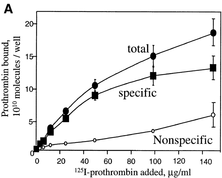
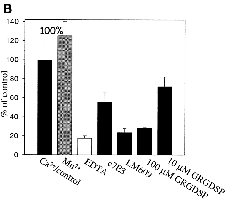
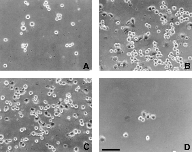
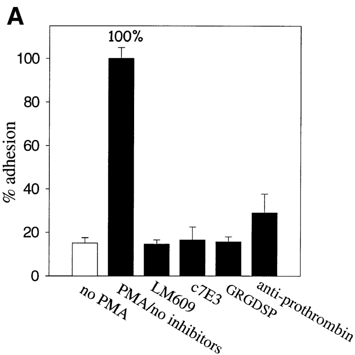
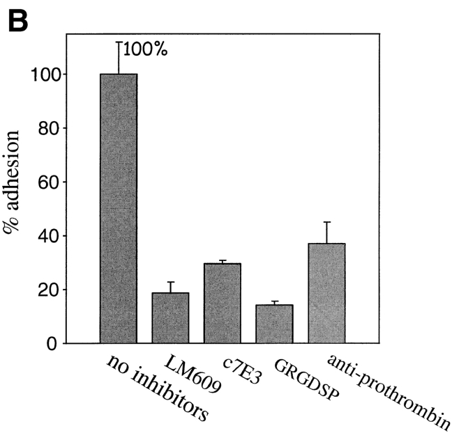
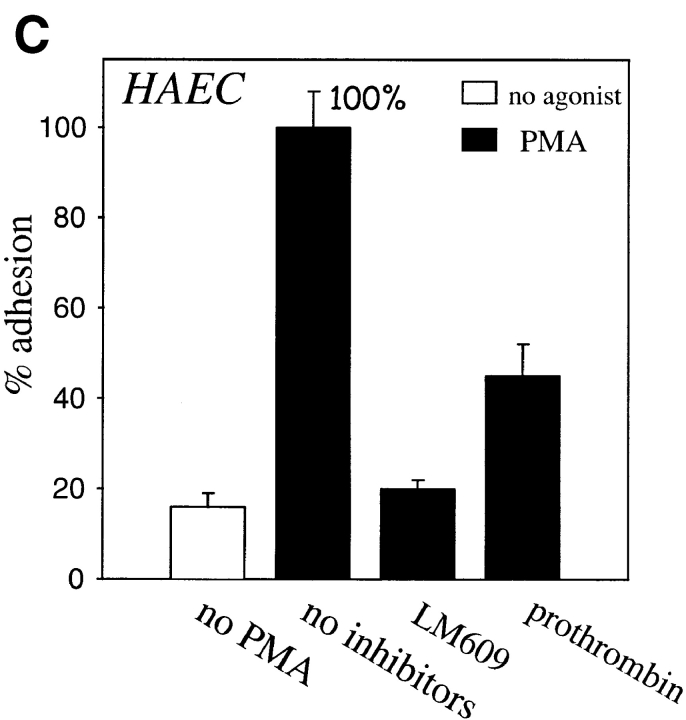
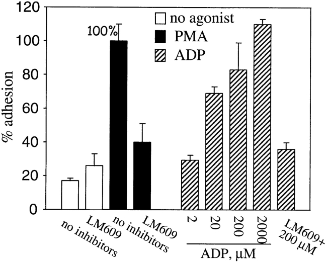
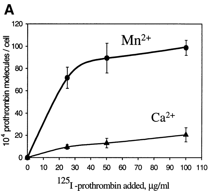
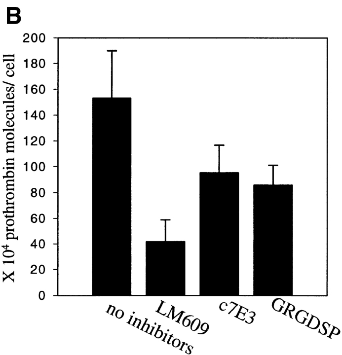
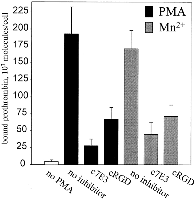
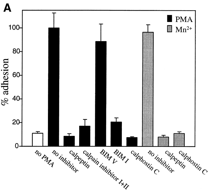
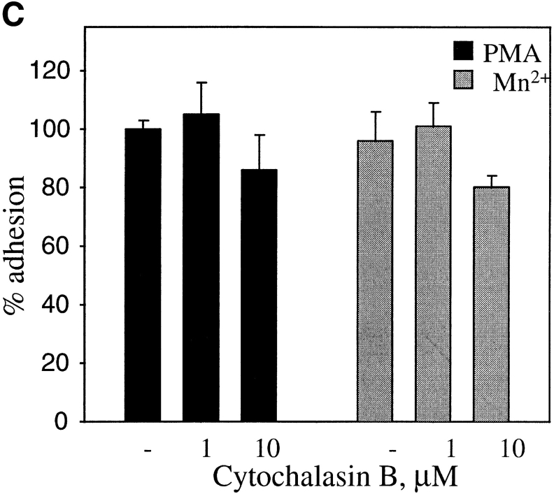
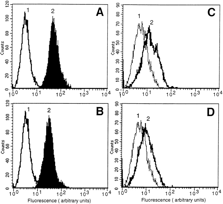
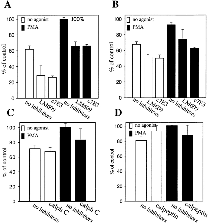
Regulation of vascular homeostasis depends upon collaboration between cells of the vessel wall and blood coagulation system. A direct interaction between integrin alphaVbeta3 on endothelial cells and smooth muscle cells and prothrombin, the pivotal proenzyme of the blood coagulation system, is demonstrated and activation of the integrin is required for receptor engagement. Evidence that prothrombin is a ligand for alphaVbeta3 on these cells include: (a) prothrombin binds to purified alphaVbeta3 via a RGD recognition specificity; (b) prothrombin supports alphaVbeta3-mediated adhesion of stimulated endothelial cells and smooth muscle cells; and (c) endothelial cells, either in suspension and in a monolayer, recognize soluble prothrombin via alphaVbeta3. alphaVbeta3-mediated cell adhesion to prothrombin, but not to fibrinogen, required activation of the receptor. Thus, the functionality of the alphaVbeta3 receptor is ligand defined, and prothrombin and fibrinogen represent activation- dependent and activation-independent ligands. Activation of alphaVbeta3 could be induced not only by model agonists, PMA and Mn2+, but also by a physiologically relevant agonist, ADP. Inhibition of protein kinase C and calpain prevented activation of alphaVbeta3 on vascular cells, suggesting that these molecules are involved in the inside-out signaling events that activate the integrin. The capacity of alphaVbeta3 to interact with prothrombin may play a significant role in the maintenance of hemostasis; and, at a general level, ligand selection by alphaVbeta3 may be controlled by the activation state of this integrin.


Figures

Figure 1
Figure 1
125I-prothrombin binding to purified αVβ3. (A) Saturation isotherms of 125I-prothrombin binding (150 min at 37°C) to purified and immobilized αVβ3. Specific binding (▪) was derived by subtracting the nonspecific binding (○), the residual binding in the presence of a 50-fold excess of nonlabeled prothrombin, from the total binding (•), no inhibitor present. Data are derived in the presence of 1 mM Ca2+. (B) 125I-prothrombin binding to immobilized αVβ3 in the presence of 1 mM Ca2+ (black bars), 1 mM Mn2+ (gray bar), and 10 mM EDTA (open bar). Wells were preincubated with mAb c7E3 or LM609 (20 μg/ml each); or GRGDSP (100 μM and 100 μM) and 125I-prothrombin was added at a concentration of 30 μg/ml. The total binding (not corrected for nonspecific background) of prothrombin to αVβ3 in the presence of 1 mM Ca2+ without inhibitors was assigned a value of 100%. The data shown are means and SD of triplicates in one experiment and are representative of three separate experiments.
Figure 1
Figure 1
125I-prothrombin binding to purified αVβ3. (A) Saturation isotherms of 125I-prothrombin binding (150 min at 37°C) to purified and immobilized αVβ3. Specific binding (▪) was derived by subtracting the nonspecific binding (○), the residual binding in the presence of a 50-fold excess of nonlabeled prothrombin, from the total binding (•), no inhibitor present. Data are derived in the presence of 1 mM Ca2+. (B) 125I-prothrombin binding to immobilized αVβ3 in the presence of 1 mM Ca2+ (black bars), 1 mM Mn2+ (gray bar), and 10 mM EDTA (open bar). Wells were preincubated with mAb c7E3 or LM609 (20 μg/ml each); or GRGDSP (100 μM and 100 μM) and 125I-prothrombin was added at a concentration of 30 μg/ml. The total binding (not corrected for nonspecific background) of prothrombin to αVβ3 in the presence of 1 mM Ca2+ without inhibitors was assigned a value of 100%. The data shown are means and SD of triplicates in one experiment and are representative of three separate experiments.
Figure 2
Figure 2
Photomicrographs of HUVEC adherent to immobilized prothrombin in the absence (A) and presence of 200 nM PMA (B) or in the presence of 1 mM MnCl2 (C). In D, cells were pretreated by mAb LM609 (20 μg/ml) to αVβ3 and then stimulated with PMA. Bar, 50 μm.
Figure 3
Figure 3
Endothelial cell adhesion to prothrombin requires stimulation. HUVEC (A and B) or HAEC were harvested, labeled by Calcein, and diluted to the 5 × 10⁵ cells per ml in DME containing 0.2% BSA and 1 mM CaCl2. In A and B, HUVEC were stimulated with 200 nM PMA or 1 mM Mn2+, respectively. Inhibitors used were: mAb LM609 or mAb c7E3 (20 μg/ml each), GRGDSP peptide (100 μg), or polyclonal antibodies to prothrombin (anti-prothrombin). In C, HAEC were stimulated with PMA. In addition to the inhibitors used in A and B, soluble prothrombin was used at a concentration of 300 μg/ml. After 50 min, adhesion was measured. Adhesion in the presence of 1 mM CaCl2 and 1 PMA (A and C) or in the presence of 1 mM MnCl2 (B) and in the absence of inhibitors was assigned a value of 100%. The data shown are means and SD of quadruplicates in one experiment and are representative of seven separate experiments.
Figure 3
Figure 3
Endothelial cell adhesion to prothrombin requires stimulation. HUVEC (A and B) or HAEC were harvested, labeled by Calcein, and diluted to the 5 × 10⁵ cells per ml in DME containing 0.2% BSA and 1 mM CaCl2. In A and B, HUVEC were stimulated with 200 nM PMA or 1 mM Mn2+, respectively. Inhibitors used were: mAb LM609 or mAb c7E3 (20 μg/ml each), GRGDSP peptide (100 μg), or polyclonal antibodies to prothrombin (anti-prothrombin). In C, HAEC were stimulated with PMA. In addition to the inhibitors used in A and B, soluble prothrombin was used at a concentration of 300 μg/ml. After 50 min, adhesion was measured. Adhesion in the presence of 1 mM CaCl2 and 1 PMA (A and C) or in the presence of 1 mM MnCl2 (B) and in the absence of inhibitors was assigned a value of 100%. The data shown are means and SD of quadruplicates in one experiment and are representative of seven separate experiments.
Figure 4
Figure 4
PMA-stimulated adhesion of HASMC to prothrombin is αVβ3 dependent. HASMC were harvested, labeled by Calcein, diluted to the 5 × 10⁵ cells per ml in DME containing 1% BSA and 1 mM CaCl2. HASMC were stimulated with either 200 nM PMA (black bars) or ADP (striped bars) at the indicated concentrations. mAb LM609 at a concentration of 20 μg/ml was included as indicated. After 40 min, adhesion was measured. Adhesion in the presence of PMA without inhibitors present was assigned a value of 100%. Nonspecific adhesion to BSA-coated wells was subtracted. The data shown are means and SD from three experiments.
Figure 5
Figure 5
HUVEC bind soluble 125I-prothrombin. (A) HUVEC in suspension were incubated with increasing concentrations of 125I-prothrombin in DME/F12 in the presence of 0.5 mM MnCl2 or 1 mM CaCl2 for 60 min, and cell-bound radioactivity was quantitated as described. Nonspecific binding was measured in the presence of 50-fold excess of nonlabeled prothrombin and subtracted to yield the specific binding data shown. Values represent the means and SD of four determinations. (B) Specificity of prothrombin binding to HUVEC. 125I-prothrombin (50 μg/ml) was incubated with HUVEC (8 × 10⁵ cells/ml) in the presence of mAb LM609, mAb c7E3 (20 μg/ml each; a similar concentration of nonimmune IgG was without effect), or 100 μM GRGDSP. MnCl2 (1 mM) was added to the DME/F12 media, and cell-bound radioactivity was measured after 60 min incubation. Nonspecific binding was measured in the presence of 50-fold excess of nonlabeled prothrombin and subtracted. The data shown are means and SD of quadruplicates in one experiment and are representative of three separate experiments.
Figure 5
Figure 5
HUVEC bind soluble 125I-prothrombin. (A) HUVEC in suspension were incubated with increasing concentrations of 125I-prothrombin in DME/F12 in the presence of 0.5 mM MnCl2 or 1 mM CaCl2 for 60 min, and cell-bound radioactivity was quantitated as described. Nonspecific binding was measured in the presence of 50-fold excess of nonlabeled prothrombin and subtracted to yield the specific binding data shown. Values represent the means and SD of four determinations. (B) Specificity of prothrombin binding to HUVEC. 125I-prothrombin (50 μg/ml) was incubated with HUVEC (8 × 10⁵ cells/ml) in the presence of mAb LM609, mAb c7E3 (20 μg/ml each; a similar concentration of nonimmune IgG was without effect), or 100 μM GRGDSP. MnCl2 (1 mM) was added to the DME/F12 media, and cell-bound radioactivity was measured after 60 min incubation. Nonspecific binding was measured in the presence of 50-fold excess of nonlabeled prothrombin and subtracted. The data shown are means and SD of quadruplicates in one experiment and are representative of three separate experiments.
Figure 6
Figure 6
125I-prothrombin binding to a HUVEC monolayer. Confluent cell monolayers were incubated with 50 μg/ml 125I-prothrombin in DME/F12-1% BSA in the presence or absence of 200 nM PMA (solid bars) or 0.5 mM MnCl2 (gray bars). Cells were preincubated with c7E3 (30 μg/ml) or a cyclic RGD peptide (10 μM) or without inhibitors for 10 min. After 70 min at 37°C, wells were washed three times with PBS, and the cells were solubilized in 1 N NaOH. Prothrombin binding to nonstimulated endothelial cells was subtracted from the total binding, and the difference is displayed. Values are means and SD of quadruplicates from one of five experiments with similar results.
Figure 8
Figure 8
Effects of protein kinase C and calpain inhibitors (A) and cytochalasin B (B and C) on adhesion of PMA- and MnCl2-treated HUVEC to immobilized prothrombin. HUVEC adhesion was measured (see legend to Fig. 3 and Materials and Methods) in the absence (open bar) or presence of 200 nM PMA (black bars) or in the presence of 0.5 mM MnCl2 (gray bars). In A, cells were pretreated with calpeptin (50 μg/ml), calpain inhibitor I and II (100 μg/ml each), bisindolylmaleimide V (BIM V) and bisindolylmaleimide I (BIM I; 20 nM each) or calphostin C light-activated; 1 μM). In B, adherent cells in the absence or presence (0.1 μm) of cytochalasin B were photographed at 40×. Bar, 50 μm. In C, adhesion of stimulated cells was measured after 50 min in the absence or presence of cytochalasin B. Adhesion in the presence of 200 nM PMA without inhibitors was assigned a value of 100%. The data shown are means and SD from three experiments.
Figure 8
Figure 8
Effects of protein kinase C and calpain inhibitors (A) and cytochalasin B (B and C) on adhesion of PMA- and MnCl2-treated HUVEC to immobilized prothrombin. HUVEC adhesion was measured (see legend to Fig. 3 and Materials and Methods) in the absence (open bar) or presence of 200 nM PMA (black bars) or in the presence of 0.5 mM MnCl2 (gray bars). In A, cells were pretreated with calpeptin (50 μg/ml), calpain inhibitor I and II (100 μg/ml each), bisindolylmaleimide V (BIM V) and bisindolylmaleimide I (BIM I; 20 nM each) or calphostin C light-activated; 1 μM). In B, adherent cells in the absence or presence (0.1 μm) of cytochalasin B were photographed at 40×. Bar, 50 μm. In C, adhesion of stimulated cells was measured after 50 min in the absence or presence of cytochalasin B. Adhesion in the presence of 200 nM PMA without inhibitors was assigned a value of 100%. The data shown are means and SD from three experiments.
Figure 7
Figure 7
Short-term PMA treatment does not increase αVβ3 expression. HUVEC (A and B) or HASMC (C and D) were grown and harvested as described. For each experimental point, 106 cells were incubated in the presence of nonimmune IgG (1) or mAb LM609 (2) for 50 min. In B and D cells were treated by 200 nM PMA. After washing, the cells were incubated with anti-mouse IgG FITC-conjugated antibody and analyzed by flow cytometry.
Figure 9
Figure 9
HUVEC adhesion to immobilized fibrinogen. HUVEC were harvested, labeled by Calcein, diluted to the 5 × 10⁵ cells per ml in DME containing 1% BSA and 1 mM CaCl2 (A) or 1 mM MnCl2 (B). Specific αVβ3-antagonists, mAb LM609, or mAb c7E3 (20 μg/ml each), were included as indicated. In C, the cells were pretreated with calphostin C at final concentration of 1 μM or calpeptin (50 μg/ml). In D, HUVEC were treated with 200 nM PMA (solid bars), and, after 50–60 min, adhesion was measured. Adhesion in the presence of 1 mM CaCl2 + 200 nM PMA was assigned a value of 100%. The data shown are means and SD of triplicates in one experiment and are representative of four separate experiments.

Similar articles

Cited by

References

    1. Al Z, Cohen CM. Phorbol 12-myristate 13-acetate-stimulated phosphorylation of erythrocyte membrane skeletal proteins is blocked by calpain inhibitors: possible role of protein kinase M. Biochem J. 1993;296:675–683. - PMC - PubMed
    1. Altieri DC, Bader R, Mannucci PM, Edgington TS. Oligospecificity of the cellular adhesion receptor MAC-1 encompasses an inducible recognition specificity for fibrinogen. J Cell Biol. 1988;107:1893–1900. - PMC - PubMed
    1. Bar-Shavit R, Sabbah V, Lampugnani MG, Marchisio PC, Fenton JW, II, Vlodavsky I, Dejana E. An arg-gly-asp sequence within thrombin promotes endothelial cell adhesion. J Cell Biol. 1991;112:335–344. - PMC - PubMed
    1. Bar-Shavit R, Eskohjido Y, Fenton JW, II, Esko JD, Vlodavsky I. Thrombin adhesive properties: induction by plasmin and heparan sulfate. J Cell Biol. 1993;123:1279–1287. - PMC - PubMed
    1. Bennett JS, Chan C, Vilaire G, Mousa SA, DeGrado WF. Agonist-activated αVβ3on platelets and lymphocytes binds to the matrix protein osteopontin. J Biol Chem. 1997;272:8137–8140. - PubMed

Publication types