. 1998 Dec 15;12(24):3900-9.
doi: 10.1101/gad.12.24.3900.

The Vestigial and Scalloped proteins act together to directly regulate wing-specific gene expression in Drosophila

Affiliations

The Vestigial and Scalloped proteins act together to directly regulate wing-specific gene expression in Drosophila

G Halder et al. Genes Dev. .

Abstract

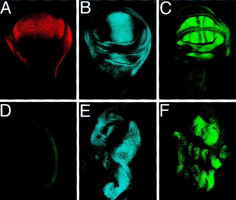
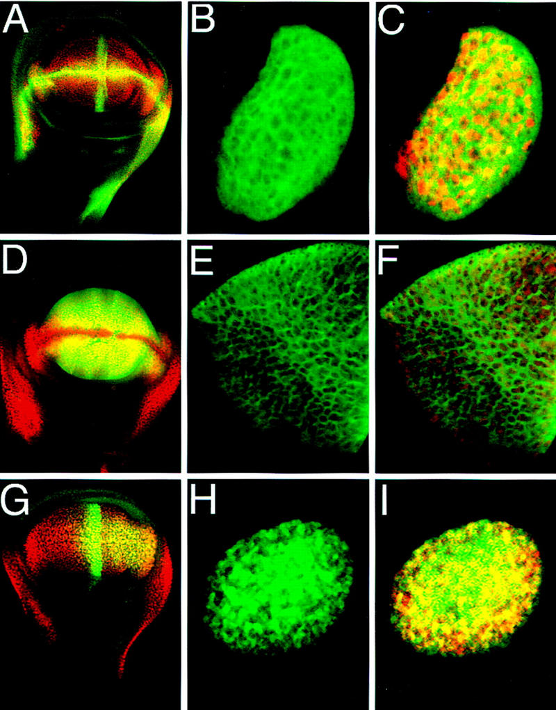
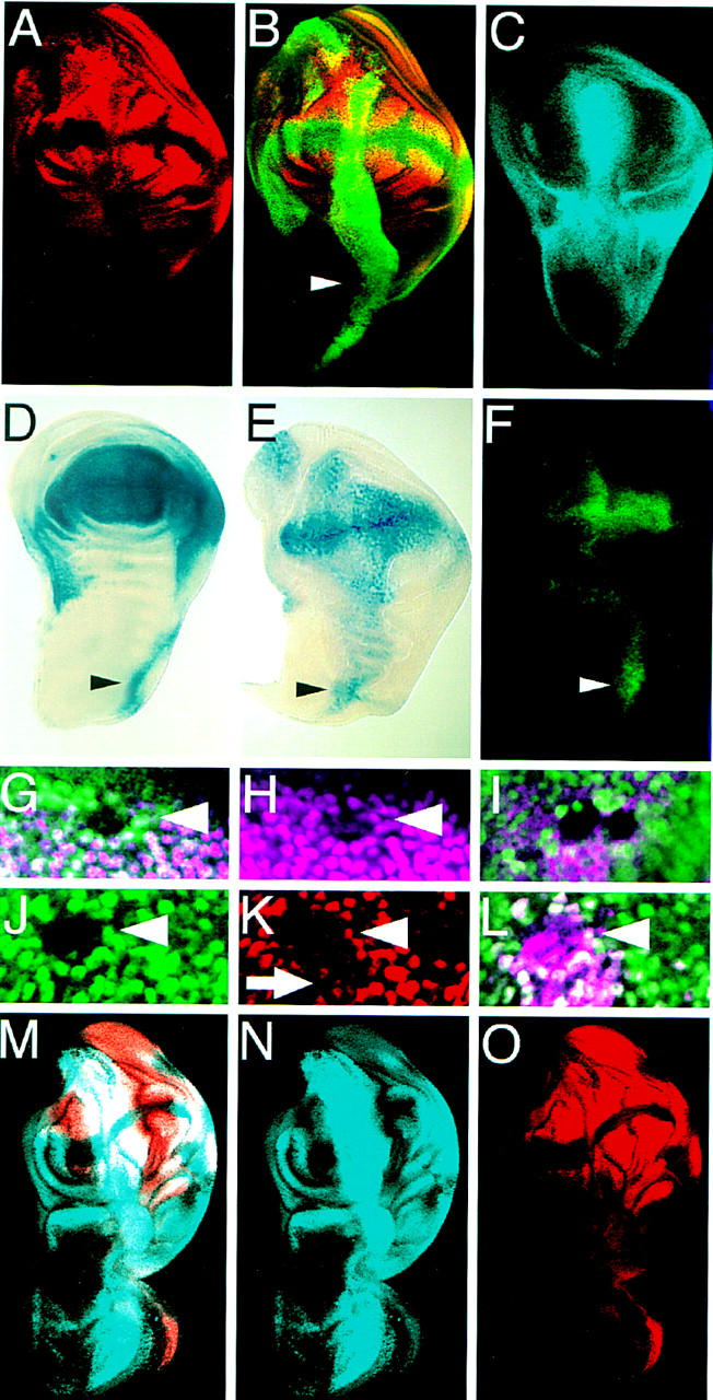
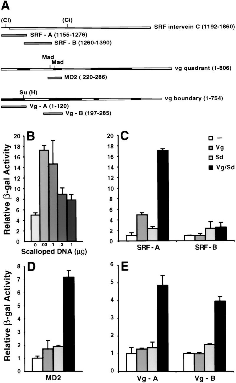
A small number of major regulatory (selector) genes have been identified in animals that control the development of particular organs or complex structures. In Drosophila, the vestigial gene is required for wing formation and is able to induce wing-like outgrowths on other structures. However, the molecular function of the nuclear Vestigial protein, which bears no informative similarities to other proteins, was unknown. Here, we show that Vestigial requires the function of the Scalloped protein, a member of the TEA family of transcriptional regulators, to directly activate the expression of genes involved in wing morphogenesis. Genetic and molecular analyses reveal that Vestigial regulates wing identity by forming a complex with the Scalloped protein that binds sequence specifically to essential sites in wing-specific enhancers. These enhancers also require the direct inputs of signaling pathways, and the response of an enhancer can be switched to another pathway through changes in signal-transducer binding sites. Combinatorial regulation by selector proteins and signal transducers is likely to be a general feature of the tissue-specific control of gene expression during organogenesis.


Figures

Figure 1
Figure 1
Targeted expression of Vg ectopically induces wing patterning genes. (AC), Endogenous expression patterns of Vg, Sal, and SRF in third instar wing discs. (A) Vg expression marks the entire developing wing field. (B) Sal is expressed in a band straddling the A/P boundary within the developing wing pouch. (C) SRF is expressed in future intervein tissue whose proximal borders are defined by Vg expression. Both Sal and SRF are also expressed in regions surrounding the wing pouch. (D) Sal (blue) and SRF (green) are not expressed in wild-type leg discs. (E) Sal and (F) SRF, are ectopically induced in leg discs in response to targeted expression of Vg by a Distal-less (Dll)–Gal4 driver. Lower levels of these genes are induced in the legs by use of the dpp–Gal4 driver (not shown). All discs are anterior to the left, in wing discs ventral is at the top, in leg discs dorsal is at the top.
Figure 2
Figure 2
Cell autonomous activation of wing-specific enhancers by Vg. (A) Wing disc double stained for β-gal (green) driven by the vg boundary enhancer and for Vg protein (red). (B) Close-up of a clone of cells in an eye disc that ectopically expressed Vg (red in C) and autonomously induced expression of the vg boundary enhancer reporter construct (green). (D) Wing disc double stained for β-gal (green) driven by the vg quadrant enhancer and Vg protein (red). (E,F) A clone of cells in a leg imaginal disc in which β-gal expression driven by the quadrant enhancer (E, green) is induced by ectopic Vg expression (red in F). (G) Wing disc double stained for β-gal expression (green) driven by the SRF intervein C element and Vg protein (red). (H,I) Clone of cells in the eye in which the intervein C element is activated (H, green) by ectopic Vg expression (red in I).
Figure 3
Figure 3
The activation of downstream genes, and autoregulation of vg requires Sd function in parallel to Vg. (AC) Wing discs in which Vg was expressed along the A/P boundary driven by dpp–Gal4. (A) SRF expression. (B) same disc as in A stained for both SRF (red) and Vg (green). Note that SRF expression does not extend into the notum portion (arrowhead). (C) Sal expression also does not extend into the notum. (D) sd expression pattern in a wing disc revealed by X-gal staining of larvae carrying a lacZ insertion into the sd gene. (E) Ectopic Vg expression driven by dpp–GAL4 induces sd expression at low levels in the notum. (F) Induction of the SRF intervein C element by ectopic Vg expression is highest where endogenous sd expression is highest in the notum (arrowheads in DF). (G,H) In sd47m mutant clones in the wing disc, Vg expression (purple) is lost in the clone. Mutant clones are illustrated by loss of Myc staining (green). (I) sd47m mutant clones in a wing disc ectopically expressing Vg along the A/P boundary. The clones are situated along the A/P boundary and are marked by loss of Myc staining (green). (JL) Expression of a lacZ reporter gene driven by the vg quadrant enhancer (purple) is lost in sd47m mutant clones, even in the presence of ectopic Vg. Homozygous sd47m mutant clones in a leg disc in which Vg is ectopically expressed. (J) The mutant clones are illustrated by the lack of Myc expression (green). SRF expression (red) is lost within the clones (K) even though Vg (purple) is still expressed (L). Just outside of the clone, ectopic Vg expression down-regulates SRF (arrow, this down-regulation is also apparent in A). (MO) Wing discs that ectopically expressed Vg as in AC with Sd ubiquitously expressed by a heat-inducible transgene. (M) Doble staining for Sal (blue) and SRF (red) (N,O) expression patterns. Coexpression of Vg with Sd induces gene expression in the notum portion of the disc.
Figure 4
Figure 4
Synergistic activation of cis-regulatory elements of wing-patterning genes by cotransfection of vg and sd. (A) Schematic of Vg-responsive enhancers and fragments analyzed in tissue culture and in vivo. The binding sites for transcriptional regulators of signaling pathways are indicated. Evolutionarily conserved regions of the vg enhancers are shaded. (B) The induction of the SRF-A element by Vg and Sd. Synergistic activation diminishes with increasing Sd concentration. (C) The SRF-A element is activated 17-fold by cotransfection of optimal amounts of Vg and Sd expression vectors, whereas either Vg or Sd alone induce five- and twofold, respectively. The SRF-B element is not activated significantly. (D) MD2, and (E) Vg-A and Vg-B are also activated synergistically by cotransfection of Sd and Vg. Error bars represent 1 s.d. of relative β-gal activity.
Figure 5
Figure 5
Sd affects the nuclear localization of Vg. Drosophila S2 cells transfected with SRF-A lacZ reporter gene (β-gal in red) and Vg (green) (AC) or the reporter, Vg, and Sd (DF). Cotransfection of Sd localizes Vg to the nucleus (E), and induces expression of the SRF-A lacZ reporter gene (D). All cells are counterstained with Topro to reveal DNA in nuclei (blue).
Figure 6
Figure 6
The Scalloped protein binds sequence specifically to the SRF-A element. (A) Sequence of the SRF-A element. Two sets of putative Sd tandem binding sites are boxed (A1, A2) and the orientation of the binding sites indicated by arrows. The extent of the region protected from DNase I digestion by the Sd–TEA domain is underlined. (B) Alignment of the Sd binding sites in the SRF enhancer with known TEF-1 binding sites. Shaded bases fit the TEF-1 consensus. (C) Gel mobility-shift experiments with the SRF-A and SRF-B fragments, the Sph fragment from the SV40 enhancer, and the wild-type and mutated SRF-A2 fragment by use of increasing amounts of purified 6-histidine-tagged Sd or TEF-1 TEA domains. Concentrations are 0.3, 1, 3, and 10 ng/25 μl, respectively. (F) Free probe; (B) complex with one TEA domain bound; (A) complex with two proteins bound. The Sd and TEF-1 TEA domains bind with high affinity to the SRF-A fragment but not to SRF-B. Both proteins bind with similar affinity to the Sph fragment (only the Sd shift is shown). Two molecules of Sd TEA domain bind to the SRF-A2 element but with much lower affinity (comparable to nonspecific binding) when the A2 sites are mutated. (D) DNase I protection assay on the SRF enhancer with increasing amounts of purified Sd TEA domain (1.7, 5, 15, 45 ng from left to right). The lane directly to the left of the G+A sequence ladder contained no Sd TEA protein. The A1 and A2 regions are shown schematically at right. Only the A2 region is protected by Sd protein.
Figure 7
Figure 7
Sd binding sites are required for target gene expression. (A) Activation of the SRF-A reporter by Vg and Sd was drastically reduced by mutating the footprinted A2 sequence to sequences no longer bound by Sd. Error bars represent 1 s.d. of relative β-gal activation. (B) The SRF-A fragment is sufficient to drive β-gal expression specifically in the developing wing pouch. (C) The activity of the SRF-A fragment is abolished when the Sd binding sites are mutated.
Figure 8
Figure 8
Vg and Sd control the wing-specific responses of target genes to signaling proteins. (A) Switching the specificity of a cis-response element. The vg boundary enhancer is activated through the N pathway by the Su(H) protein along the D/V boundary of the wing disc (left; Kim et al. 1996). Replacing the Su(H) site with two binding sites for the Ci protein changes the response of the enhancer from the N to the Hh pathway and the lacZ reporter gene is now expressed along the anterior side of the A/P boundary in the wing (middle), but not in the leg (right). (B) The selector and signal model for combinatorial control of gene expression in the wing field. Schematics of wing imaginal discs showing the expression patterns of the Dpp, Ser, and Hh signaling proteins (blue). Target gene responses (orange) are restricted to the circular wing field (outlined in black) by the Vg/Sd selector function, which acts together with the DNA-binding transducers of respective signaling pathways on cis-regulatory elements.

References

    1. Bailey A, Posakony JW. Suppressor of hairless directly activates transcription of enhancer of split complex genes in response to Notch receptor activity. Genes & Dev. 1995;9:2609–2622. - PubMed
    1. Brand A, Perrimon N. Targeted gene expression as a means of altering cell fates and generating dominant phenotypes. Development. 1993;118:401–415. - PubMed
    1. Brook W, Diaz-Benjumea F, Cohen S. Organizing spatial pattern in limb development. Annu Rev Cell Dev Biol. 1996;12:161–180. - PubMed
    1. Calleja M, Moreno E, Pelaz S, Morata G. Visualization of gene expression in living adult Drosophila. Science. 1996;274:252–254. - PubMed
    1. Campbell S, Duttaroy A, Katzen A, Chovnick A. Cloning and characterization of the scalloped region of Drosophila melanogaster. Genetics. 1991;127:367–380. - PMC - PubMed

Publication types

MeSH terms