. 1999 Jan 1;19(1):21-33.
doi: 10.1523/JNEUROSCI.19-01-00021.1999.

The mammalian brain high-affinity L-proline transporter is enriched preferentially in synaptic vesicles in a subpopulation of excitatory nerve terminals in rat forebrain

Affiliations

The mammalian brain high-affinity L-proline transporter is enriched preferentially in synaptic vesicles in a subpopulation of excitatory nerve terminals in rat forebrain

S E Renick et al. J Neurosci. .

Abstract

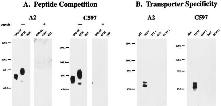
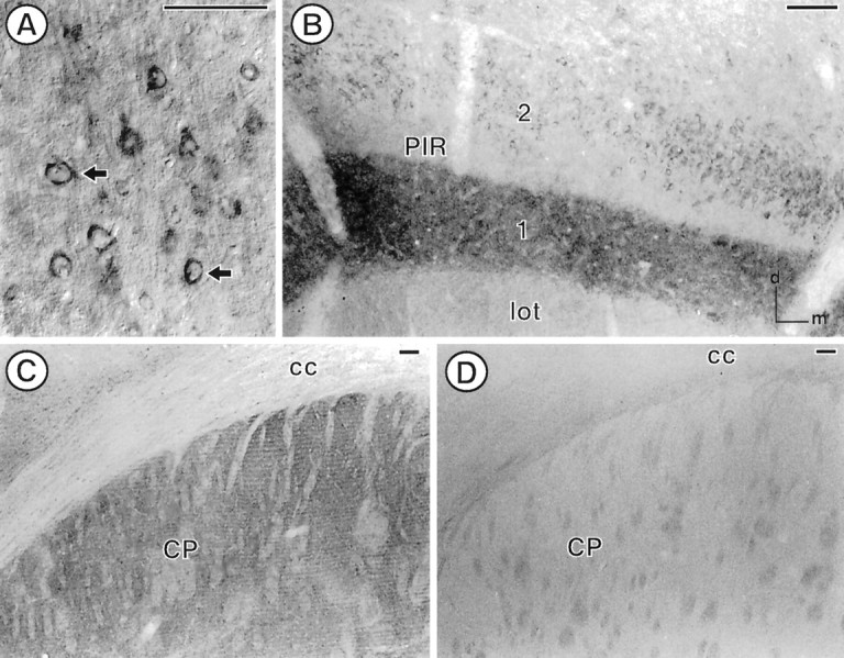
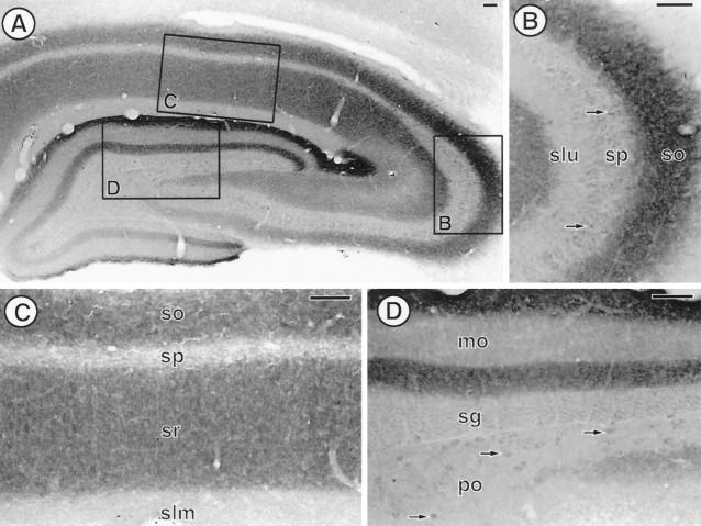
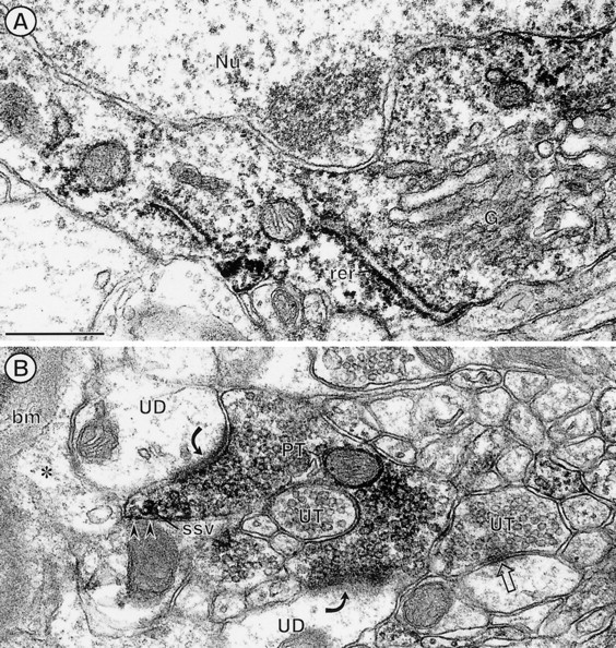
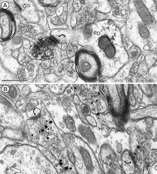
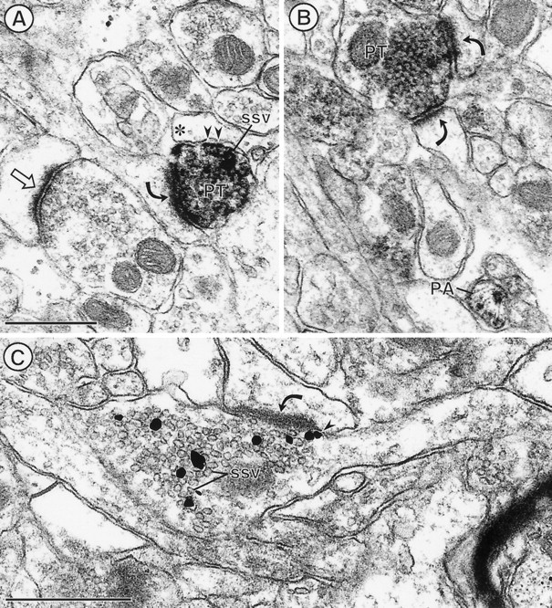
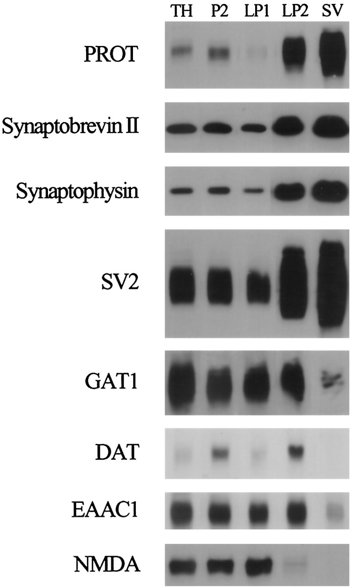
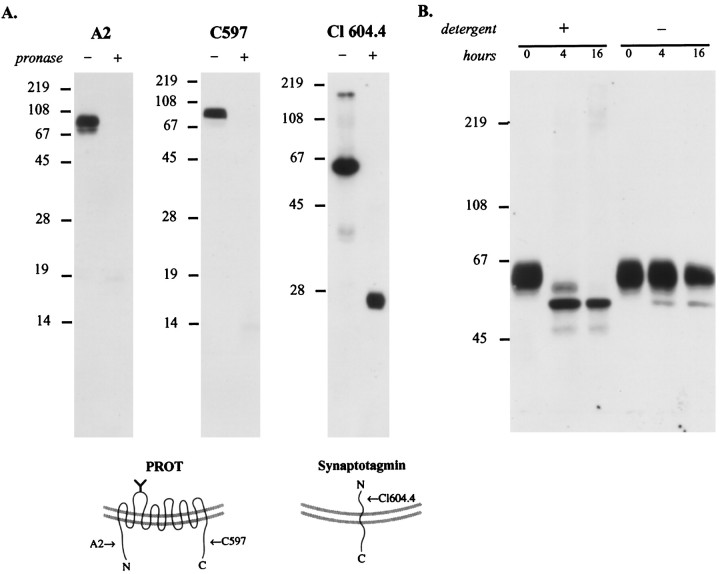
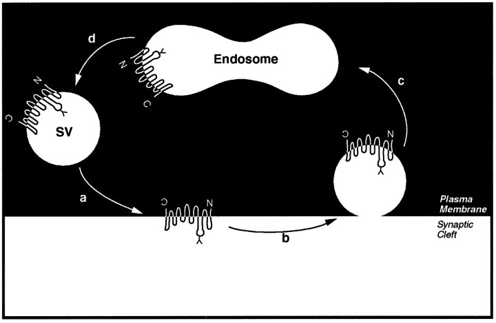
The expression of a brain-specific high-affinity Na+-dependent (and Cl--dependent) L-proline transporter (PROT) in subpopulations of putative glutamatergic neurons in mammalian brain suggests a physiological role for this carrier in excitatory neurotransmission (). To gain insights into potential sites where PROT may function, we used a C-terminal domain antipeptide antibody to determine the regional distribution and subcellular localization of PROT in rat forebrain. PROT immunoreactivity was seen in processes having a regional light microscopic distribution comparable to that of known glutamatergic projections within the cortex, caudate putamen nucleus (CPN), hippocampal formation, and other forebrain regions. In all regions examined by electron microscopy (cortex, CPN, and the stratum oriens of CA1), PROT labeling was observed primarily within subpopulations of axon terminals forming asymmetric excitatory-type synapses. Immunogold labeling for PROT was detected in close contact with membranes of small synaptic vesicles (SSVs) and more rarely with the plasma membrane in these axon terminals. Subcellular fractionation studies confirmed the preferential distribution of PROT to synaptic vesicles. The topology of PROT in synaptic vesicles was found to be inverted with respect to the plasma membrane, suggesting that PROT-containing vesicles are generated by a process involving endocytosis from the plasma membrane. Because PROT lacks any of the known characteristics of other vesicular transporters, these results suggest that certain excitatory terminals have a reserve pool of PROT associated with SSVs. The delivery of PROT to the plasma membrane by exocytosis could play a critical role in the plasticity of certain glutamatergic pathways.


Figures

Fig. 1.
Fig. 1.
Molecular specificity of the anti-PROT antibodies.A, Peptide competition. Membranes from controlled pore glass bead-purified synaptic vesicles (CPG-SV), whole-cell detergent lysates of HEK-293 cells stably expressing the rat PROT cDNA (HP-21), or untransfected HEK-293 cells (HEK) were subjected to SDS-PAGE (8%; 5 μg of protein per lane) and immunoblotted with affinity-purified antibodies directed against the N terminus (A2; 1:10,000 dilution) or C terminus (C597; 1:40,000 dilution) of rat PROT with (+) or without (−) preabsorption with a 100 nmconcentration of the peptide used for its affinity purification.B, Transporter specificity. HeLa cells were transfected with the pBluescript II SK vector or with the rPROT, rGAT1, hDAT, or hGlyT1b cDNAs. Whole-cell detergent lysates were prepared and subjected to SDS-PAGE (8%; 5 μg of protein per lane) and immunoblotted with the A2 (1:10,000) or C597 (1:40,000) antibodies. The mobilities of prestained protein molecular weight standards are shown on the left of each panel in kilodaltons.
Fig. 2.
Fig. 2.
PROT immunoreactivity is found in select regions in the forebrain of the adult rat. A, In the somatosensory cortex (corresponding to level 18 in Swanson), PROT immunoreactivity is found in the cytoplasm of perikarya (arrows) located in layer 5. B, A dense array of PROT-labeled processes is found in layer 1 of the piriform cortex (PIR; level 14 in Swanson). Several perikarya with PROT immunoreactivity are found in layer 2, whereas negligible PROT labeling is found in the lateral olfactory tract (lot). C, In the dorsal quadrant of the caudate putamen nucleus (CP), PROT-immunoreactive processes are found in the neuropil surrounding bundles of white matter. D, No PROT immunoreactivity is observed in the dorsal caudate putamen nucleus after preadsorption of the antibody with a 100 nm concentration of the cognate peptide.cc, Corpus callosum. Scale bar, 100 μm.
Fig. 3.
Fig. 3.
PROT immunoreactivity has a laminar distribution in the adult rat hippocampal formation. A, Low-magnification photomicrograph showing the distribution of PROT immunoreactivity in a coronal section of a mid-rostrocaudal level of the hippocampal formation (level 32 in Swanson). Boxed regions are enlarged in B–D. B, In the CA3 region of the hippocampus a dense array of PROT-immunoreactive processes is found in stratum oriens (so). A few PROT-labeled perikarya (arrows) are observed in stratum pyramidale (sp), whereas negligible PROT immunoreactivity is found in stratum lucidum (slu). C, In the CA1 region of the hippocampus, PROT-immunoreactive processes are densest in strata oriens (so) and radiatum (sr).D, In the dentate gyrus a dense plexus of PROT-immunoreactive processes is found in the inner and outer third of the molecular layer (mo). A few PROT-labeled perikarya (arrows) are detected in the polymorph layer (po). sg, Stratum granulosum;slm, stratum lacunosum moleculare. Scale bar, 100 μm.
Fig. 4.
Fig. 4.
Electron micrographs showing immunoperoxidase labeling for PROT in the cytoplasm of a neuronal perikaryon (A) and axon terminal (B) within the deep layers of the somatosensory cortex. A, The peroxidase labeling is distributed diffusely throughout the cytoplasm surrounding an unlabeled nucleus (Nu). The peroxidase product is associated most intensively with saccules of rough endoplasmic reticulum (rer). The trans-Golgi lamellae (G) show comparatively little immunoreactivity. B, PROT immunoperoxidase labeling is seen in a nonuniform distribution throughout an axon terminal (PT). Many clusters of small synaptic vesicles (ssv) near portions of the plasma membrane (arrowheads) that face an astrocytic process (*) also show peroxidase immunoreactivity. This astrocytic process is continuous with the glial profile contacting the basement membrane (bm) of a small blood vessel. The labeled terminal forms asymmetric synapses (curved arrows) with two separate unlabeled dendrites (UD) and also is apposed to another unlabeled axon terminal (UT). Scale bar, 0.5 μm.
Fig. 5.
Fig. 5.
Electron micrographs showing immunoperoxidase (A) and immunogold silver (B) labeling for PROT in axon terminals (PT) in the dorsal striatum. A, Immunoperoxidase reaction product (black precipitate) is distributed intensely along membranes of small synaptic vesicles (ssv) in an axon terminal forming an asymmetric axospinous synapse (curved arrow). Sparse labeling also is seen within the dendrite but is absent from other morphologically similar terminals. B, Immunogold particles (straight arrows) mainly contact membranes of SSVs. Several particles are present along the plasma membrane (arrowheads). UT, Unlabeled axon terminal. PD, Postsynaptic dendrite. Scale bar, 0.5 μm.
Fig. 6.
Fig. 6.
PROT is localized prominently to synaptic vesicles in axon terminals that form asymmetric excitatory-type synapses in the CA1 region of the hippocampal formation (curved arrows). In A and B, peroxidase labeling is distributed intensely throughout the cytoplasm of specific axon terminals (PT) that form asymmetric synapses. Certain SSVs, particularly those near the plasma membrane (small arrows) apposed to a glial process, appear to be rimmed with peroxidase labeling. A, An unlabeled terminal forms a similar asymmetric synapse (straight arrow).B, A labeled terminal forms dual contacts (curved arrows) on two spines. Additionally, a small unmyelinated axon is labeled intensely for PROT (PA). Immunogold silver particles in C are seen in direct contact with membranes of many SSVs in an axon terminal, forming an asymmetric synapse. One gold particle is also in contact with the plasma membrane (arrowhead) adjacent to the synaptic specialization. Scale bar, 0.5 μm.
Fig. 7.
Fig. 7.
PROT is enriched substantially in highly purified synaptic vesicles. We isolated synaptic vesicles from nerve terminals by subcellular fractionation and monitored the distribution of PROT in the various fractions in comparison to several synaptic vesicle and plasma membrane markers. Briefly, crude synaptosomes (P2) were prepared from a rat forebrain homogenate (TH) by differential centrifugation and lysed to release synaptic vesicles and other internal membrane compartments. Then most large membranes, including synaptic plasma membranes, were removed by centrifugation at 25,000 × g for 20 min (LP1). Synaptic vesicles were collected from the synaptosomal lysate supernatant by centrifugation at 165,000 ×g for 2 hr (LP2) and purified further by rate-zonal sucrose density gradient centrifugation and size exclusion chromatography on a controlled pore glass bead column (SV). Samples from each subcellular fraction were subjected to SDS-PAGE (5 μg of protein per lane) and immunoblotted with antibodies to the indicated proteins. Note that PROT is enriched substantially in the SV fraction like the synaptic vesicle proteins synaptobrevin II, synaptophysin, and SV2. In contrast, the plasma membrane neurotransmitter transporters and receptors that have been examined are deenriched in the highly purified SV fraction.
Fig. 8.
Fig. 8.
Mapping the topology of PROT in the synaptic vesicle membrane. A, PROT N and C termini are oriented cytoplasmically. The location of PROT termini was mapped by subjecting a synaptic vesicle-enriched fraction (LP2) to limited proteolysis. After a 20 min incubation with or without Pronase the samples were separated by SDS-PAGE (15 or 12%) and subjected to immunoblot analysis by using domain-specific antibodies. PROT immunoreactivity is present only in the Pronase-negative reaction when probed with antibodies specific for the N terminus (A2; 1:10,000 dilution) or C terminus (C597; 1:40,000 dilution), indicating that these structures are present on the external (cytoplasmic) face of the vesicle membrane. In contrast, a monoclonal antibody against the intraluminal N-terminal domain of the integral synaptic vesicle protein synaptotagmin (Cl 604.4; 1:1000 dilution) recognized the protected epitope in the partially digested protein. This indicates that during the protease reaction the synaptic vesicles were intact, and loss of PROT immunoreactivity was not attributable to vesicle rupture. The arrows in the models indicate the positions of the epitopes of the antibodies used for immunoblotting.B, The N-glycosylated loop is intraluminal. A synaptic vesicle-enriched fraction, LP2, was subjected to deglycosylation by PNGase F in the presence or absence of detergents (2.5% NP-40/1% SDS), separated by SDS-PAGE (8%), and immunoblotted with anti-PROT antibody C597 (1:40,000 dilution). In the presence of detergent the native PROT protein is reduced progressively to a single band of ∼53 kDa. However, in the absence of detergent a significant loss of glycosylation fails to occur, indicating that the N-linked glycosylation site is located within the vesicle lumen.
Fig. 9.
Fig. 9.
Model depicting the exo-/endocytotic recycling of PROT-containing synaptic vesicles. a, According to this model the exocytosis of PROT-containing vesicles in response to a signaling event transiently stimulates high-affinityl-proline uptake into specific excitatory nerve terminals.b, Then PROT is retrieved from the nerve terminal membrane by a process that may involve clathrin-dependent endocytosis.c, Coated vesicles containing PROT are translocated to early endosomes. d, PROT-containing synaptic vesicles are regenerated by budding from endosomes.

References

    1. Alfonso A, Grundahl K, Duerr JS, Han HP, Rand JB. The Caenorhabditis elegans unc-17 gene: a putative vesicular acetylcholine transporter. Science. 1993;261:617–619. - PubMed
    1. Amara SG, Kuhar MJ. Neurotransmitter transporters: recent progress. Annu Rev Neurosci. 1993;16:73–93. - PubMed
    1. Balcar VJ, Johnston GAR, Stephenson AL. Transport of l-proline by rat brain slices. Brain Res. 1976;102:143–151. - PubMed
    1. Bennett JP, Logan WJ, Snyder SH. Amino acid neurotransmitter candidates: sodium-dependent high-affinity uptake by unique synaptosomal fractions. Science. 1972;178:997–999. - PubMed
    1. Birnbaum MJ. Identification of a novel gene encoding an insulin-responsive glucose transporter protein. Cell. 1989;57:305–315. - PubMed

Publication types

MeSH terms

LinkOut - more resources