. 1999 Jan 1;19(1):64-71.
doi: 10.1523/JNEUROSCI.19-01-00064.1999.

Mechanisms and structural determinants of HIV-1 coat protein, gp41-induced neurotoxicity

Affiliations

Mechanisms and structural determinants of HIV-1 coat protein, gp41-induced neurotoxicity

D C Adamson et al. J Neurosci. .

Abstract

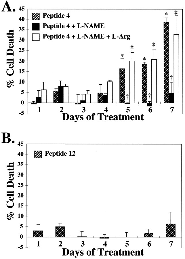
Of the individuals with human immunodeficiency virus type 1 (HIV-1) infection, 20-30% will develop the neurological complication of HIV-associated dementia (HAD). The mechanisms underlying HAD are unknown; however, indirect immunologically mediated mechanisms are theorized to play a role. Recently, the HIV-1 coat protein gp41 has been implicated as a major mediator of HAD through induction of neurocytokines and subsequent neuronal cell death. Using primary mixed cortical cultures from neuronal nitric oxide synthase (NOS) null (nNOS-/-) mice and immunological NOS null (iNOS-/-) mice, we establish iNOS-derived NO as a major mediator of gp41 neurotoxicity. Neurotoxicity elicited by gp41 is markedly attenuated in iNOS-/- cultures compared with wild-type and nNOS-/- cultures. The NOS inhibitor L-nitroarginine methyl ester is neuroprotective in wild-type and nNOS-/- cultures, confirming the role of iNOS-derived NO in gp41 neurotoxicity. Confirming that iNOS-/- cultures lack iNOS, gp41 did not induce iNOS in iNOS-/- cultures, but it markedly induced iNOS in wild-type and nNOS-/- cultures. We elucidate the region of gp41 that is critical for iNOS induction and neuronal cell death by monitoring iNOS induction with overlapping peptides spanning gp41. We show that the N-terminal region of gp41, which we designate as the neurotoxic domain, induces iNOS protein activity and iNOS-dependent neurotoxicity at picomolar concentrations in a manner similar to recombinant gp41 protein. Our experiments suggest that gp41 is eliciting the induction of iNOS through potential cell surface receptors or binding sites because the induction of iNOS is dose dependent and saturable and occurs at physiologically relevant concentrations. These data confirm that the induction of iNOS by gp41 and the production of NO are primary mediators of neuronal damage and identify a neurotoxic domain of gp41 that may play an important role in HAD.


Figures

Fig. 1.
Fig. 1.
Neuronal cortical cultures from iNOS−/− mice are resistant to gp41 neurotoxicity.A, Wild-type mice cultures were exposed to 100 nm recombinant gp41IIIB in the presence or absence of 500 μm L-NAME, a competitive NOS inhibitor, ± 5 mm L-Arg. Cell death was assayed by microscopic examination with computer-assisted cell counting after staining of all nuclei with 1 μg/ml Hoescht 33342 and staining of dead cell nuclei with 7 μm propidium iodide (Gonzalez-Zulueta et al., 1998). Percentage of cell death was assessed on days 3, 5, and 7 and was normalized to percentage of cell death in untreated control cultures. The presence of iNOS protein in gp41-treated (41) and untreated control cultures was assessed on the same days via immunoblot analysis. B, Neuronal NOS−/−cultures were similarly treated and assessed for percentage of cell death and the presence of iNOS protein on days 3, 5, and 7.C, iNOS−/− cultures were similarly treated and assessed for percentage of cell death and the presence of iNOS protein on days 3, 5, and 7. For all conditions, at least two separate experiments with four separate wells were performed, with a minimum of 15,000–20,000 neurons counted per data point. Data are means ± SEM for n ≥ 8 from at least two experiments. For each set of culture data, significance was determined by a 3 × 3 ANOVA repeated measures, with differences between groups ascertained by Fisher’s PLSD ANOVA post hoctest. Wild-type (F = 43.05) and nNOS−/− (F = 12.03) culture data: *p ≤ 0.0001 for gp41 (Day 3) versus gp41 (Day 5), gp41 (Day 3) versus gp41 (Day 7); †p ≤ 0.0001 for gp41 (Day 5) versus gp41 + L-NAME (Day 5), gp41 (Day 7) versus gp41 + L-NAME (Day 7); ‡p ≤ 0.0001 for gp41 + L-NAME (Day 5) versus gp41 + L-NAME + LArg (Day 5), gp41 + L-NAME (Day 7) versus gp41 + L-NAME + LArg (Day 7). iNOS−/− culture data: F = 0.34 (p = 0.85).
Fig. 2.
Fig. 2.
Epitope mapping of gp41 induction of iNOS reveals a number of regions in the extracellular domain of gp41 capable of inducing iNOS. A, A number of overlapping peptides (1–16) spanning the region of gp41 from the gp120/gp41 junction (slash seen to the left of peptide 1) to the transmembrane domain (TMD) of gp41 were obtained.B, Rodent mixed cortical cultures were exposed to 100 nm each peptide, harvested on day 7, and assessed for iNOS protein via immunoblot analysis. Positive controls (PC) were obtained from rodent glial cultures stimulated with 100 ng/ml lipopolysaccharide.
Fig. 3.
Fig. 3.
Epitope mapping of gp41 induction of iNOS reveals a neurotoxic domain. Based on the dose–response relationships of induction of iNOS protein, we have designated amino acids 530–559, which are contained within peptides 2 (amino acids 534–524), 3 (amino acids 543–533), and 4 (amino acids 559–539), as the neurotoxic domain (•). This domain lies adjacent to the putative fusion peptide domain of gp41.
Fig. 4.
Fig. 4.
Active gp41 peptide 4 induces iNOS protein and activity. A, Rodent mixed cortical cultures were treated with 0.1, 1, 10, and 100 nm peptide 4 or peptide 12 (B) and assessed for NOS activity and protein on day 7 of exposure. C, Similar cultures were treated with 100 nm peptide 4 or peptide 12 (D) and assessed for NOS activity and protein each day for a 7 d period. Untreated control cultures (C) were also examined. Catalytic activity (cpm) assessments are means ± SEM for n ≥ 3 experiments. For each set of data, significance was determined by a one-way ANOVA with differences between groups ascertained by Fisher’s PLSD ANOVApost hoc test. A, F = 9.00, *p ≤ 0.02. B,F = 0.18, p = 0.95.C, F = 3.96, *p≤ 0.02. D, F = 1.92,p = 0.13.
Fig. 5.
Fig. 5.
Alternatively synthesized gp41 peptide 4 induces iNOS protein. Rodent mixed cortical cultures were treated with 100 nm peptide 4 (F, forward sequence) from a different peptide synthesis facility or a reverse peptide 4 (B, backward sequence), harvested on day 7 of exposure and assessed for iNOS protein via immunoblot analysis. Untreated control cultures (C) were also assessed. Positive controls (PC) were obtained from rodent glial cultures stimulated with 100 ng/ml lipopolysaccharide.
Fig. 6.
Fig. 6.
NOS inhibitors block neuronal cell death elicited by a gp41 neurotoxic domain peptide. A, Rodent mixed cortical cultures were treated with 100 nm peptide 4 in the presence or absence of 500 μm L-NAME, a competitive NOS inhibitor, ± 5 mm L-Arg. Cell death was assayed by microscopic examination with computer-assisted cell counting after staining of all nuclei with 1 μg/ml Hoescht 33342 and staining of dead cell nuclei with 7 μm propidium iodide (Gonzalez-Zulueta et al., 1998). Percentage of cell death was assessed on each day for a 7 d period and normalized to untreated control cultures. B, Similar cultures were treated with 100 nm peptide 12 and similarly assayed for cell death over a 7 d period. For all conditions, at least three separate experiments with four separate wells were performed, with a minimum of 15,000–20,000 neurons counted per data point. Data are means ± SEM for n ≥ 3 experiments. For each set of data, significance was determined by ANOVA repeated measures, with differences between groups ascertained by Fisher’s PLSD ANOVApost hoc test. A, F = 3.63, *p ≤ 0.01 for Peptide 4 (day1) versus Peptide 4 (days 5, 6, 7); †p ≤ 0.01 for Peptide 4 (day5) versus Peptide 4 + L-NAME (day 5), Peptide 4 (day 6) versus Peptide 4 + L-NAME (day6), Peptide 4 (day 7) versus Peptide 4 + L-NAME (day 7); ‡p ≤ 0.01 for Peptide 4 + L-NAME (day5) versus Peptide 4 + L-NAME + L-Arg (day5), Peptide 4 + L-NAME (day 6) versus Peptide 4 + L-NAME + L-Arg (day 6), Peptide 4 + L-NAME (day 7) versus Peptide 4 + L-NAME + L-Arg (day 7).
Fig. 7.
Fig. 7.
Schematic diagram of the relationship of HIV-1 coat proteins to extracellular receptors. The close proximity of the putative neurotoxic domain of gp41 to the fusion peptide places it in an ideal position for cell/cell interaction.

References

    1. Adamson DC, Dawson TM, Zink MC, Clements JE, Dawson VL. Neurovirulent simian immunodeficiency virus infection induces neuronal, endothelial and glial apoptosis. Mol Med. 1996a;2:417–428. - PMC - PubMed
    1. Adamson DC, Wildemann B, Sasaki M, Glass JD, McArthur JC, Christov VI, Dawson TM, Dawson VL. Immunologic NO synthase: elevation in severe AIDS dementia and induction by HIV-1 coat protein, gp41. Science. 1996b;274:1917–1921. - PubMed
    1. Adamson DC, McArthur JC, Dawson TM, Dawson VL (1999) Course of HIV-associated dementia: correlations with gp41, iNOS and macrophage/microglial activation. Mol Med, in press. - PMC - PubMed
    1. Alkhatib G, Combadiere C, Broder CC, Feng Y, Kennedy PE, Murphy PM, Berger EA. CC CKR5: a RANTES, MIP-1alpha, MIP-1beta receptor as a fusion cofactor for macrophage-tropic HIV-1. Science. 1996;272:1955–1958. - PubMed
    1. Barcova M, Kacani L, Speth C, Dierich MP. gp41 envelope protein of human immunodeficiency virus induces interleukin (IL)-10 in monocytes, but not in B, T, or NK cells, leading to reduced IL-2 and interferon-gamma production. J Infect Dis. 1998;177:905–913. - PubMed

Publication types

LinkOut - more resources