. 1999 Jan 1;19(1):72-84.
doi: 10.1523/JNEUROSCI.19-01-00072.1999.

The cat-1 gene of Caenorhabditis elegans encodes a vesicular monoamine transporter required for specific monoamine-dependent behaviors

Affiliations

The cat-1 gene of Caenorhabditis elegans encodes a vesicular monoamine transporter required for specific monoamine-dependent behaviors

J S Duerr et al. J Neurosci. .

Abstract

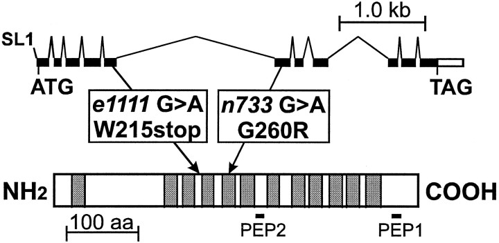
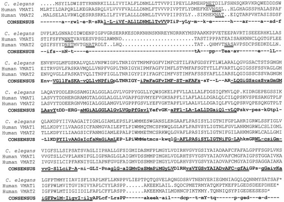
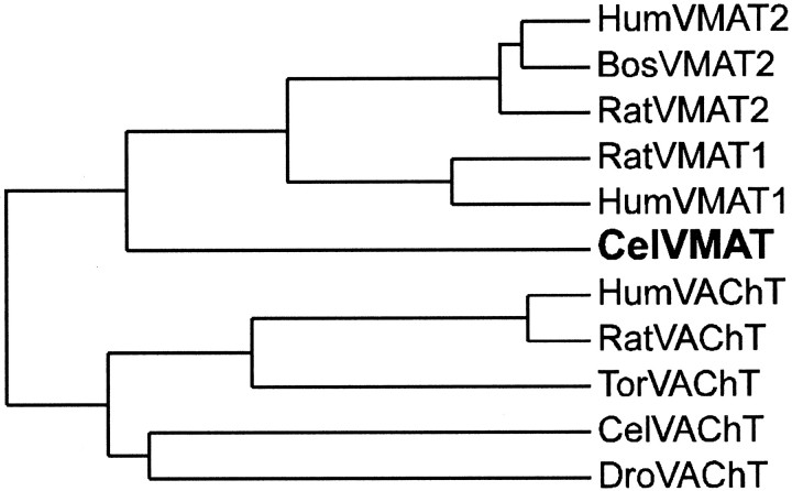
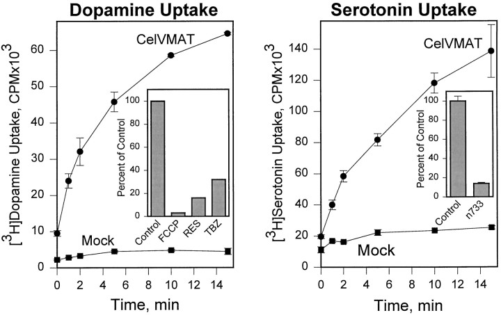
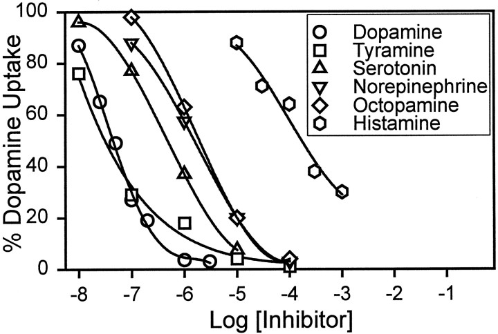
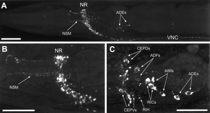
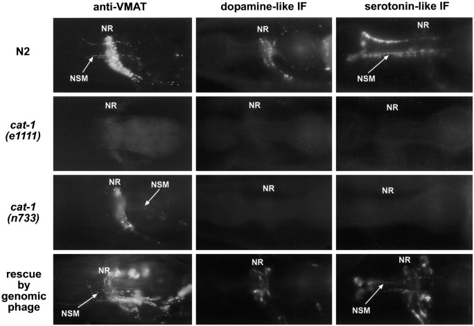
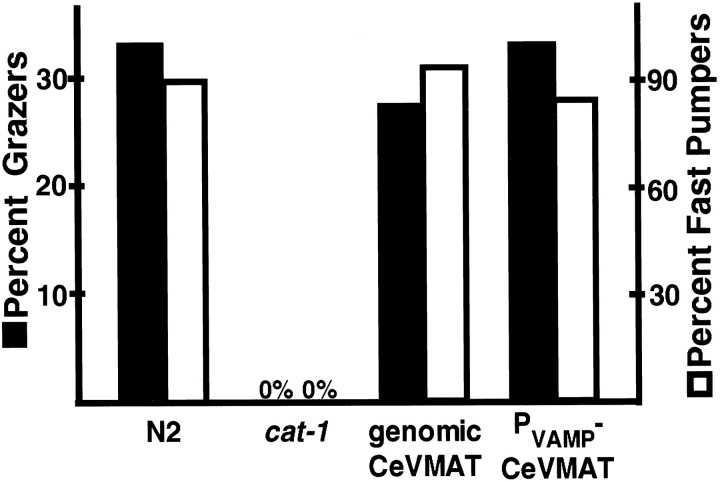
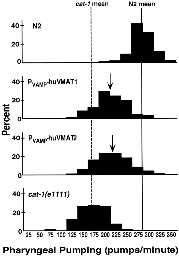
We have identified the Caenorhabditis elegans homolog of the mammalian vesicular monoamine transporters (VMATs); it is 47% identical to human VMAT1 and 49% identical to human VMAT2. C. elegans VMAT is associated with synaptic vesicles in approximately 25 neurons, including all of the cells reported to contain dopamine and serotonin, plus a few others. When C. elegans VMAT is expressed in mammalian cells, it has serotonin and dopamine transport activity; norepinephrine, tyramine, octopamine, and histamine also have high affinity for the transporter. The pharmacological profile of C. elegans VMAT is closer to mammalian VMAT2 than VMAT1. The C. elegans VMAT gene is cat-1; cat-1 knock-outs are totally deficient for VMAT immunostaining and for dopamine-mediated sensory behaviors, yet they are viable and grow relatively well. The cat-1 mutant phenotypes can be rescued by C. elegans VMAT constructs and also (at least partially) by human VMAT1 or VMAT2 transgenes. It therefore appears that the function of amine neurotransmitters can be completely dependent on their loading into synaptic vesicles.


Figures

Fig. 1.
Fig. 1.
Structure of the C. elegans VMAT gene and protein. At the top are shown the splicing pattern and exon structure of the VMAT (cat-1) genomic region. Protein-coding regions are filled; the 5′- and 3′-untranslated regions are white. The positions of the initiation (ATG) and termination (TAG) codons are indicated, as well as the addition site of the trans-spliced SL1 leader RNA (Krause and Hirsh, 1987; Bektesh et al., 1988). The VMAT protein sequence is represented by the long rectangle at thebottom. The sites of the e1111 andn733 mutations are indicated, along with the associated sequence alterations. The 12 putative transmembrane domains of the protein are indicated by shading, and the locations of the two peptides (PEP1 and PEP2) used to raise anti-VMAT antiserum are shown below the rectangle.
Fig. 2.
Fig. 2.
Deduced protein sequence of C. elegans VMAT and alignment with the human VMAT1 (GenBank accession number U39905) and VMAT2 (GenBank accession number L23205) deduced proteins. The protein sequences were aligned using the PILEUP program (Genetics Computer Group Wisconsin package, version 8). TheCONSENSUS sequence uses uppercase letterswhen all three sequences agree and lowercase letterswhen two of the three sequences are the same. Dots are used in the three VMAT sequences to indicate gaps introduced by the alignment program; hyphens are used in the consensus sequence for sites where all three sequences differ. The 12 putative transmembrane domains, as suggested for the mammalian proteins (Erickson et al., 1992; Liu et al., 1992) are shown as double underlines below the consensus sequence. Putative glycosylation sites (in the large intravesicular loop between transmembrane domains 1 and 2) are shown as double underlines below each of the VMAT sequences.
Fig. 3.
Fig. 3.
Dendrogram of published VMAT and VAChT sequences. The PILEUP program (Genetics Computer Group Wisconsin package, version 8) was used. Bos, Bovine; Cel, C. elegans; Dro, Drosophila;Hum, human; Tor, Torpedo. The individual GenBank Accession numbers are HumVMAT2, L23205 (Erickson and Eiden, 1993); BosVMAT2, U02876 (Howell et al., 1994); RatVMAT2,L00603 (Erickson et al., 1992); RatVMAT1, M97380 (Liu et al., 1992); HumVMAT1, U39905 (Erickson et al., 1996); CelVMAT, present study; HumVAChT, U10554 (Erickson et al., 1994); RatVAChT, U09211 (Erickson et al., 1994); TorVAChT, U05591 (Varoqui et al., 1994); CelVAChT, L19621(Alfonso et al., 1993); and DroVAChT, AF030197 (Kitamoto et al., 1998).
Fig. 4.
Fig. 4.
Transport of [3H]dopamine and [3H]serotonin mediated by C. elegans VMAT. A C. elegans VMAT cDNA or pBluescript with no insert (Mock) was used in a permeablized cell uptake assay as previously described (Erickson and Eiden, 1993). Data points represent the means of duplicate determinations; error bars represent the range of the duplicates.A, time course of [3H]dopamine uptake. Inset, Relative inhibition of specific [3H]dopamine uptake (i.e., with the mock values subtracted) at 4 min by 5 μm FCCP, 100 nm reserpine (RES), or 1 μmtetrabenazine (TBZ). B, Time course of [3H]serotonin uptake. Inset, Decreased [3H]serotonin uptake by the n733 mutant VMAT (means of four measurements ± SEM, measured at 15 min). In this experiment, the wild-type CelVMAT control corresponded to 0.59 ± .03 pmol of serotonin/15 min per well. The mutant uptake is significantly different from the control with p< 0.0001. Uptake values for CelVMAT and CelVMAT/n733 were not corrected for transfection efficiency in these experiments, because there were no consistent differences in efficiency of transfection of CV-1 cells with CelVMAT compared with CelVMAT/n733 as judged by immunohistochemical staining of VMAT-expressing CV-1 cells.
Fig. 5.
Fig. 5.
Inhibition of [3H]dopamine uptake by biogenic amines. Digitonin-permeabilized CV-1 cells expressing CelVMAT cDNA were used to measure transport of [3H]dopamine in the absence or presence of the indicated concentration of unlabeled inhibitor. Assays were for 4 min, and data points represent the means of one to five assays of duplicate samples.
Fig. 6.
Fig. 6.
Immunolocalization of C. elegansVMAT. Hermaphrodites were stained with indirect immunofluorescence using affinity-purified anti-VMAT antibodies and were imaged with a Leica TCS NT confocal microscope. Images are maximum projections ofz-series. Anterior is to the left; ventral is down. A, Lateral view of the anterior third of a wild-type adult; B, lateral view of a wild-type head; C, lateral view ofunc-104(e1265), in which synaptic vesicles are mislocalized to cell somas; individual identified neuronal somas are indicated by arrows. NR, Nerve ring;VNC, ventral nerve cord. The processes of the NSM cells are indicated. Cell bodies of other identified immunopositive neurons include the ADE, ADF, AIM, CEPD, CEPV, and RIC bilateral homologs and the unpaired RIH cell. Scale bar, 20 μm.
Fig. 7.
Fig. 7.
Cellular phenotypes of cat-1mutants. Young adult hermaphrodites were immunostained for VMAT (left column) or were prepared for induced fluorescence (see Materials and Methods) and visualized for dopamine-like fluorescence (middle column) and serotonin-like fluorescence (right column). Lateral views: anterior is to the left, and ventral is down.Top row, Wild type (N2). Second row, cat-1(e1111); the animals are completely deficient for VMAT immunoreactivity, and the induced fluorescence for both transmitters is completely absent from neuronal processes and significantly (often totally) absent from cell somas. Third row, cat-1(n733); the dopamine-specific and serotonin-specific induced fluorescence are comparable to those ine1111, but the VMAT immunostaining is almost wild type.Bottom row, Transgenic animals containing the genomic phage RM#424L in a cat-1(e1111) host. Note the restoration and overexpression of all three cellular markers.NR, Nerve ring; NSM, processes of the pharyngeal NSM cells.
Fig. 8.
Fig. 8.
Pharyngeal pumping and grazing behavior in response to a bacterial lawn. Grazers (dark bars) are individuals taking >1 min to enter a bacterial lawn, as described in Materials and Methods. White bars represent the percent of individuals of each phenotype that pumped at a rate of at least 250/min in the presence of bacteria. Values represent the percent for 100 individual animals for each behavior. The transgenic lines were significantly different from cat-1 for both behaviors with p < .01.
Fig. 9.
Fig. 9.
Synaptic localization and function of transgenic human VMAT2. cat-1(e1111) mutants were transformed with human VMAT2 cDNA driven by the neuron-specific VAMP (synaptobrevin) promoter (see Materials and Methods). A, Immunostaining with anti-human VMAT2 reveals that the protein is expressed in neurons and is properly localized to synaptic regions, including the nerve ring and the dorsal nerve cord (arrows). B, Induced fluorescence specific for dopamine reveals that the human transgene restores proper wild-type staining pattern in the correct neurons. C, Serotonin-like immunofluorescence is also restored, including that of the NSM cells of the pharynx. Comparable photographs of untransformed (control) cat-1(e1111)mutants are shown in Figure 7, second row. Lateral views: anterior is to the left, and ventral isdown.
Fig. 10.
Fig. 10.
Human VMAT transgenes partially rescuecat-1 deficits in pharyngeal pumping. Pumps per minute in a bacterial lawn were measured in 100 individual animals of each genotype. Data are plotted in bins of 25. The mean for N2 is 275 (indicated with a solid line); the cat-1mean is 160 (dashed line). Arrowsindicate the means for Pvamp-huVMAT1 (202) and Pvamp-huVMAT2 (208). Transgenic lines were significantly different fromcat-1(e1111) with p < 0.01 (Mann–Whitney–Wilcoxson test).

References

    1. Albertson DG. Formation of the first cleavage spindle in nematode embryos. Dev Biol. 1984;101:61–72. - PubMed
    1. Albertson DG, Thomson JN. The pharynx of Caenorhabditis elegans. Philos Trans R Soc Lond B Biol Sci. 1976;275:299–325. - PubMed
    1. Alfonso A, Grundahl K, Duerr JS, Han H-P, Rand JB. The Caenorhabditis elegans unc-17 gene: a putative vesicular acetylcholine transporter. Science. 1993;261:617–619. - PubMed
    1. Avery L, Horvitz HR. Effects of starvation and neuroactive drugs on feeding in Caenorhabditis elegans. J Exp Zool. 1990;253:263–270. - PubMed
    1. Bargmann CI, Horvitz HR. Chemosensory neurons with overlapping functions direct chemotaxis to multiple chemicals in C. elegans. Neuron. 1991a;7:729–742. - PubMed

Publication types

MeSH terms

LinkOut - more resources